
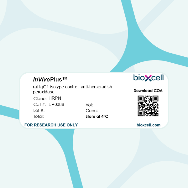
InVivoPlus rat IgG1 isotype control, anti-horseradish peroxidase
Product Description
Specifications
| Isotype | Rat IgG1, κ |
|---|---|
| Recommended Dilution Buffer | InVivoPure pH 7.0 Dilution Buffer |
| Conjugation | This product is unconjugated. Conjugation is available via our Antibody Conjugation Services. |
| Formulation |
PBS, pH 7.0 Contains no stabilizers or preservatives |
| Endotoxin* |
≤0.5EU/mg (≤0.0005EU/μg) Determined by LAL assay |
| Aggregation* |
<5% Determined by SEC |
| Purity |
≥95% Determined by SDS-PAGE |
| Sterility | 0.2 µm filtration |
| Production | Purified from cell culture supernatant in an animal-free facility |
| Purification | Protein G |
| RRID | AB_1107775 |
| Molecular Weight | 150 kDa |
| Murine Pathogen Tests* |
Ectromelia/Mousepox Virus: Negative Hantavirus: Negative K Virus: Negative Lactate Dehydrogenase-Elevating Virus: Negative Lymphocytic Choriomeningitis virus: Negative Mouse Adenovirus: Negative Mouse Cytomegalovirus: Negative Mouse Hepatitis Virus: Negative Mouse Minute Virus: Negative Mouse Norovirus: Negative Mouse Parvovirus: Negative Mouse Rotavirus: Negative Mycoplasma Pulmonis: Negative Pneumonia Virus of Mice: Negative Polyoma Virus: Negative Reovirus Screen: Negative Sendai Virus: Negative Theiler’s Murine Encephalomyelitis: Negative |
| Storage | The antibody solution should be stored at the stock concentration at 4°C. Do not freeze. |
| Need a Custom Formulation? | See All Antibody Customization Options |
Application References
-
Sell, S., et al (2015). "Control of murine cytomegalovirus infection by gammadelta T cells" PLoS Pathog 11(2): e1004481.
PubMed
Infections with cytomegalovirus (CMV) can cause severe disease in immunosuppressed patients and infected newborns. Innate as well as cellular and humoral adaptive immune effector functions contribute to the control of CMV in immunocompetent individuals. None of the innate or adaptive immune functions are essential for virus control, however. Expansion of gammadelta T cells has been observed during human CMV (HCMV) infection in the fetus and in transplant patients with HCMV reactivation but the protective function of gammadelta T cells under these conditions remains unclear. Here we show for murine CMV (MCMV) infections that mice that lack CD8 and CD4 alphabeta-T cells as well as B lymphocytes can control a MCMV infection that is lethal in RAG-1(-/-) mice lacking any T- and B-cells. gammadelta T cells, isolated from infected mice can kill MCMV infected target cells in vitro and, importantly, provide long-term protection in infected RAG-1(-/-) mice after adoptive transfer. gammadelta T cells in MCMV infected hosts undergo a prominent and long-lasting phenotypic change most compatible with the view that the majority of the gammadelta T cell population persists in an effector/memory state even after resolution of the acute phase of the infection. A clonotypically focused Vgamma1 and Vgamma2 repertoire was observed at later stages of the infection in the organs where MCMV persists. These findings add gammadelta T cells as yet another protective component to the anti-CMV immune response. Our data provide clear evidence that gammadelta T cells can provide an effective control mechanism of acute CMV infections, particularly when conventional adaptive immune mechanisms are insufficient or absent, like in transplant patient or in the developing immune system in utero. The findings have implications in the stem cell transplant setting, as antigen recognition by gammadelta T cells is not MHC-restricted and dual reactivity against CMV and tumors has been described.
-
Grinberg-Bleyer, Y., et al (2015). "Cutting edge: NF-kappaB p65 and c-Rel control epidermal development and immune homeostasis in the skin" J Immunol 194(6): 2472-2476.
PubMed
Psoriasis is an inflammatory skin disease in which activated immune cells and the proinflammatory cytokine TNF are well-known mediators of pathogenesis. The transcription factor NF-kappaB is a key regulator of TNF production and TNF-induced proinflammatory gene expression, and both the psoriatic transcriptome and genetic susceptibility further implicate NF-kappaB in psoriasis etiopathology. However, the role of NF-kappaB in psoriasis remains controversial. We analyzed the function of canonical NF-kappaB in the epidermis using CRE-mediated deletion of p65 and c-Rel in keratinocytes. In contrast to animals lacking p65 or c-Rel alone, mice lacking both subunits developed severe dermatitis after birth. Consistent with its partial histological similarity to human psoriasis, this condition could be prevented by anti-TNF treatment. Moreover, regulatory T cells in lesional skin played an important role in disease remission. Our results demonstrate that canonical NF-kappaB in keratinocytes is essential for the maintenance of skin immune homeostasis and is protective against spontaneous dermatitis.
-
Beug, S. T., et al (2014). "Smac mimetics and innate immune stimuli synergize to promote tumor death" Nat Biotechnol 32(2): 182-190.
PubMed
Smac mimetic compounds (SMC), a class of drugs that sensitize cells to apoptosis by counteracting the activity of inhibitor of apoptosis (IAP) proteins, have proven safe in phase 1 clinical trials in cancer patients. However, because SMCs act by enabling transduction of pro-apoptotic signals, SMC monotherapy may be efficacious only in the subset of patients whose tumors produce large quantities of death-inducing proteins such as inflammatory cytokines. Therefore, we reasoned that SMCs would synergize with agents that stimulate a potent yet safe “cytokine storm.” Here we show that oncolytic viruses and adjuvants such as poly(I:C) and CpG induce bystander death of cancer cells treated with SMCs that is mediated by interferon beta (IFN-beta), tumor necrosis factor alpha (TNF-alpha) and/or TNF-related apoptosis-inducing ligand (TRAIL). This combinatorial treatment resulted in tumor regression and extended survival in two mouse models of cancer. As these and other adjuvants have been proven safe in clinical trials, it may be worthwhile to explore their clinical efficacy in combination with SMCs.
-
DeBerge, M. P., et al (2014). "Soluble, but not transmembrane, TNF-alpha is required during influenza infection to limit the magnitude of immune responses and the extent of immunopathology" J Immunol 192(12): 5839-5851.
PubMed
TNF-alpha is a pleotropic cytokine that has both proinflammatory and anti-inflammatory functions during influenza infection. TNF-alpha is first expressed as a transmembrane protein that is proteolytically processed to release a soluble form. Transmembrane TNF-alpha (memTNF-alpha) and soluble TNF-alpha (solTNF-alpha) have been shown to exert distinct tissue-protective or tissue-pathologic effects in several disease models. However, the relative contributions of memTNF-alpha or solTNF-alpha in regulating pulmonary immunopathology following influenza infection are unclear. Therefore, we performed intranasal influenza infection in mice exclusively expressing noncleavable memTNF-alpha or lacking TNF-alpha entirely and examined the outcomes. We found that solTNF-alpha, but not memTNF-alpha, was required to limit the size of the immune response and the extent of injury. In the absence of solTNF-alpha, there was a significant increase in the CD8(+) T cell response, including virus-specific CD8(+) T cells, which was due in part to an increased resistance to activation-induced cell death. We found that solTNF-alpha mediates these immunoregulatory effects primarily through TNFR1, because mice deficient in TNFR1, but not TNFR2, exhibited dysregulated immune responses and exacerbated injury similar to that observed in mice lacking solTNF-alpha. We also found that solTNF-alpha expression was required early during infection to regulate the magnitude of the CD8(+) T cell response, indicating that early inflammatory events are critical for the regulation of the effector phase. Taken together, these findings suggest that processing of memTNF-alpha to release solTNF-alpha is a critical event regulating the immune response during influenza infection.
Product Citations
-
IL-10 secretion by CD8 T cells orchestrates early NK cell recruitment and antitumor immunity after anti-VEGFR-2/PD-1 therapy.
In J Immunother Cancer on 22 June 2026 by Geindreau, M., Pignol, C., et al.
PubMed
The combination of antiangiogenic therapy with immune checkpoint blockade has demonstrated significant clinical benefits in various cancers, however, the precise mechanisms of action on the immune system are not fully understood. In particular, the early intratumoral immune responses induced by angiogenesis inhibition remain unclear.
-
An early protective effect of IL-9 in murine systemic lupus erythematosus.
In Immunohorizons on 16 June 2026 by Krishnan, M. S., Zhou, B., et al.
PubMed
Systemic lupus erythematosus (SLE) is a progressive antibody-mediated autoimmune disease characterized by systemic immune complex deposition. A subset of SLE patients has elevated CD4+IL-9+ T cells as well as increased levels of secreted interleukin (IL)-9 and IL9 messenger RNA compared with healthy control subjects. However, because IL-9 can have both pro- and anti-inflammatory effects in autoimmune disease, its function in SLE is unclear. We use the MRL/lpr murine model of SLE to demonstrate that IL-9 exhibits protective activity in the early stages of disease. Treatment of these mice with an IL-9 neutralizing antibody from 6 to 12 wk of age results in an expansion of immune cells, leading to exacerbation of disease. In contrast, treatment with anti-IL-9 from 6 to 18 wk of age does not significantly alter disease course compared with isotype control. Anti-IL-9 antibody treatment of these mice results in reduced systemic IL-2 levels, IL-9+ type 2 innate lymphoid cells, and regulatory T cells in the kidney, suggesting an IL-9-dependent suppressive cellular circuit similar to that observed in rheumatoid arthritis. Importantly, supplementation of IL-2 during IL-9 blockade recovers regulatory T cell numbers and limits disease. Together, these data demonstrate an IL-9-dependent suppressive circuit that is evident early in the development of SLE which may be amenable to manipulation to achieve a therapeutic benefit.
-
Limited nesting stress in mice as a model to study neuroimmune relationships in postpartum depression.
In J Neuroendocrinol on 1 June 2026 by Armbruster, M., Mann-Nüttel, R., et al.
PubMed
Postpartum depression (PPD) is a mood disorder that affects the ability of mothers to engage with and care for their infants. Stress exposure is known to be a major predisposing factor for PPD, while immune factors such as T regulatory cells (Tregs) are also hypothesized to influence the development of the disorder. Here we assessed limited nesting (LN) in mice, with or without depletion of Tregs through anti-CD25 antibody treatment, as a potential model to investigate the relationship between stress and the immune system in PPD. Dams and their pups were exposed to LN from post-natal day 3-10 and maternal behaviour was evaluated over this period followed by assessment of maternal self-care using the splash test. Gene expression in selected brain regions and the peripheral lymphocyte profile were also assessed. LN exposure led to a significant increase in aggressive behaviour towards the pups and altered grooming in the splash test. These effects on behaviour were associated with brain region specific changes in expression of several genes related to depression and maternal behaviour and with increased levels of Tregs in the periphery. When LN was combined with anti-CD25 antibody treatment, there were further deficits in nursing behaviour and changes in the brain circuitry related to lactation and stress. Our study indicates that LN in mice may be a useful model for studying neuroimmune relationships in certain aspects of PPD. Our data also suggest that post-partum regulatory immune changes may mitigate effects of stress on brain circuitry associated with maternal behaviour, lactation and stress.
-
IL1R2 Deficiency Unleashes Neutrophil-Mediated Antitumor Potential in Sarcoma.
In Cancer Immunol Res on 4 May 2026 by Mariancini, A., Supino, D., et al.
PubMed
Interleukin 1 (IL1) plays dual functions in cancer. It promotes cancer-related inflammation and progression but also influences leukocyte functional activation. IL1 receptor 2 (IL1R2) functions as an IL1 decoy receptor, inhibiting IL1 activity. In this study, we investigated the contribution of IL1R2 in tuning IL1-dependent effects in mouse models of cancer, including colorectal cancer, lung cancer, and primary and metastatic transplantable and chemically induced sarcoma. Even though the prominent role of IL1 is protumoral, IL1R2 deficiency was selectively associated with reduced sarcoma growth, whereas it was irrelevant in other preclinical models investigated. IL1R2 deficiency was associated with a massive infiltration of neutrophils in the tumor, neutrophilia, and increased extramedullary emergency granulopoiesis. Neutrophils were crucial for tumor control in IL1R2-deficient mice. Immunophenotypic and transcriptional profiling of sarcoma-infiltrating neutrophils revealed that IL1R2 deficiency was associated with higher expression of activation or maturation markers and gene expression reprogramming, with downregulation of pathways associated with protumoral functions. In patients with sarcoma, the IL1R2 deficiency gene signature correlated with better clinical outcomes. Thus, this study shows that IL1R2 tunes IL1-driven cancer-associated emergency granulopoiesis and neutrophil functional activation to an antitumor mode in sarcomas and reveals the antitumor potential of neutrophils in this tumor.