InVivoMAb anti-mouse CD4
Product Description
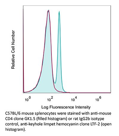
Specifications
| Isotype | Rat IgG2b, κ |
|---|---|
| Recommended Isotype Control(s) | InVivoMAb rat IgG2b isotype control, anti-keyhole limpet hemocyanin |
| Recommended Dilution Buffer | InVivoPure pH 6.5 Dilution Buffer |
| Conjugation | This product is unconjugated. Conjugation is available via our Antibody Conjugation Services. |
| Immunogen | Mouse CTL clone V4 |
| Reported Applications |
in vivo CD4+ T cell depletion Flow cytometry Western blot |
| Formulation |
PBS, pH 6.5 Contains no stabilizers or preservatives |
| Endotoxin |
≤1EU/mg (≤0.001EU/μg) Determined by LAL assay |
| Purity |
≥95% Determined by SDS-PAGE |
| Sterility | 0.2 µm filtration |
| Production | Purified from cell culture supernatant in an animal-free facility |
| Purification | Protein G |
| RRID | AB_1107636 |
| Molecular Weight | 150 kDa |
| Storage | The antibody solution should be stored at the stock concentration at 4°C. Do not freeze. |
| Need a Custom Formulation? | See All Antibody Customization Options |
Application References
-
Moynihan, K. D., et al (2016). "Eradication of large established tumors in mice by combination immunotherapy that engages innate and adaptive immune responses" Nat Med. doi : 10.1038/nm.4200.
PubMed
Checkpoint blockade with antibodies specific for cytotoxic T lymphocyte-associated protein (CTLA)-4 or programmed cell death 1 (PDCD1; also known as PD-1) elicits durable tumor regression in metastatic cancer, but these dramatic responses are confined to a minority of patients. This suboptimal outcome is probably due in part to the complex network of immunosuppressive pathways present in advanced tumors, which are unlikely to be overcome by intervention at a single signaling checkpoint. Here we describe a combination immunotherapy that recruits a variety of innate and adaptive immune cells to eliminate large tumor burdens in syngeneic tumor models and a genetically engineered mouse model of melanoma; to our knowledge tumors of this size have not previously been curable by treatments relying on endogenous immunity. Maximal antitumor efficacy required four components: a tumor-antigen-targeting antibody, a recombinant interleukin-2 with an extended half-life, anti-PD-1 and a powerful T cell vaccine. Depletion experiments revealed that CD8+ T cells, cross-presenting dendritic cells and several other innate immune cell subsets were required for tumor regression. Effective treatment induced infiltration of immune cells and production of inflammatory cytokines in the tumor, enhanced antibody-mediated tumor antigen uptake and promoted antigen spreading. These results demonstrate the capacity of an elicited endogenous immune response to destroy large, established tumors and elucidate essential characteristics of combination immunotherapies that are capable of curing a majority of tumors in experimental settings typically viewed as intractable.
-
Uddin, M. N., et al (2014). "TNF-alpha-dependent hematopoiesis following Bcl11b deletion in T cells restricts metastatic melanoma" J Immunol 192(4): 1946-1953.
PubMed
Using several tumor models, we demonstrate that mice deficient in Bcl11b in T cells, although having reduced numbers of T cells in the peripheral lymphoid organs, developed significantly less tumors compared with wild-type mice. Bcl11b(-/-) CD4(+) T cells, with elevated TNF-alpha levels, but not the Bcl11b(-/-) CD8(+) T cells, were required for the reduced tumor burden, as were NK1.1(+) cells, found in increased numbers in Bcl11b(F/F)/CD4-Cre mice. Among NK1.1(+) cells, the NK cell population was predominant in number and was the only population displaying elevated granzyme B levels and increased degranulation, although not increased proliferation. Although the number of myeloid-derived suppressor cells was increased in the lungs with metastatic tumors of Bcl11b(F/F)/CD4-Cre mice, their arginase-1 levels were severely reduced. The increase in NK cell and myeloid-derived suppressor cell numbers was associated with increased bone marrow and splenic hematopoiesis. Finally, the reduced tumor burden, increased numbers of NK cells in the lung, and increased hematopoiesis in Bcl11b(F/F)/CD4-Cre mice were all dependent on TNF-alpha. Moreover, TNF-alpha treatment of wild-type mice also reduced the tumor burden and increased hematopoiesis and the numbers and activity of NK cells in the lung. In vitro treatment with TNF-alpha of lineage-negative hematopoietic progenitors increased NK and myeloid differentiation, further supporting a role of TNF-alpha in promoting hematopoiesis. These studies reveal a novel role for TNF-alpha in the antitumor immune response, specifically in stimulating hematopoiesis and increasing the numbers and activity of NK cells.
-
Xin, L., et al (2014). "Commensal microbes drive intestinal inflammation by IL-17-producing CD4+ T cells through ICOSL and OX40L costimulation in the absence of B7-1 and B7-2" Proc Natl Acad Sci U S A 111(29): 10672-10677.
PubMed
The costimulatory B7-1 (CD80)/B7-2 (CD86) molecules, along with T-cell receptor stimulation, together facilitate T-cell activation. This explains why in vivo B7 costimulation neutralization efficiently silences a variety of human autoimmune disorders. Paradoxically, however, B7 blockade also potently moderates accumulation of immune-suppressive regulatory T cells (Tregs) essential for protection against multiorgan systemic autoimmunity. Here we show that B7 deprivation in mice overrides the necessity for Tregs in averting systemic autoimmunity and inflammation in extraintestinal tissues, whereas peripherally induced Tregs retained in the absence of B7 selectively mitigate intestinal inflammation caused by Th17 effector CD4(+) T cells. The need for additional immune suppression in the intestine reflects commensal microbe-driven T-cell activation through the accessory costimulation molecules ICOSL and OX40L. Eradication of commensal enteric bacteria mitigates intestinal inflammation and IL-17 production triggered by Treg depletion in B7-deficient mice, whereas re-establishing intestinal colonization with Candida albicans primes expansion of Th17 cells with commensal specificity. Thus, neutralizing B7 costimulation uncovers an essential role for Tregs in selectively averting intestinal inflammation by Th17 CD4(+) T cells with commensal microbe specificity.
-
Zander, R. A., et al (2015). "PD-1 Co-inhibitory and OX40 Co-stimulatory Crosstalk Regulates Helper T Cell Differentiation and Anti-Plasmodium Humoral Immunity" Cell Host Microbe 17(5): 628-641.
PubMed
The differentiation and protective capacity of Plasmodium-specific T cells are regulated by both positive and negative signals during malaria, but the molecular and cellular details remain poorly defined. Here we show that malaria patients and Plasmodium-infected rodents exhibit atypical expression of the co-stimulatory receptor OX40 on CD4 T cells and that therapeutic enhancement of OX40 signaling enhances helper CD4 T cell activity, humoral immunity, and parasite clearance in rodents. However, these beneficial effects of OX40 signaling are abrogated following coordinate blockade of PD-1 co-inhibitory pathways, which are also upregulated during malaria and associated with elevated parasitemia. Co-administration of biologics blocking PD-1 and promoting OX40 signaling induces excessive interferon-gamma that directly limits helper T cell-mediated support of humoral immunity and decreases parasite control. Our results show that targeting OX40 can enhance Plasmodium control and that crosstalk between co-inhibitory and co-stimulatory pathways in pathogen-specific CD4 T cells can impact pathogen clearance.
Product Citations
-
Immunogenic tumor cell death and T-cell-derived IFN-γ elicit tumoricidal macrophages to potentiate OX40 immunotherapy.
In Cell Rep Med on 21 April 2026 by Liu, Y., Zhao, J., et al.
PubMed
Understanding the mechanisms limiting OX40 agonist antibody efficacy is critical for developing more effective combination immunotherapies. Tumor microenvironment (TME) analysis revealed that OX40-antibody-responsive mice harbored tumor-associated macrophages (TAMs) with elevated NOS2 expression and heightened pattern recognition receptor (PRR) activation and interferon gamma (IFN-γ) signaling. In addition, patients with more favorable treatment responses to OX40 antibody therapy exhibited increased NOS2 expression. Mechanistically, tumor-infiltrating T-cell-derived IFN-γ synergizes with endogenous ligands of PRR released during immunogenic cell death to drive NOS2+ TAMs reprogramming. Translating these insights into therapeutic strategy, a Combo approach composing of MPLA, IFN-γ, and OX40 agonist antibody is designed to actively polarize TAMs to express NOS2, which mediate tumor clearance through an NOS2-dependent cytotoxicity. Moreover, OX40-antibody-mediated regulatory T cell (Treg) depletion potentiated NOS2+ macrophage induction. This multimodal strategy offers a promising solution to overcome the limitations of OX40 antibody monotherapy and enhance outcomes of the OX40-targeted immunotherapies.
-
Immune-induced TCR-like antibodies regulate specific T cell response in mice.
In Nat Commun on 16 April 2026 by Kishida, K., Kawakami, K., et al.
PubMed
Antigen-specific regulation of T cell response is crucial for limiting hyperimmune response. However, the molecular mechanisms governing specific immune regulation remain unclear. In this study, we discover that antibodies specific to the antigen peptide-MHC class II complex are produced during helper T cell responses to various antigens, including hen egg lysozyme and proteolipid protein peptide. These antibodies specifically inhibit T cell receptor (TCR) recognition of MHC class II molecules presenting specific antigen peptide. We term these antibodies 'immune-induced TCR-like antibodies' or iTabs. Immunization with peptides containing flanking residues induces iTabs whereas immunization with peptides lacking flanking residues does not. Furthermore, we show that immunization with iTab-inducible peptide or iTab treatment suppress autoimmune disease development in a mouse model of experimental autoimmune encephalomyelitis. Thus, our findings provide a strategy for suppressing antigen-specific helper T cell responses using specific peptides, potentially controlling autoimmune diseases.
-
Lymphodepleting preconditioning impairs host antitumor immunity induced by adoptive T cell therapy in mouse models.
In Nat Commun on 31 March 2026 by Figueroa, D., Vega, J. P., et al.
PubMed
Adoptive T cell therapy (ACT) is effective against hematologic cancers, but the mechanisms underlying durable responses in solid tumors remain unclear. We show that adoptively transferred CD8+ T cells that eradicate established murine tumors promote expansion of host CD8+ T cells exhibiting tumor-reactive and tissue-resident phenotypes that contribute to tumor elimination. Mechanistically, tumor necrosis factor (TNF) from transferred cells induces dendritic cell (DC)-dependent expansion of host CD8+ T cells, conferring protection against ACT-resistant tumor cells lacking the targeted antigen. Lymphodepleting preconditioning promotes expansion of transferred cells and primary tumor eradication but impairs host antitumor immunity and abrogates protection against ACT-resistant tumors. In human tumors, increased TNF/DC/CD8+ T cell profiles correlate with favorable ACT responses and improved survival. These findings reveal a TNF-dependent interplay between transferred and host CD8+ T cells underlying durable antitumor immunity that is impaired by lymphodepleting preconditioning in mouse models, suggesting an underappreciated mechanism of ACT resistance.
-
IL-12-secreting CAR-T cells reprogram the tumor microenvironment and improve efficacy against heterogeneous models of glioblastoma.
In J Immunother Cancer on 24 March 2026 by Shen, S., Mohan, A. A., et al.
PubMed
Glioblastoma (GBM) remains uniformly lethal due to pronounced intratumoral heterogeneity and a highly immunosuppressive microenvironment that limits the efficacy of targeted therapies.