InVivoMAb anti-mouse CTLA-4 (CD152)
Product Description
Specifications
| Isotype | Armenian hamster IgG |
|---|---|
| Recommended Isotype Control(s) | InVivoMAb polyclonal Armenian hamster IgG |
| Recommended Dilution Buffer | InVivoPure pH 6.5 Dilution Buffer |
| Conjugation | This product is unconjugated. Conjugation is available via our Antibody Conjugation Services. |
| Immunogen | Mouse CTLA-4 IgG2a fusion protein |
| Reported Applications |
in vivo CTLA-4 neutralization in vitro CTLA-4 neutralization Flow cytometry Western blot |
| Formulation |
PBS, pH 6.5 Contains no stabilizers or preservatives |
| Endotoxin |
≤1EU/mg (≤0.001EU/μg) Determined by LAL assay |
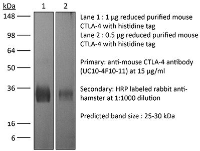
| Purity |
≥95% Determined by SDS-PAGE |
| Sterility | 0.2 µm filtration |
| Production | Purified from cell culture supernatant in an animal-free facility |
| Purification | Protein G |
| RRID | AB_1107598 |
| Molecular Weight | 150 kDa |
| Storage | The antibody solution should be stored at the stock concentration at 4°C. Do not freeze. |
| Need a Custom Formulation? | See All Antibody Customization Options |
Application References
-
Makkouk, A., et al (2015). "Three steps to breaking immune tolerance to lymphoma: a microparticle approach" Cancer Immunol Res 3(4): 389-398.
PubMed
In situ immunization aims at generating antitumor immune responses through manipulating the tumor microenvironment. On the basis of recent advances in the understanding of antitumor immunity, we designed a three-step approach to in situ immunization to lymphoma: (i) inducing immunogenic tumor cell death with the chemotherapeutic drug doxorubicin. Doxorubicin enhances the expression of “eat-me” signals by dying tumor cells, facilitating their phagocytosis by dendritic cells (DC). Because of the vesicant activity of doxorubicin, microparticles made of biodegradable polymer poly(lactide-co-glycolide) or PLGA can safely deliver doxorubicin intratumorally and are effective vaccine adjuvants, (ii) enhancing T-cell activation using anti-OX40 and (iii) sustaining T-cell responses by checkpoint blockade using anti-CTLA-4. In vitro, doxorubicin microparticles were less cytotoxic to DCs than to B lymphoma cells, did not require internalization by tumor cells, and significantly enhanced phagocytosis of tumor cells by DCs as compared with soluble doxorubicin. In mice, this three-step therapy induced CD4- and CD8-dependent systemic immune responses that enhanced T-cell infiltration into distant tumors, leading to their eradication and significantly improving survival. Our findings demonstrate that systemic antitumor immune responses can be generated locally by three-step therapy and merit further investigation as an immunotherapy for patients with lymphoma.
-
Triplett, T. A., et al (2018). "Reversal of indoleamine 2,3-dioxygenase-mediated cancer immune suppression by systemic kynurenine depletion with a therapeutic enzyme" Nat Biotechnol 36(8): 758-764.
PubMed
Increased tryptophan (Trp) catabolism in the tumor microenvironment (TME) can mediate immune suppression by upregulation of interferon (IFN)-gamma-inducible indoleamine 2,3-dioxygenase (IDO1) and/or ectopic expression of the predominantly liver-restricted enzyme tryptophan 2,3-dioxygenase (TDO). Whether these effects are due to Trp depletion in the TME or mediated by the accumulation of the IDO1 and/or TDO (hereafter referred to as IDO1/TDO) product kynurenine (Kyn) remains controversial. Here we show that administration of a pharmacologically optimized enzyme (PEGylated kynureninase; hereafter referred to as PEG-KYNase) that degrades Kyn into immunologically inert, nontoxic and readily cleared metabolites inhibits tumor growth. Enzyme treatment was associated with a marked increase in the tumor infiltration and proliferation of polyfunctional CD8(+) lymphocytes. We show that PEG-KYNase administration had substantial therapeutic effects when combined with approved checkpoint inhibitors or with a cancer vaccine for the treatment of large B16-F10 melanoma, 4T1 breast carcinoma or CT26 colon carcinoma tumors. PEG-KYNase mediated prolonged depletion of Kyn in the TME and reversed the modulatory effects of IDO1/TDO upregulation in the TME.
-
Pletinckx, K., et al (2015). "Immature dendritic cells convert anergic nonregulatory T cells into Foxp3- IL-10+ regulatory T cells by engaging CD28 and CTLA-4" Eur J Immunol 45(2): 480-491.
PubMed
Anergic T cells can survive for long time periods passively in a hyporesponsive state without obvious active functions. Thus, the immunological reason for their maintenance is unclear. Here, we induced peptide-specific anergy in T cells from mice by coculturing these cells with immature murine dendritic cells (DCs). We found that these anergic, nonsuppressive IL-10(-) Foxp3(-) CTLA-4(+) CD25(low) Egr2(+) T cells could be converted into suppressive IL-10(+) Foxp3(-) CTLA-4(+) CD25(high) Egr2(+) cells resembling type-1 Treg cells (Tr1) when stimulated a second time by immature DCs in vitro. Addition of TGF-beta during anergy induction favored Foxp3(+) Treg-cell induction, while TGF-beta had little effect when added to the second stimulation. Expression of both CD28 and CTLA-4 molecules on anergic T cells was required to allow their conversion into Tr1-like cells. Suppressor activity was enabled via CD28-mediated CD25 upregulation, acting as an IL-2 sink, together with a CTLA-4-mediated inhibition of NFATc1/alpha activation to shut down IL-2-mediated proliferation. Together, these data provide evidence and mechanistical insights into how persistent anergic T cells may serve as a resting memory pool for Tr1-like cells.
-
Welten, S. P., et al (2015). "The viral context instructs the redundancy of costimulatory pathways in driving CD8(+) T cell expansion" Elife 4. doi : 10.7554/eLife.07486.
PubMed
Signals delivered by costimulatory molecules are implicated in driving T cell expansion. The requirements for these signals, however, vary from dispensable to essential in different infections. We examined the underlying mechanisms of this differential T cell costimulation dependence and found that the viral context determined the dependence on CD28/B7-mediated costimulation for expansion of naive and memory CD8(+) T cells, indicating that the requirement for costimulatory signals is not imprinted. Notably, related to the high-level costimulatory molecule expression induced by lymphocytic choriomeningitis virus (LCMV), CD28/B7-mediated costimulation was dispensable for accumulation of LCMV-specific CD8(+) T cells because of redundancy with the costimulatory pathways induced by TNF receptor family members (i.e., CD27, OX40, and 4-1BB). Type I IFN signaling in viral-specific CD8(+) T cells is slightly redundant with costimulatory signals. These results highlight that pathogen-specific conditions differentially and uniquely dictate the utilization of costimulatory pathways allowing shaping of effector and memory antigen-specific CD8(+) T cell responses.
Product Citations
-
Cryoablation Plus Immune Checkpoint Inhibitors Enhanced Dendritic Cell and T Cell Activation in TNBC Murine Model.
In Immunotargets Ther on 14 April 2026 by Sardela de Miranda, F., Babcock, R. L., et al.
PubMed
Cryoablation eradicates tumors through repeated freeze-thaw cycles and preserves tumor-associated antigens, triggering inflammatory signals capable of priming anti-tumor immunity, yet its therapeutic potential in triple-negative breast cancer (TNBC) remains largely unexplored. Immune checkpoint inhibitors (ICIs) have shown clinical benefit in TNBC but come with significant immune-related toxicities. Combining cryoablation with ICIs in TNBC may amplify the efficacy of cryoablation, which is significantly less toxic than ICIs, thereby providing opportunities for lowering the doses of ICIs in clinical practice. Here, we investigated the therapeutic impact of cryoablation with ICIs in an orthotopic bilateral murine TNBC model.
-
ZBTB21 Is a Dual Suppressor of Pyroptosis and MHC-I Antigen Presentation That Promotes Tumor Immune Evasion.
In Adv Sci (Weinh) on 1 April 2026 by Zhao, L., Sheng, L., et al.
PubMed
Immune checkpoint blockade (ICB) efficacy is limited by tumor-intrinsic immune escape mechanisms. This study identifies the transcription factor ZBTB21 as a central orchestrator of dual immunosuppressive programs. ZBTB21 epigenetically silences gasdermin D (GSDMD)-dependent pyroptosis by restricting STAT1-mediated chromatin accessibility via H3K27ac modulation at the GSDMD locus. Simultaneously, it represses MHC-I antigen presentation by attenuating IRF1 expression and its transactivation capacity. Genetic ablation of ZBTB21 unleashes pyroptotic cell death and enhances tumor antigen presentation, establishing a self-reinforcing cycle that recruits and activates CD8+ T cells. This dual activation overcomes ICB resistance in murine models, while B2M deletion ablates efficacy, confirming MHC-I dependency. Pharmacological inhibition of ZBTB21 with dobutamine disrupts its DNA-binding domain, which triggers pyroptotic inflammation and MHC-I upregulation to synergize with anti-PD-1 therapy. Thus, ZBTB21 represents a druggable nexus coordinating pyroptosis resistance and antigen presentation escape, providing a combinatorial strategy to reinvigorate antitumor immunity.
-
CRISPR screens in the context of immune selection identify CHD1 and MAP3K7 as mediators of cancer immunotherapy resistance.
In Cell Rep Med on 20 January 2026 by Watterson, A., Picco, G., et al.
PubMed
Cancer immunotherapy is only effective in a subset of patients, highlighting the need for effective biomarkers and combination therapies. Here, we systematically identify genetic determinants of cancer cell sensitivity to anti-tumor immunity by performing whole-genome CRISPR-Cas9 knockout screens in autologous tumoroid-T cell co-cultures, isogenic cancer cell models deficient in interferon signaling, and in the context of four cytokines. We discover that loss of CHD1 and MAP3K7 (encoding TAK1) potentiates the transcriptional response to IFN-γ, thereby creating an acquired vulnerability by sensitizing cancer cells to tumor-reactive T cells. Immune checkpoint blockade is more effective in a syngeneic mouse model of melanoma deficient in Chd1 and Map3k7 and is associated with elevated intra-tumoral CD8+ T cell numbers and activation. CHD1 and MAP3K7 are recurrently mutated in cancer, and reduced expression in tumors correlates with response to immune checkpoint inhibitors in patients, nominating these genes as potential biomarkers of immunotherapy response.