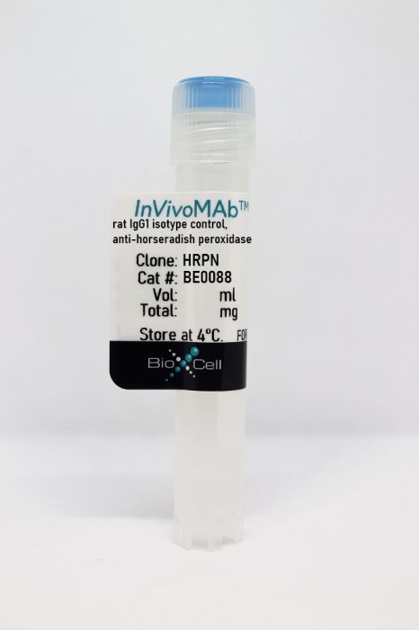
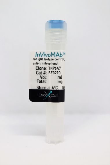
InVivoMAb rat IgG1 isotype control, anti-horseradish peroxidase
Product Details
The HRPN monoclonal antibody reacts with horseradish peroxidase (HRP). Because HRP is not expressed by mammals this antibody is ideal for use as an isotype-matched control for rat IgG1 antibodies in most in vivo and in vitro applications. This antibody can interfere with HRP detection based assays. If using downstream HRP based assays to analyze samples derived from treated animals, please consider using our alternative rat IgG1 isotype control antibody BP0290.Specifications
| Isotype | Rat IgG1, κ |
|---|---|
| Recommended Dilution Buffer | InVivoPure pH 7.0 Dilution Buffer |
| Conjugation | This product is unconjugated. Conjugation is available via our Antibody Conjugation Services. |
| Formulation |
PBS, pH 7.0 Contains no stabilizers or preservatives |
| Endotoxin |
<2EU/mg (<0.002EU/μg) Determined by LAL gel clotting assay |
| Purity |
>95% Determined by SDS-PAGE |
| Sterility | 0.2 µm filtration |
| Production | Purified from cell culture supernatant in an animal-free facility |
| Purification | Protein G |
| RRID | AB_1107775 |
| Molecular Weight | 150 kDa |
| Storage | The antibody solution should be stored at the stock concentration at 4°C. Do not freeze. |
Additional Formats
Recommended Products
Goschl, L., et al. (2018). "A T cell-specific deletion of HDAC1 protects against experimental autoimmune encephalomyelitis" J Autoimmun 86: 51-61. PubMed
Multiple sclerosis (MS) is a human neurodegenerative disease characterized by the invasion of autoreactive T cells from the periphery into the CNS. Application of pan-histone deacetylase inhibitors (HDACi) ameliorates experimental autoimmune encephalomyelitis (EAE), an animal model for MS, suggesting that HDACi might be a potential therapeutic strategy for MS. However, the function of individual HDAC members in the pathogenesis of EAE is not known. In this study we report that mice with a T cell-specific deletion of HDAC1 (using the Cd4-Cre deleter strain; HDAC1-cKO) were completely resistant to EAE despite the ability of HDAC1cKO CD4(+) T cells to differentiate into Th17 cells. RNA sequencing revealed STAT1 as a prominent upstream regulator of differentially expressed genes in activated HDAC1-cKO CD4(+) T cells and this was accompanied by a strong increase in phosphorylated STAT1 (pSTAT1). This suggests that HDAC1 controls STAT1 activity in activated CD4(+) T cells. Increased pSTAT1 levels correlated with a reduced expression of the chemokine receptors Ccr4 and Ccr6, which are important for the migration of T cells into the CNS. Finally, EAE susceptibility was restored in WT:HDAC1-cKO mixed BM chimeric mice, indicating a cell-autonomous defect. Our data demonstrate a novel pathophysiological role for HDAC1 in EAE and provide evidence that selective inhibition of HDAC1 might be a promising strategy for the treatment of MS.
Clemente-Casares, X., et al. (2016). "Expanding antigen-specific regulatory networks to treat autoimmunity" Nature 530(7591): 434-440. PubMed
Regulatory T cells hold promise as targets for therapeutic intervention in autoimmunity, but approaches capable of expanding antigen-specific regulatory T cells in vivo are currently not available. Here we show that systemic delivery of nanoparticles coated with autoimmune-disease-relevant peptides bound to major histocompatibility complex class II (pMHCII) molecules triggers the generation and expansion of antigen-specific regulatory CD4(+) T cell type 1 (TR1)-like cells in different mouse models, including mice humanized with lymphocytes from patients, leading to resolution of established autoimmune phenomena. Ten pMHCII-based nanomedicines show similar biological effects, regardless of genetic background, prevalence of the cognate T-cell population or MHC restriction. These nanomedicines promote the differentiation of disease-primed autoreactive T cells into TR1-like cells, which in turn suppress autoantigen-loaded antigen-presenting cells and drive the differentiation of cognate B cells into disease-suppressing regulatory B cells, without compromising systemic immunity. pMHCII-based nanomedicines thus represent a new class of drugs, potentially useful for treating a broad spectrum of autoimmune conditions in a disease-specific manner.
Sell, S., et al. (2015). "Control of murine cytomegalovirus infection by gammadelta T cells" PLoS Pathog 11(2): e1004481. PubMed
Infections with cytomegalovirus (CMV) can cause severe disease in immunosuppressed patients and infected newborns. Innate as well as cellular and humoral adaptive immune effector functions contribute to the control of CMV in immunocompetent individuals. None of the innate or adaptive immune functions are essential for virus control, however. Expansion of gammadelta T cells has been observed during human CMV (HCMV) infection in the fetus and in transplant patients with HCMV reactivation but the protective function of gammadelta T cells under these conditions remains unclear. Here we show for murine CMV (MCMV) infections that mice that lack CD8 and CD4 alphabeta-T cells as well as B lymphocytes can control a MCMV infection that is lethal in RAG-1(-/-) mice lacking any T- and B-cells. gammadelta T cells, isolated from infected mice can kill MCMV infected target cells in vitro and, importantly, provide long-term protection in infected RAG-1(-/-) mice after adoptive transfer. gammadelta T cells in MCMV infected hosts undergo a prominent and long-lasting phenotypic change most compatible with the view that the majority of the gammadelta T cell population persists in an effector/memory state even after resolution of the acute phase of the infection. A clonotypically focused Vgamma1 and Vgamma2 repertoire was observed at later stages of the infection in the organs where MCMV persists. These findings add gammadelta T cells as yet another protective component to the anti-CMV immune response. Our data provide clear evidence that gammadelta T cells can provide an effective control mechanism of acute CMV infections, particularly when conventional adaptive immune mechanisms are insufficient or absent, like in transplant patient or in the developing immune system in utero. The findings have implications in the stem cell transplant setting, as antigen recognition by gammadelta T cells is not MHC-restricted and dual reactivity against CMV and tumors has been described.
Grinberg-Bleyer, Y., et al. (2015). "Cutting edge: NF-kappaB p65 and c-Rel control epidermal development and immune homeostasis in the skin" J Immunol 194(6): 2472-2476. PubMed
Psoriasis is an inflammatory skin disease in which activated immune cells and the proinflammatory cytokine TNF are well-known mediators of pathogenesis. The transcription factor NF-kappaB is a key regulator of TNF production and TNF-induced proinflammatory gene expression, and both the psoriatic transcriptome and genetic susceptibility further implicate NF-kappaB in psoriasis etiopathology. However, the role of NF-kappaB in psoriasis remains controversial. We analyzed the function of canonical NF-kappaB in the epidermis using CRE-mediated deletion of p65 and c-Rel in keratinocytes. In contrast to animals lacking p65 or c-Rel alone, mice lacking both subunits developed severe dermatitis after birth. Consistent with its partial histological similarity to human psoriasis, this condition could be prevented by anti-TNF treatment. Moreover, regulatory T cells in lesional skin played an important role in disease remission. Our results demonstrate that canonical NF-kappaB in keratinocytes is essential for the maintenance of skin immune homeostasis and is protective against spontaneous dermatitis.
Park, H. J., et al. (2015). "PD-1 upregulated on regulatory T cells during chronic virus infection enhances the suppression of CD8+ T cell immune response via the interaction with PD-L1 expressed on CD8+ T cells" J Immunol 194(12): 5801-5811. PubMed
Regulatory T (Treg) cells act as terminators of T cell immuniy during acute phase of viral infection; however, their role and suppressive mechanism in chronic viral infection are not completely understood. In this study, we compared the phenotype and function of Treg cells during acute or chronic infection with lymphocytic choriomeningitis virus. Chronic infection, unlike acute infection, led to a large expansion of Treg cells and their upregulation of programmed death-1 (PD-1). Treg cells from chronically infected mice (chronic Treg cells) displayed greater suppressive capacity for inhibiting both CD8(+) and CD4(+) T cell proliferation and subsequent cytokine production than those from naive or acutely infected mice. A contact between Treg and CD8(+) T cells was necessary for the potent suppression of CD8(+) T cell immune response. More importantly, the suppression required cell-specific expression and interaction of PD-1 on chronic Treg cells and PD-1 ligand on CD8(+) T cells. Our study defines PD-1 upregulated on Treg cells and its interaction with PD-1 ligand on effector T cells as one cause for the potent T cell suppression and proposes the role of PD-1 on Treg cells, in addition to that on exhausted T cells, during chronic viral infection.
Meisen, W. H., et al. (2015). "The Impact of Macrophage- and Microglia-Secreted TNFalpha on Oncolytic HSV-1 Therapy in the Glioblastoma Tumor Microenvironment" Clin Cancer Res 21(14): 3274-3285. PubMed
PURPOSE: Oncolytic herpes simplex viruses (oHSV) represent a promising therapy for glioblastoma (GBM), but their clinical success has been limited. Early innate immune responses to viral infection reduce oHSV replication, tumor destruction, and efficacy. Here, we characterized the antiviral effects of macrophages and microglia on viral therapy for GBM. EXPERIMENTAL DESIGN: Quantitative flow cytometry of mice with intracranial gliomas (+/-oHSV) was used to examine macrophage/microglia infiltration and activation. In vitro coculture assays of infected glioma cells with microglia/macrophages were used to test their impact on oHSV replication. Macrophages from TNFalpha-knockout mice and blocking antibodies were used to evaluate the biologic effects of TNFalpha on virus replication. TNFalpha blocking antibodies were used to evaluate the impact of TNFalpha on oHSV therapy in vivo. RESULTS: Flow-cytometry analysis revealed a 7.9-fold increase in macrophage infiltration after virus treatment. Tumor-infiltrating macrophages/microglia were polarized toward a M1, proinflammatory phenotype, and they expressed high levels of CD86, MHCII, and Ly6C. Macrophages/microglia produced significant amounts of TNFalpha in response to infected glioma cells in vitro and in vivo. Using TNFalpha-blocking antibodies and macrophages derived from TNFalpha-knockout mice, we discovered TNFalpha-induced apoptosis in infected tumor cells and inhibited virus replication. Finally, we demonstrated the transient blockade of TNFalpha from the tumor microenvironment with TNFalpha-blocking antibodies significantly enhanced virus replication and survival in GBM intracranial tumors. CONCLUSIONS: The results of these studies suggest that FDA approved TNFalpha inhibitors may significantly improve the efficacy of oncolytic virus therapy.
Ellis, G. T., et al. (2015). "TRAIL+ monocytes and monocyte-related cells cause lung damage and thereby increase susceptibility to influenza-Streptococcus pneumoniae coinfection" EMBO Rep 16(9): 1203-1218. PubMed
Streptococcus pneumoniae coinfection is a major cause of influenza-associated mortality; however, the mechanisms underlying pathogenesis or protection remain unclear. Using a clinically relevant mouse model, we identify immune-mediated damage early during coinfection as a new mechanism causing susceptibility. Coinfected CCR2(-/-) mice lacking monocytes and monocyte-derived cells control bacterial invasion better, show reduced epithelial damage and are overall more resistant than wild-type controls. In influenza-infected wild-type lungs, monocytes and monocyte-derived cells are the major cell populations expressing the apoptosis-inducing ligand TRAIL. Accordingly, anti-TRAIL treatment reduces bacterial load and protects against coinfection if administered during viral infection, but not following bacterial exposure. Post-influenza bacterial outgrowth induces a strong proinflammatory cytokine response and massive inflammatory cell infiltrate. Depletion of neutrophils or blockade of TNF-alpha facilitate bacterial outgrowth, leading to increased mortality, demonstrating that these factors aid bacterial control. We conclude that inflammatory monocytes recruited early, during the viral phase of coinfection, induce TRAIL-mediated lung damage, which facilitates bacterial invasion, while TNF-alpha and neutrophil responses help control subsequent bacterial outgrowth. We thus identify novel determinants of protection versus pathology in influenza-Streptococcus pneumoniae coinfection.
Walsh, K. B., et al. (2014). "Animal model of respiratory syncytial virus: CD8+ T cells cause a cytokine storm that is chemically tractable by sphingosine-1-phosphate 1 receptor agonist therapy" J Virol 88(11): 6281-6293. PubMed
The cytokine storm is an intensified, dysregulated, tissue-injurious inflammatory response driven by cytokine and immune cell components. The cytokine storm during influenza virus infection, whereby the amplified innate immune response is primarily responsible for pulmonary damage, has been well characterized. Now we describe a novel event where virus-specific T cells induce a cytokine storm. The paramyxovirus pneumonia virus of mice (PVM) is a model of human respiratory syncytial virus (hRSV). Unexpectedly, when C57BL/6 mice were infected with PVM, the innate inflammatory response was undetectable until day 5 postinfection, at which time CD8(+) T cells infiltrated into the lung, initiating a cytokine storm by their production of gamma interferon (IFN-gamma) and tumor necrosis factor alpha (TNF-alpha). Administration of an immunomodulatory sphingosine-1-phosphate (S1P) receptor 1 (S1P1R) agonist significantly inhibited PVM-elicited cytokine storm by blunting the PVM-specific CD8(+) T cell response, resulting in diminished pulmonary disease and enhanced survival. IMPORTANCE: A dysregulated overly exuberant immune response, termed a “cytokine storm,” accompanies virus-induced acute respiratory diseases (VARV), is primarily responsible for the accompanying high morbidity and mortality, and can be controlled therapeutically in influenza virus infection of mice and ferrets by administration of sphingosine-1-phosphate 1 receptor (S1P1R) agonists. Here, two novel findings are recorded. First, in contrast to influenza infection, where the cytokine storm is initiated early by the innate immune system, for pneumonia virus of mice (PVM), a model of RSV, the cytokine storm is initiated late in infection by the adaptive immune response: specifically, by virus-specific CD8 T cells via their release of IFN-gamma and TNF-alpha. Blockading these cytokines with neutralizing antibodies blunts the cytokine storm and protects the host. Second, PVM infection is controlled by administration of an S1P1R agonist.
Beug, S. T., et al. (2014). "Smac mimetics and innate immune stimuli synergize to promote tumor death" Nat Biotechnol 32(2): 182-190. PubMed
Smac mimetic compounds (SMC), a class of drugs that sensitize cells to apoptosis by counteracting the activity of inhibitor of apoptosis (IAP) proteins, have proven safe in phase 1 clinical trials in cancer patients. However, because SMCs act by enabling transduction of pro-apoptotic signals, SMC monotherapy may be efficacious only in the subset of patients whose tumors produce large quantities of death-inducing proteins such as inflammatory cytokines. Therefore, we reasoned that SMCs would synergize with agents that stimulate a potent yet safe “cytokine storm.” Here we show that oncolytic viruses and adjuvants such as poly(I:C) and CpG induce bystander death of cancer cells treated with SMCs that is mediated by interferon beta (IFN-beta), tumor necrosis factor alpha (TNF-alpha) and/or TNF-related apoptosis-inducing ligand (TRAIL). This combinatorial treatment resulted in tumor regression and extended survival in two mouse models of cancer. As these and other adjuvants have been proven safe in clinical trials, it may be worthwhile to explore their clinical efficacy in combination with SMCs.
DeBerge, M. P., et al. (2014). "Soluble, but not transmembrane, TNF-alpha is required during influenza infection to limit the magnitude of immune responses and the extent of immunopathology" J Immunol 192(12): 5839-5851. PubMed
TNF-alpha is a pleotropic cytokine that has both proinflammatory and anti-inflammatory functions during influenza infection. TNF-alpha is first expressed as a transmembrane protein that is proteolytically processed to release a soluble form. Transmembrane TNF-alpha (memTNF-alpha) and soluble TNF-alpha (solTNF-alpha) have been shown to exert distinct tissue-protective or tissue-pathologic effects in several disease models. However, the relative contributions of memTNF-alpha or solTNF-alpha in regulating pulmonary immunopathology following influenza infection are unclear. Therefore, we performed intranasal influenza infection in mice exclusively expressing noncleavable memTNF-alpha or lacking TNF-alpha entirely and examined the outcomes. We found that solTNF-alpha, but not memTNF-alpha, was required to limit the size of the immune response and the extent of injury. In the absence of solTNF-alpha, there was a significant increase in the CD8(+) T cell response, including virus-specific CD8(+) T cells, which was due in part to an increased resistance to activation-induced cell death. We found that solTNF-alpha mediates these immunoregulatory effects primarily through TNFR1, because mice deficient in TNFR1, but not TNFR2, exhibited dysregulated immune responses and exacerbated injury similar to that observed in mice lacking solTNF-alpha. We also found that solTNF-alpha expression was required early during infection to regulate the magnitude of the CD8(+) T cell response, indicating that early inflammatory events are critical for the regulation of the effector phase. Taken together, these findings suggest that processing of memTNF-alpha to release solTNF-alpha is a critical event regulating the immune response during influenza infection.
Perng, O. A., et al. (2014). "The degree of CD4+ T cell autoreactivity determines cellular pathways underlying inflammatory arthritis" J Immunol 192(7): 3043-3056. PubMed
Although therapies targeting distinct cellular pathways (e.g., anticytokine versus anti-B cell therapy) have been found to be an effective strategy for at least some patients with inflammatory arthritis, the mechanisms that determine which pathways promote arthritis development are poorly understood. We have used a transgenic mouse model to examine how variations in the CD4(+) T cell response to a surrogate self-peptide can affect the cellular pathways that are required for arthritis development. CD4(+) T cells that are highly reactive with the self-peptide induce inflammatory arthritis that affects male and female mice equally. Arthritis develops by a B cell-independent mechanism, although it can be suppressed by an anti-TNF treatment, which prevented the accumulation of effector CD4(+) Th17 cells in the joints of treated mice. By contrast, arthritis develops with a significant female bias in the context of a more weakly autoreactive CD4(+) T cell response, and B cells play a prominent role in disease pathogenesis. In this setting of lower CD4(+) T cell autoreactivity, B cells promote the formation of autoreactive CD4(+) effector T cells (including Th17 cells), and IL-17 is required for arthritis development. These studies show that the degree of CD4(+) T cell reactivity for a self-peptide can play a prominent role in determining whether distinct cellular pathways can be targeted to prevent the development of inflammatory arthritis.
Weinlich, R., et al. (2013). "Protective roles for caspase-8 and cFLIP in adult homeostasis" Cell Rep 5(2): 340-348. PubMed
Caspase-8 or cellular FLICE-like inhibitor protein (cFLIP) deficiency leads to embryonic lethality in mice due to defects in endothelial tissues. Caspase-8(-/-) and receptor-interacting protein kinase-3 (RIPK3)(-/-), but not cFLIP(-/-) and RIPK3(-/-), double-knockout animals develop normally, indicating that caspase-8 antagonizes the lethal effects of RIPK3 during development. Here, we show that the acute deletion of caspase-8 in the gut of adult mice induces enterocyte death, disruption of tissue homeostasis, and inflammation, resulting in sepsis and mortality. Likewise, acute deletion of caspase-8 in a focal region of the skin induces local keratinocyte death, tissue disruption, and inflammation. Strikingly, RIPK3 ablation rescues both phenotypes. However, acute loss of cFLIP in the skin produces a similar phenotype that is not rescued by RIPK3 ablation. TNF neutralization protects from either acute loss of caspase-8 or cFLIP. These results demonstrate that caspase-8-mediated suppression of RIPK3-induced death is required not only during development but also for adult homeostasis. Furthermore, RIPK3-dependent inflammation is dispensable for the skin phenotype.
Mohr, E., et al. (2010). "IFN-{gamma} produced by CD8 T cells induces T-bet-dependent and -independent class switching in B cells in responses to alum-precipitated protein vaccine" Proc Natl Acad Sci U S A 107(40): 17292-17297. PubMed
Alum-precipitated protein (alum protein) vaccines elicit long-lasting neutralizing antibody responses that prevent bacterial exotoxins and viruses from entering cells. Typically, these vaccines induce CD4 T cells to become T helper 2 (Th2) cells that induce Ig class switching to IgG1. We now report that CD8 T cells also respond to alum proteins, proliferating extensively and producing IFN-gamma, a key Th1 cytokine. These findings led us to question whether adoptive transfer of antigen-specific CD8 T cells alters the characteristic CD4 Th2 response to alum proteins and the switching pattern in responding B cells. To this end, WT mice given transgenic ovalbumin (OVA)-specific CD4 (OTII) or CD8 (OTI) T cells, or both, were immunized with alum-precipitated OVA. Cotransfer of antigen-specific CD8 T cells skewed switching patterns in responding B cells from IgG1 to IgG2a and IgG2b. Blocking with anti-IFN-gamma antibody largely inhibited this altered B-cell switching pattern. The transcription factor T-bet is required in B cells for IFN-gamma-dependent switching to IgG2a. By contrast, we show that this transcription factor is dispensable in B cells both for IFN-gamma-induced switching to IgG2b and for inhibition of switching to IgG1. Thus, T-bet dependence identifies distinct transcriptional pathways in B cells that regulate IFN-gamma-induced switching to different IgG isotypes.
Ultra-low volume intradermal administration of radiation-attenuated sporozoites with the glycolipid adjuvant 7DW8-5 completely protects mice against malaria.
In Scientific Reports on 4 February 2024 by Watson, F. N., Shears, M. J., et al.
PubMed
Radiation-attenuated sporozoite (RAS) vaccines can completely prevent blood stage Plasmodium infection by inducing liver-resident memory CD8+ T cells to target parasites in the liver. Such T cells can be induced by 'Prime-and-trap' vaccination, which here combines DNA priming against the P. yoelii circumsporozoite protein (CSP) with a subsequent intravenous (IV) dose of liver-homing RAS to "trap" the activated and expanding T cells in the liver. Prime-and-trap confers durable protection in mice, and efforts are underway to translate this vaccine strategy to the clinic. However, it is unclear whether the RAS trapping dose must be strictly administered by the IV route. Here we show that intradermal (ID) RAS administration can be as effective as IV administration if RAS are co-administrated with the glycolipid adjuvant 7DW8-5 in an ultra-low inoculation volume. In mice, the co-administration of RAS and 7DW8-5 in ultra-low ID volumes (2.5 µL) was completely protective and dose sparing compared to standard volumes (10-50 µL) and induced protective levels of CSP-specific CD8+ T cells in the liver. Our finding that adjuvants and ultra-low volumes are required for ID RAS efficacy may explain why prior reports about higher volumes of unadjuvanted ID RAS proved less effective than IV RAS. The ID route may offer significant translational advantages over the IV route and could improve sporozoite vaccine development. © 2024. The Author(s).
- Mus musculus (House mouse),
- Cancer Research
Type 2 innate lymphoid cells are not involved in mouse bladder tumor development.
In Frontiers in Immunology on 29 January 2024 by Schneider, A. K., Domingos-Pereira, S., et al.
PubMed
Therapies for bladder cancer patients are limited by side effects and failures, highlighting the need for novel targets to improve disease management. Given the emerging evidence highlighting the key role of innate lymphoid cell subsets, especially type 2 innate lymphoid cells (ILC2s), in shaping the tumor microenvironment and immune responses, we investigated the contribution of ILC2s in bladder tumor development. Using the orthotopic murine MB49 bladder tumor model, we found a strong enrichment of ILC2s in the bladder under steady-state conditions, comparable to that in the lung. However, as tumors grew, we observed an increase in ILC1s but no changes in ILC2s. Targeting ILC2s by blocking IL-4/IL-13 signaling pathways, IL-5, or IL-33 receptor, or using IL-33-deficient or ILC2-deficient mice, did not affect mice survival following bladder tumor implantation. Overall, these results suggest that ILC2s do not contribute significantly to bladder tumor development, yet further investigations are required to confirm these results in bladder cancer patients. Copyright © 2024 Schneider, Domingos-Pereira, Cesson, Polak, Fallon, Zhu, Roth, Nardelli-Haefliger and Derré.
- Immunology and Microbiology
Elimination of Chlamydia muridarum from the female reproductive tract is IL-12p40 dependent, but independent of Th1 and Th2 cells.
In PLoS Pathogens on 1 January 2024 by Rixon, J. A., Fong, K. D., et al.
PubMed
Chlamydia vaccine approaches aspire to induce Th1 cells for optimal protection, despite the fact that there is no direct evidence demonstrating Th1-mediated Chlamydia clearance from the female reproductive tract (FRT). We recently reported that T-bet-deficient mice can resolve primary Chlamydia infection normally, undermining the potentially protective role of Th1 cells in Chlamydia immunity. Here, we show that T-bet-deficient mice develop robust Th17 responses and that mice deficient in Th17 cells exhibit delayed bacterial clearance, demonstrating that Chlamydia-specific Th17 cells represent an underappreciated protective population. Additionally, Th2-deficient mice competently clear cervicovaginal infection. Furthermore, we show that sensing of IFN-γ by non-hematopoietic cells is essential for Chlamydia immunity, yet bacterial clearance in the FRT does not require IFN-γ secretion by CD4 T cells. Despite the fact that Th1 cells are not necessary for Chlamydia clearance, protective immunity to Chlamydia is still dependent on MHC class-II-restricted CD4 T cells and IL-12p40. Together, these data point to IL-12p40-dependent CD4 effector maturation as essential for Chlamydia immunity, and Th17 cells to a lesser extent, yet neither Th1 nor Th2 cell development is critical. Future Chlamydia vaccination efforts will be more effective if they focus on induction of this protective CD4 T cell population. Copyright: © 2024 Rixon et al. This is an open access article distributed under the terms of the Creative Commons Attribution License, which permits unrestricted use, distribution, and reproduction in any medium, provided the original author and source are credited.
- Mus musculus (House mouse),
- Immunology and Microbiology
Interferon gamma immunoPET imaging to evaluate response to immune checkpoint inhibitors.
In Frontiers in Oncology on 22 December 2023 by Hackett, J. B., Ramos, N., et al.
PubMed
We previously developed a 89Zr-labeled antibody-based immuno-positron emission tomography (immunoPET) tracer targeting interferon gamma (IFNγ), a cytokine produced predominantly by activated T and natural killer (NK) cells during pathogen clearance, anti-tumor immunity, and various inflammatory and autoimmune conditions. The current study investigated [89Zr]Zr-DFO-anti-IFNγ PET as a method to monitor response to immune checkpoint inhibitors (ICIs). BALB/c mice bearing CT26 colorectal tumors were treated with combined ICI (anti-cytotoxic T-lymphocyte-associated protein 4 (CTLA-4) and anti-programmed death 1 (PD-1)). The [89Zr]Zr-DFO-anti-IFNγ PET tracer, generated with antibody clone AN18, was administered on the day of the second ICI treatment, with PET imaging 72 hours later. Tumor mRNA was analyzed by quantitative reverse-transcribed PCR (qRT-PCR). We detected significantly higher intratumoral localization of [89Zr]Zr-DFO-anti-IFNγ in ICI-treated mice compared to untreated controls, while uptake of an isotype control tracer remained similar between treated and untreated mice. Interestingly, [89Zr]Zr-DFO-anti-IFNγ uptake was also elevated relative to the isotype control in untreated mice, suggesting that the IFNγ-specific tracer might be able to detect underlying immune activity in situ in this immunogenic model. In an efficacy experiment, a significant inverse correlation between tracer uptake and tumor burden was also observed. Because antibodies to cytokines often exhibit neutralizing effects which might alter cellular communication within the tumor microenvironment, we also evaluated the impact of AN18 on downstream IFNγ signaling and ICI outcomes. Tumor transcript analysis using interferon regulatory factor 1 (IRF1) expression as a readout of IFNγ signaling suggested there may be a marginal disruption of this pathway. However, compared to a 250 µg dose known to neutralize IFNγ, which diminished ICI efficacy, a tracer-equivalent 50 µg dose did not reduce ICI response rates. These results support the use of IFNγ PET as a method to monitor immune activity in situ after ICI, which may also extend to additional T cell-activating immunotherapies. Copyright © 2023 Hackett, Ramos, Barr, Bross, Viola and Gibson.
- Immunology and Microbiology
Recently activated CD4 T cells in tuberculosis express OX40 as a target for host-directed immunotherapy.
In Nature Communications on 19 December 2023 by Gress, A. R., Ronayne, C. E., et al.
PubMed
After Mycobacterium tuberculosis (Mtb) infection, many effector T cells traffic to the lungs, but few become activated. Here we use an antigen receptor reporter mouse (Nur77-GFP) to identify recently activated CD4 T cells in the lungs. These Nur77-GFPHI cells contain expanded TCR clonotypes, have elevated expression of co-stimulatory genes such as Tnfrsf4/OX40, and are functionally more protective than Nur77-GFPLO cells. By contrast, Nur77-GFPLO cells express markers of terminal exhaustion and cytotoxicity, and the trafficking receptor S1pr5, associated with vascular localization. A short course of immunotherapy targeting OX40+ cells transiently expands CD4 T cell numbers and shifts their phenotype towards parenchymal protective cells. Moreover, OX40 agonist immunotherapy decreases the lung bacterial burden and extends host survival, offering an additive benefit to antibiotics. CD4 T cells from the cerebrospinal fluid of humans with HIV-associated tuberculous meningitis commonly express surface OX40 protein, while CD8 T cells do not. Our data thus propose OX40 as a marker of recently activated CD4 T cells at the infection site and a potential target for immunotherapy in tuberculosis. © 2023. The Author(s).
- Mus musculus (House mouse)
Oral administration of Bifidobacterium breve improves anti-angiogenic drugs-derived oral mucosal wound healing impairment via upregulation of interleukin-10.
In International Journal of Oral Science on 11 December 2023 by Li, Q., Li, Y., et al.
PubMed
Recent studies have suggested that long-term application of anti-angiogenic drugs may impair oral mucosal wound healing. This study investigated the effect of sunitinib on oral mucosal healing impairment in mice and the therapeutic potential of Bifidobacterium breve (B. breve). A mouse hard palate mucosal defect model was used to investigate the influence of sunitinib and/or zoledronate on wound healing. The volume and density of the bone under the mucosal defect were assessed by micro-computed tomography (micro-CT). Inflammatory factors were detected by protein microarray analysis and enzyme-linked immunosorbent assay (ELISA). The senescence and biological functions were tested in oral mucosal stem cells (OMSCs) treated with sunitinib. Ligated loop experiments were used to investigate the effect of oral B. breve. Neutralizing antibody for interleukin-10 (IL-10) was used to prove the critical role of IL-10 in the pro-healing process derived from B. breve. Results showed that sunitinib caused oral mucosal wound healing impairment in mice. In vitro, sunitinib induced cellular senescence in OMSCs and affected biological functions such as proliferation, migration, and differentiation. Oral administration of B. breve reduced oral mucosal inflammation and promoted wound healing via intestinal dendritic cells (DCs)-derived IL-10. IL-10 reversed cellular senescence caused by sunitinib in OMSCs, and IL-10 neutralizing antibody blocked the ameliorative effect of B. breve on oral mucosal wound healing under sunitinib treatment conditions. In conclusion, sunitinib induces cellular senescence in OMSCs and causes oral mucosal wound healing impairment and oral administration of B. breve could improve wound healing impairment via intestinal DCs-derived IL-10. © 2023. The Author(s).
- Cancer Research,
- Immunology and Microbiology
Differential requirements for CD4+ T cells in the efficacy of the anti-PD-1+LAG-3 and anti-PD-1+CTLA-4 combinations in melanoma flank and brain metastasis models.
In Journal for Immunotherapy of Cancer on 6 December 2023 by Phadke, M. S., Li, J., et al.
PubMed
Although the anti-PD-1+LAG-3 and the anti-PD-1+CTLA-4 combinations are effective in advanced melanoma, it remains unclear whether their mechanisms of action overlap. We used single cell (sc) RNA-seq, flow cytometry and IHC analysis of responding SM1, D4M-UV2 and B16 melanoma flank tumors and SM1 brain metastases to explore the mechanism of action of the anti-PD-1+LAG-3 and the anti-PD-1+CTLA-4 combination. CD4+ and CD8+ T cell depletion, tetramer binding assays and ELISPOT assays were used to demonstrate the unique role of CD4+T cell help in the antitumor effects of the anti-PD-1+LAG-3 combination. The anti-PD-1+CTLA-4 combination was associated with the infiltration of FOXP3+regulatory CD4+ cells (Tregs), fewer activated CD4+T cells and the accumulation of a subset of IFNγ secreting cytotoxic CD8+T cells, whereas the anti-PD-1+LAG-3 combination led to the accumulation of CD4+T helper cells that expressed CXCR4, TNFSF8, IL21R and a subset of CD8+T cells with reduced expression of cytotoxic markers. T cell depletion studies showed a requirement for CD4+T cells for the anti-PD-1+LAG-3 combination, but not the PD-1-CTLA-4 combination at both flank and brain tumor sites. In anti-PD-1+LAG-3 treated tumors, CD4+T cell depletion was associated with fewer activated (CD69+) CD8+T cells and impaired IFNγ release but, conversely, increased numbers of activated CD8+T cells and IFNγ release in anti-PD-1+CTLA-4 treated tumors. Together these studies suggest that these two clinically relevant immune checkpoint inhibitor (ICI) combinations have differential effects on CD4+T cell polarization, which in turn, impacted cytotoxic CD8+T cell function. Further insights into the mechanisms of action/resistance of these clinically-relevant ICI combinations will allow therapy to be further personalized. © Author(s) (or their employer(s)) 2023. Re-use permitted under CC BY-NC. No commercial re-use. See rights and permissions. Published by BMJ.
- Cancer Research
Leukemia-intrinsic determinants of CAR-T response revealed by iterative in vivo genome-wide CRISPR screening.
In Nature Communications on 5 December 2023 by Ramos, A., Koch, C. E., et al.
PubMed
CAR-T therapy is a promising, novel treatment modality for B-cell malignancies and yet many patients relapse through a variety of means, including loss of CAR-T cells and antigen escape. To investigate leukemia-intrinsic CAR-T resistance mechanisms, we performed genome-wide CRISPR-Cas9 loss-of-function screens in an immunocompetent murine model of B-cell acute lymphoblastic leukemia (B-ALL) utilizing a modular guide RNA library. We identified IFNγR/JAK/STAT signaling and components of antigen processing and presentation pathway as key mediators of resistance to CAR-T therapy in vivo; intriguingly, loss of this pathway yielded the opposite effect in vitro (sensitized leukemia to CAR-T cells). Transcriptional characterization of this model demonstrated upregulation of these pathways in tumors relapsed after CAR-T treatment, and functional studies showed a surprising role for natural killer (NK) cells in engaging this resistance program. Finally, examination of data from B-ALL patients treated with CAR-T revealed an association between poor outcomes and increased expression of JAK/STAT and MHC-I in leukemia cells. Overall, our data identify an unexpected mechanism of resistance to CAR-T therapy in which tumor cell interaction with the in vivo tumor microenvironment, including NK cells, induces expression of an adaptive, therapy-induced, T-cell resistance program in tumor cells. © 2023. The Author(s).
- Cancer Research,
- Immunology and Microbiology
ID1 expressing macrophages support cancer cell stemness and limit CD8+ T cell infiltration in colorectal cancer.
In Nature Communications on 23 November 2023 by Shang, S., Yang, C., et al.
PubMed
Elimination of cancer stem cells (CSCs) and reinvigoration of antitumor immunity remain unmet challenges for cancer therapy. Tumor-associated macrophages (TAMs) constitute the prominant population of immune cells in tumor tissues, contributing to the formation of CSC niches and a suppressive immune microenvironment. Here, we report that high expression of inhibitor of differentiation 1 (ID1) in TAMs correlates with poor outcome in patients with colorectal cancer (CRC). ID1 expressing macrophages maintain cancer stemness and impede CD8+ T cell infiltration. Mechanistically, ID1 interacts with STAT1 to induce its cytoplasmic distribution and inhibits STAT1-mediated SerpinB2 and CCL4 transcription, two secretory factors responsible for cancer stemness inhibition and CD8+ T cell recruitment. Reducing ID1 expression ameliorates CRC progression and enhances tumor sensitivity to immunotherapy and chemotherapy. Collectively, our study highlights the pivotal role of ID1 in controlling the protumor phenotype of TAMs and paves the way for therapeutic targeting of ID1 in CRC. © 2023. The Author(s).
- Immunology and Microbiology
Patient phenotypes and their relation to TNFα signaling and immune cell composition in critical illness and autoimmune disease
Preprint on BioRxiv : the Preprint Server for Biology on 1 November 2023 by Krishna, V., Banie, H., et al.
PubMed
Rationale TNFα inhibitors have shown promise in reducing mortality in hospitalized COVID-19 patients; one hypothesis explaining the limited clinical efficacy is patient heterogeneity in the TNFα pathway. Methods We evaluated the effect of TNFα inhibitors in a mouse model of LPS-induced acute lung injury. Using machine learning we attempted predictive enrichment of TNFα signaling in patients with either ARDS or sepsis. We examined biological factors that drive heterogeneity in host responses to critical infection and their relation to clinical outcomes. Results In mice, LPS induced TNFα–dependent neutrophilia, alveolar permeability and endothelial injury. In humans, TNFα pathway activation was significantly increased in peripheral blood of patients with critical illnesses and associated with the presence of mature neutrophils across critical illnesses and several autoimmune conditions. Machine learning using a gene signature separated patients into 5 phenotypes; one was a hyper-inflammatory, interferon-associated phenotype enriched for increased TNFα pathway activation and conserved across critical illnesses and autoimmune diseases. Cell subset profiles segregated severely ill patients into neutrophil-subset-dependent groups that were enriched for disease severity, demonstrating the importance of neutrophils in the immune response in critical illness. Conclusions TNFα signaling and mature neutrophils are associated with a hyper-inflammatory phenotype of patients, shared across critical illness and autoimmune disease. This phenotyping provides a personalized medicine hypothesis to test anti-TNFα therapy in severe respiratory illness. Graphical abstract
Candida-induced granulocytic myeloid-derived suppressor cells are protective against polymicrobial sepsis.
In mBio on 31 October 2023 by Esher, S. K., Harriett, A. J., et al.
PubMed
Polymicrobial intra-abdominal infections are serious clinical infections that can lead to life-threatening sepsis, which is difficult to treat in part due to the complex and dynamic inflammatory responses involved. Our prior studies demonstrated that immunization with low-virulence Candida species can provide strong protection against lethal polymicrobial sepsis challenge in mice. This long-lived protection was found to be mediated by trained Gr-1+ polymorphonuclear leukocytes with features resembling myeloid-derived suppressor cells (MDSCs). Here we definitively characterize these cells as MDSCs and demonstrate that their mechanism of protection involves the abrogation of lethal inflammation, in part through the action of the anti-inflammatory cytokine interleukin (IL)-10. These studies highlight the role of MDSCs and IL-10 in controlling acute lethal inflammation and give support for the utility of trained tolerogenic immune responses in the clinical treatment of sepsis.
- Immunology and Microbiology
Interferon-γ couples CD8+ T cell avidity and differentiation during infection.
In Nature Communications on 23 October 2023 by Uhl, L. F. K., Cai, H., et al.
PubMed
Effective responses to intracellular pathogens are characterized by T cell clones with a broad affinity range for their cognate peptide and diverse functional phenotypes. How T cell clones are selected throughout the response to retain a breadth of avidities remains unclear. Here, we demonstrate that direct sensing of the cytokine IFN-γ by CD8+ T cells coordinates avidity and differentiation during infection. IFN-γ promotes the expansion of low-avidity T cells, allowing them to overcome the selective advantage of high-avidity T cells, whilst reinforcing high-avidity T cell entry into the memory pool, thus reducing the average avidity of the primary response and increasing that of the memory response. IFN-γ in this context is mainly provided by virtual memory T cells, an antigen-inexperienced subset with memory features. Overall, we propose that IFN-γ and virtual memory T cells fulfil a critical immunoregulatory role by enabling the coordination of T cell avidity and fate. © 2023. Springer Nature Limited.
- Immunology and Microbiology
Integrated Organ Immunity: Antigen-specific CD4-T cell-derived IFN-γ induced by BCG imprints prolonged lung innate resistance against respiratory viruses
Preprint on BioRxiv : the Preprint Server for Biology on 2 August 2023 by Lee, A., Floyd, K., et al.
PubMed
ABSTRACT Bacille Calmette-Guérin (BCG) vaccination can confer non-specific protection against heterologous pathogens. However, the underlying mechanisms remain mysterious. Here, we show that mice immunized intravenously with BCG exhibited reduced weight loss and/or improved viral clearance when challenged with SARS-CoV-2 and influenza. Protection was first evident between 14 - 21 days post vaccination, and lasted for at least 42 days. Remarkably, BCG induced a biphasic innate response in the lung, initially at day 1 and a subsequent prolonged phase starting at ∼15 days post vaccination, and robust antigen-specific Th1 responses. MyD88-dependent TLR signaling was essential for the induction of the innate and Th1 responses, and protection against SARS-CoV-2. Depletion of CD4 + T cells or IFN-γ activity prior to infection obliterated innate activation and protection. Single cell and spatial transcriptomics revealed CD4-dependent expression of interferon-stimulated genes (ISGs) in myeloid, type II alveolar and lung epithelial cells. Thus, BCG elicits “integrated organ immunity” where CD4+ T cells act on local myeloid and epithelial cells to imprint prolonged antiviral innate resistance.
- Biochemistry and Molecular biology,
- Cancer Research,
- Cell Biology
Absence of CD47 in the tumor microenvironment modulates tumor metabolism and immunosuppressive signatures limiting breast cancer progression
Preprint on BioRxiv : the Preprint Server for Biology on 14 July 2023 by Stirling, E. R., Tsai, Y., et al.
PubMed
The majority of breast cancers are generally considered immune-deprived tumors. This lack of immunogenicity severely hinders effectiveness of current immunotherapy approaches limiting therapeutic options to control disease. Therefore, we need new biomarkers to determine and enhance immune responses to improve the outcome of cancer patients experiencing invasive disease. Our data in matched human patient biopsies show that CD47 expression increases from primary to metastatic tumors. CD47 is an integral membrane protein that impairs antitumor immunosurveillance and influences normal tissue metabolism. However, whether CD47 plays a role in regulating tumor bioenergetics is unknown. A carcinogen-induced mouse mammary carcinogenesis model demonstrates that the absence of CD47 reduces tumor burden, which is associated with a distinct metabolic signature compared to WT tumors. Depletion of several lipid metabolites was observed in the absence of CD47, and metabolic dependency experiments suggest that anti-sense blockade of CD47 limits reliance on fatty acid oxidation as a fuel supporting cellular respiration on cancer cells. Our global metabolomics analysis also implicated the absence of CD47 in downregulation of immunosuppressive metabolites of the tryptophan and prostaglandin pathways. Spatial proteomic analysis revealed increased immune infiltrate and substantial reduction in immunosuppressive immune checkpoint proteins in the absence of CD47 with the highest reduction in intra-tumoral PD-L1 expression. Since anti-PD-L1 therapy is used in the current strategy to treat triple-negative breast cancer (TNBC), we targeted CD47 in an EMT-6 syngeneic TNBC model. The in vivo knockdown of CD47 sensitized tumors to anti-PD-L1 therapy to decrease tumor burden and increase intratumoral cytotoxic T cells. Therefore, targeting CD47 may be a suitable immunotherapeutic option to limit immunosuppression and enhance the efficacy of immune checkpoint blockade.
- Immunology and Microbiology
Early Infiltration of Innate Immune Cells to the Liver Depletes HNF4α and Promotes Extrahepatic Carcinogenesis.
In Cancer Discovery on 7 July 2023 by Goldman, O., Adler, L. N., et al.
PubMed
Multiple studies have identified metabolic changes within the tumor and its microenvironment during carcinogenesis. Yet, the mechanisms by which tumors affect the host metabolism are unclear. We find that systemic inflammation induced by cancer leads to liver infiltration of myeloid cells at early extrahepatic carcinogenesis. The infiltrating immune cells via IL6-pSTAT3 immune-hepatocyte cross-talk cause the depletion of a master metabolic regulator, HNF4α, consequently leading to systemic metabolic changes that promote breast and pancreatic cancer proliferation and a worse outcome. Preserving HNF4α levels maintains liver metabolism and restricts carcinogenesis. Standard liver biochemical tests can identify early metabolic changes and predict patients' outcomes and weight loss. Thus, the tumor induces early metabolic changes in its macroenvironment with diagnostic and potentially therapeutic implications for the host. Cancer growth requires a permanent nutrient supply starting from early disease stages. We find that the tumor extends its effect to the host's liver to obtain nutrients and rewires the systemic and tissue-specific metabolism early during carcinogenesis. Preserving liver metabolism restricts tumor growth and improves cancer outcomes. This article is highlighted in the In This Issue feature, p. 1501. ©2023 The Authors; Published by the American Association for Cancer Research.
- Cancer Research
Overcoming cancer-associated fibroblast-induced immunosuppression by anti-interleukin-6 receptor antibody.
In Cancer Immunology, Immunotherapy : CII on 1 July 2023 by Nishiwaki, N., Noma, K., et al.
PubMed
Cancer-associated fibroblasts (CAFs) are a critical component of the tumor microenvironment and play a central role in tumor progression. Previously, we reported that CAFs might induce tumor immunosuppression via interleukin-6 (IL-6) and promote tumor progression by blocking local IL-6 in the tumor microenvironment with neutralizing antibody. Here, we explore whether an anti-IL-6 receptor antibody could be used as systemic therapy to treat cancer, and further investigate the mechanisms by which IL-6 induces tumor immunosuppression. In clinical samples, IL-6 expression was significantly correlated with α-smooth muscle actin expression, and high IL-6 cases showed tumor immunosuppression. Multivariate analysis showed that IL-6 expression was an independent prognostic factor. In vitro, IL-6 contributed to cell proliferation and differentiation into CAFs. Moreover, IL-6 increased hypoxia-inducible factor 1α (HIF1α) expression and induced tumor immunosuppression by enhancing glucose uptake by cancer cells and competing for glucose with immune cells. MR16-1, a rodent analog of anti-IL-6 receptor antibody, overcame CAF-induced immunosuppression and suppressed tumor progression in immunocompetent murine cancer models by regulating HIF1α activation in vivo. The anti-IL-6 receptor antibody could be systemically employed to overcome tumor immunosuppression and improve patient survival with various cancers. Furthermore, the tumor immunosuppression was suggested to be induced by IL-6 via HIF1α activation. © 2023. The Author(s).
- Immunology and Microbiology
Anticancer effects of ikarugamycin and astemizole identified in a screen for stimulators of cellular immune responses.
In Journal for Immunotherapy of Cancer on 1 July 2023 by Zhang, S., Zhao, L., et al.
PubMed
Most immunotherapies approved for clinical use rely on the use of recombinant proteins and cell-based approaches, rendering their manufacturing expensive and logistics onerous. The identification of novel small molecule immunotherapeutic agents might overcome such limitations. For immunopharmacological screening campaigns, we built an artificial miniature immune system in which dendritic cells (DCs) derived from immature precursors present MHC (major histocompatibility complex) class I-restricted antigen to a T-cell hybridoma that then secretes interleukin-2 (IL-2). The screening of three drug libraries relevant to known signaling pathways, FDA (Food and Drug Administration)-approved drugs and neuroendocrine factors yielded two major hits, astemizole and ikarugamycin. Mechanistically, ikarugamycin turned out to act on DCs to inhibit hexokinase 2, hence stimulating their antigen presenting potential. In contrast, astemizole acts as a histamine H1 receptor (H1R1) antagonist to activate T cells in a non-specific, DC-independent fashion. Astemizole induced the production of IL-2 and interferon-γ (IFN-γ) by CD4+ and CD8+ T cells both in vitro and in vivo. Both ikarugamycin and astemizole improved the anticancer activity of the immunogenic chemotherapeutic agent oxaliplatin in a T cell-dependent fashion. Of note, astemizole enhanced the CD8+/Foxp3+ ratio in the tumor immune infiltrate as well as IFN-γ production by local CD8+ T lymphocytes. In patients with cancer, high H1R1 expression correlated with low infiltration by TH1 cells, as well as with signs of T-cell exhaustion. The combination of astemizole and oxaliplatin was able to cure the majority of mice bearing orthotopic non-small cell lung cancers (NSCLC), then inducing a state of protective long-term immune memory. The NSCLC-eradicating effect of astemizole plus oxaliplatin was lost on depletion of either CD4+ or CD8+ T cells, as well as on neutralization of IFN-γ. These findings underscore the potential utility of this screening system for the identification of immunostimulatory drugs with anticancer effects. © Author(s) (or their employer(s)) 2023. Re-use permitted under CC BY-NC. No commercial re-use. See rights and permissions. Published by BMJ.
- Cancer Research,
- Immunology and Microbiology,
- Genetics
DNA hypomethylation silences anti-tumor immune genes in early prostate cancer and CTCs.
In Cell on 22 June 2023 by Guo, H., Vuille, J. A., et al.
PubMed
Cancer is characterized by hypomethylation-associated silencing of large chromatin domains, whose contribution to tumorigenesis is uncertain. Through high-resolution genome-wide single-cell DNA methylation sequencing, we identify 40 core domains that are uniformly hypomethylated from the earliest detectable stages of prostate malignancy through metastatic circulating tumor cells (CTCs). Nested among these repressive domains are smaller loci with preserved methylation that escape silencing and are enriched for cell proliferation genes. Transcriptionally silenced genes within the core hypomethylated domains are enriched for immune-related genes; prominent among these is a single gene cluster harboring all five CD1 genes that present lipid antigens to NKT cells and four IFI16-related interferon-inducible genes implicated in innate immunity. The re-expression of CD1 or IFI16 murine orthologs in immuno-competent mice abrogates tumorigenesis, accompanied by the activation of anti-tumor immunity. Thus, early epigenetic changes may shape tumorigenesis, targeting co-located genes within defined chromosomal loci. Hypomethylation domains are detectable in blood specimens enriched for CTCs. Copyright © 2023 The Authors. Published by Elsevier Inc. All rights reserved.
- Cancer Research,
- Immunology and Microbiology
PD-L1 methylation restricts PD-L1/PD-1 interactions to control cancer immune surveillance.
In Science Advances on 26 May 2023 by Changsheng, H., Ren, S., et al.
PubMed
Immune checkpoint inhibitors targeting programmed cell death protein 1 (PD-1) or programmed cell death 1 ligand 1 (PD-L1) have enabled some patients with cancer to experience durable, complete treatment responses; however, reliable anti-PD-(L)1 treatment response biomarkers are lacking. Our research found that PD-L1 K162 was methylated by SETD7 and demethylated by LSD2. Furthermore, PD-L1 K162 methylation controlled the PD-1/PD-L1 interaction and obviously enhanced the suppression of T cell activity controlling cancer immune surveillance. We demonstrated that PD-L1 hypermethylation was the key mechanism for anti-PD-L1 therapy resistance, investigated that PD-L1 K162 methylation was a negative predictive marker for anti-PD-1 treatment in patients with non-small cell lung cancer, and showed that the PD-L1 K162 methylation:PD-L1 ratio was a more accurate biomarker for predicting anti-PD-(L)1 therapy sensitivity. These findings provide insights into the regulation of the PD-1/PD-L1 pathway, identify a modification of this critical immune checkpoint, and highlight a predictive biomarker of the response to PD-1/PD-L1 blockade therapy.
- Cancer Research,
- Immunology and Microbiology
Immune checkpoint B7-H3 is a therapeutic vulnerability in prostate cancer harboring PTEN and TP53 deficiencies.
In Science Translational Medicine on 10 May 2023 by Shi, W., Wang, Y., et al.
PubMed
Checkpoint immunotherapy has yielded meaningful responses across many cancers but has shown modest efficacy in advanced prostate cancer. B7 homolog 3 protein (B7-H3/CD276) is an immune checkpoint molecule and has emerged as a promising therapeutic target. However, much remains to be understood regarding B7-H3's role in cancer progression, predictive biomarkers for B7-H3-targeted therapy, and combinatorial strategies. Our multi-omics analyses identified B7-H3 as one of the most abundant immune checkpoints in prostate tumors containing PTEN and TP53 genetic inactivation. Here, we sought in vivo genetic evidence for, and mechanistic understanding of, the role of B7-H3 in PTEN/TP53-deficient prostate cancer. We found that loss of PTEN and TP53 induced B7-H3 expression by activating transcriptional factor Sp1. Prostate-specific deletion of Cd276 resulted in delayed tumor progression and reversed the suppression of tumor-infiltrating T cells and NK cells in Pten/Trp53 genetically engineered mouse models. Furthermore, we tested the efficacy of the B7-H3 inhibitor in preclinical models of castration-resistant prostate cancer (CRPC). We demonstrated that enriched regulatory T cells and elevated programmed cell death ligand 1 (PD-L1) in myeloid cells hinder the therapeutic efficacy of B7-H3 inhibition in prostate tumors. Last, we showed that B7-H3 inhibition combined with blockade of PD-L1 or cytotoxic T lymphocyte-associated protein 4 (CTLA-4) achieved durable antitumor effects and had curative potential in a PTEN/TP53-deficient CRPC model. Given that B7-H3-targeted therapies have been evaluated in early clinical trials, our studies provide insights into the potential of biomarker-driven combinatorial immunotherapy targeting B7-H3 in prostate cancer, among other malignancies.

