
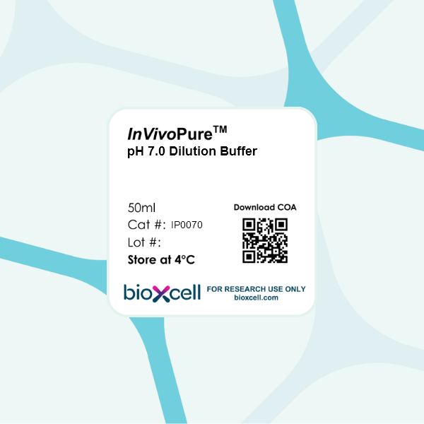
InVivoPure pH 7.0 Dilution Buffer
Product Description
Specifications
| Endotoxin |
≤0.5 EU/mL (≤0.0005EU/μL) Endotoxin level is determined using an LAL gel clotting test |
|---|---|
| Sterility | 0.2 μM filtered |
| Murine Pathogen Tests |
Mouse Norovirus: Negative Mouse Parvovirus: Negative Mouse Minute Virus: Negative Mouse Hepatitis Virus: Negative Reovirus Screen: Negative Lymphocytic Choriomeningitis virus: Negative Lactate Dehydrogenase-Elevating Virus: Negative Mouse Rotavirus: Negative Theiler’s Murine Encephalomyelitis: Negative Ectromelia/Mousepox Virus: Negative Hantavirus: Negative Polyoma Virus: Negative Mouse Adenovirus: Negative Sendai Virus: Negative Mycoplasma Pulmonis: Negative Pneumonia Virus of Mice: Negative Mouse Cytomegalovirus: Negative K Virus: Negative |
| Toxicity Test Results | Nontoxic and nonantigenic in animal models |
| Concentration | 1X |
| Volume | 50 ml |
| Composition |
22 mM Na2HPO4 9.9 mM NaH2PO4 136 mM NaCl This buffer does not contain calcium, magnesium, phenol red, or preservatives such as azide. Keep contents sterile. Open only in a biological safety cabinet. |
| Storage | 4°C |
Product Citations
-
Interferon-γ orchestrates leptomeningeal anti-tumour response.
In Nature on 14 May 2025 by Remšík, J., Tong, X., et al.
PubMed
Metastasis to the cerebrospinal-fluid-filled leptomeninges, or leptomeningeal metastasis, represents a fatal complication of solid tumours1. Multimodal analyses of clinical specimens reveal substantial inflammatory infiltrate in leptomeningeal metastases with enrichment of IFNγ and resulting downstream signalling. Here, to investigate and overcome this futile anti-tumour response within the leptomeninges, we developed syngeneic lung cancer, breast cancer and melanoma leptomeningeal-metastasis mouse models. We show that transgenic host mice lacking IFNγ or its receptor fail to control the growth of leptomeningeal metastases growth. Leptomeningeal overexpression of Ifng through a targeted adeno-associated-virus-based system controls cancer cell growth independent of adaptive immunity. Using a suite of transgenic hosts, we demonstrate that leptomeningeal T cells generate IFNγ to actively recruit and activate peripheral myeloid cells, generating a diverse spectrum of dendritic cell subsets. Independent of antigen presentation, migratory CCR7+ dendritic cells orchestrate the influx, proliferation and cytotoxic action of natural killer cells to control cancer cell growth in the leptomeninges. This study identifies unique, leptomeninges-specific IFNγ signalling and suggests an immune-therapeutic approach against tumours within this space.
-
Lack of consistent effect of dietary fiber on immune checkpoint blockade efficacy across diverse murine tumor models
In bioRxiv on 2 April 2025 by Roichman, A., Reyes-Castellanos, G., et al.
-
Depletion of tumor-derived CXCL5 improves T cell infiltration and anti-PD-1 therapy response in an obese model of pancreatic cancer.
In J Immunother Cancer on 22 March 2025 by Walsh, R. M., Ambrose, J., et al.
PubMed
CXCR1/2 inhibitors are being implemented with immunotherapies in PDAC clinical trials. CXC-ligands are a family of cytokines responsible for stimulating these receptors; while typically secreted by activated immune cells, fibroblasts, and even adipocytes, they are also secreted by immune-evasive cancer cells. CXC-ligand release is known to occur in response to inflammatory stimuli. Adipose tissue is an endocrine organ and a source of inflammatory signaling peptides. Importantly, adipose-derived cytokines and chemokines are implicated as potential drivers of tumor cell immune evasion; cumulatively, these findings suggest that targeting CXC-ligands may be beneficial in the context of obesity.
-
Propionyl-CoA carboxylase subunit B regulates anti-tumor T cells in a pancreatic cancer mouse model.
In Elife on 11 March 2025 by Han, H. V., Efem, R., et al.
PubMed
Most human pancreatic ductal adenocarcinoma (PDAC) are not infiltrated with cytotoxic T cells and are highly resistant to immunotherapy. Over 90% of PDAC have oncogenic KRAS mutations, and phosphoinositide 3-kinases (PI3Ks) are direct effectors of KRAS. Our previous study demonstrated that ablation of Pik3ca in KPC (KrasG12D; Trp53R172H; Pdx1-Cre) pancreatic cancer cells induced host T cells to infiltrate and completely eliminate the tumors in a syngeneic orthotopic implantation mouse model. Now, we show that implantation of Pik3ca-/- KPC (named αKO) cancer cells induces clonal enrichment of cytotoxic T cells infiltrating the pancreatic tumors. To identify potential molecules that can regulate the activity of these anti-tumor T cells, we conducted an in vivo genome-wide gene-deletion screen using αKO cells implanted in the mouse pancreas. The result shows that deletion of propionyl-CoA carboxylase subunit B gene (Pccb) in αKO cells (named p-αKO) leads to immune evasion, tumor progression, and death of host mice. Surprisingly, p-αKO tumors are still infiltrated with clonally enriched CD8+ T cells but they are inactive against tumor cells. However, blockade of PD-L1/PD1 interaction reactivated these clonally enriched T cells infiltrating p-αKO tumors, leading to slower tumor progression and improve survival of host mice. These results indicate that Pccb can modulate the activity of cytotoxic T cells infiltrating some pancreatic cancers and this understanding may lead to improvement in immunotherapy for this difficult-to-treat cancer.