InVivoMAb anti-mouse/human/rat PD-L1
Product Description
Specifications
| Isotype | Mouse IgG1, κ |
|---|---|
| Recommended Isotype Control(s) | InVivoMAb mouse IgG1 isotype control, unknown specificity |
| Recommended Dilution Buffer | InVivoPure pH 7.0 Dilution Buffer |
| Conjugation | This product is unconjugated. Conjugation is available via our Antibody Conjugation Services. |
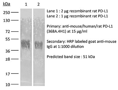
| Immunogen | Human PD-L1-Fc fusion protein |
| Reported Applications |
in vivo PD-L1 blockade in vitro PD-L1 blockade Flow cytometry |
| Formulation |
PBS, pH 7.0 Contains no stabilizers or preservatives |
| Endotoxin |
<2EU/mg (<0.002EU/μg) Determined by LAL assay |
| Purity |
≥95% Determined by SDS-PAGE |
| Sterility | 0.2 µm filtration |
| Production | Purified from tissue culture supernatant in an animal free facility |
| Purification | Protein G |
| RRID | AB_2927520 |
| Molecular Weight | 150 kDa |
| Storage | The antibody solution should be stored at the stock concentration at 4°C. Do not freeze. |
| Need a Custom Formulation? | See All Antibody Customization Options |
Application References
-
Gil Del Alcazar, C. R., et al (2022). "Insights into Immune Escape During Tumor Evolution and Response to Immunotherapy Using a Rat Model of Breast Cancer" Cancer Immunol Res 10(6): 680-697.
PubMed
Animal models are critical for the preclinical validation of cancer immunotherapies. Unfortunately, mouse breast cancer models do not faithfully reproduce the molecular subtypes and immune environment of the human disease. In particular, there are no good murine models of estrogen receptor-positive (ER+) breast cancer, the predominant subtype in patients. Here, we show that Nitroso-N-methylurea-induced mammary tumors in outbred Sprague-Dawley rats recapitulate the heterogeneity for mutational profiles, ER expression, and immune evasive mechanisms observed in human breast cancer. We demonstrate the utility of this model for preclinical studies by dissecting mechanisms of response to immunotherapy using combination TGFBR inhibition and PD-L1 blockade. Short-term treatment of early-stage tumors induced durable responses. Gene expression profiling and spatial mapping classified tumors as inflammatory and noninflammatory, and identified IFNgamma, T-cell receptor (TCR), and B-cell receptor (BCR) signaling, CD74/MHC II, and epithelium-interacting CD8+ T cells as markers of response, whereas the complement system, M2 macrophage phenotype, and translation in mitochondria were associated with resistance. We found that the expression of CD74 correlated with leukocyte fraction and TCR diversity in human breast cancer. We identified a subset of rat ER+ tumors marked by expression of antigen-processing genes that had an active immune environment and responded to treatment. A gene signature characteristic of these tumors predicted disease-free survival in patients with ER+ Luminal A breast cancer and overall survival in patients with metastatic breast cancer receiving anti-PD-L1 therapy. We demonstrate the usefulness of this preclinical model for immunotherapy and suggest examination to expand immunotherapy to a subset of patients with ER+ disease.
-
Gil Del Alcazar CR, Trinh A, Alečković M, Rojas Jimenez E, Harper NW, Oliphant MUJ, Xie S, Krop ED, Lulseged B, Murphy KC, Keenan TE, Van Allen EM, Tolaney SM, Freeman GJ, Dillon DA, Muthuswamy SK, Polyak K (2022). "Insights into Immune Escape During
PubMed
Animal models are critical for the preclinical validation of cancer immunotherapies. Unfortunately, mouse breast cancer models do not faithfully reproduce the molecular subtypes and immune environment of the human disease. In particular, there are no good murine models of estrogen receptor-positive (ER+) breast cancer, the predominant subtype in patients. Here, we show that Nitroso-N-methylurea-induced mammary tumors in outbred Sprague-Dawley rats recapitulate the heterogeneity for mutational profiles, ER expression, and immune evasive mechanisms observed in human breast cancer. We demonstrate the utility of this model for preclinical studies by dissecting mechanisms of response to immunotherapy using combination TGFBR inhibition and PD-L1 blockade. Short-term treatment of early-stage tumors induced durable responses. Gene expression profiling and spatial mapping classified tumors as inflammatory and noninflammatory, and identified IFNγ, T-cell receptor (TCR), and B-cell receptor (BCR) signaling, CD74/MHC II, and epithelium-interacting CD8+ T cells as markers of response, whereas the complement system, M2 macrophage phenotype, and translation in mitochondria were associated with resistance. We found that the expression of CD74 correlated with leukocyte fraction and TCR diversity in human breast cancer. We identified a subset of rat ER+ tumors marked by expression of antigen-processing genes that had an active immune environment and responded to treatment. A gene signature characteristic of these tumors predicted disease-free survival in patients with ER+ Luminal A breast cancer and overall survival in patients with metastatic breast cancer receiving anti-PD-L1 therapy. We demonstrate the usefulness of this preclinical model for immunotherapy and suggest examination to expand immunotherapy to a subset of patients with ER+ disease. See related Spotlight by Roussos Torres, p. 672.
-
Gil Del Alcazar, C. R., et al (2022). "Insights into Immune Escape During Tumor Evolution and Response to Immunotherapy Using a Rat Model of Breast Cancer" Cancer Immunol Res 10(6): 680-697.
PubMed
Animal models are critical for the preclinical validation of cancer immunotherapies. Unfortunately, mouse breast cancer models do not faithfully reproduce the molecular subtypes and immune environment of the human disease. In particular, there are no good murine models of estrogen receptor-positive (ER+) breast cancer, the predominant subtype in patients. Here, we show that Nitroso-N-methylurea-induced mammary tumors in outbred Sprague-Dawley rats recapitulate the heterogeneity for mutational profiles, ER expression, and immune evasive mechanisms observed in human breast cancer. We demonstrate the utility of this model for preclinical studies by dissecting mechanisms of response to immunotherapy using combination TGFBR inhibition and PD-L1 blockade. Short-term treatment of early-stage tumors induced durable responses. Gene expression profiling and spatial mapping classified tumors as inflammatory and noninflammatory, and identified IFNgamma, T-cell receptor (TCR), and B-cell receptor (BCR) signaling, CD74/MHC II, and epithelium-interacting CD8+ T cells as markers of response, whereas the complement system, M2 macrophage phenotype, and translation in mitochondria were associated with resistance. We found that the expression of CD74 correlated with leukocyte fraction and TCR diversity in human breast cancer. We identified a subset of rat ER+ tumors marked by expression of antigen-processing genes that had an active immune environment and responded to treatment. A gene signature characteristic of these tumors predicted disease-free survival in patients with ER+ Luminal A breast cancer and overall survival in patients with metastatic breast cancer receiving anti-PD-L1 therapy. We demonstrate the usefulness of this preclinical model for immunotherapy and suggest examination to expand immunotherapy to a subset of patients with ER+ disease.
Product Citations
-
Idarubicin-loaded degradable hydrogel for TACE therapy enhances anti-tumor immunity in hepatocellular carcinoma.
In Mater Today Bio on 1 December 2024 by Zhang, X., Deng, X., et al.
PubMed
Hepatocellular carcinoma (HCC) is a common and deadly cancer, often diagnosed at advanced stages, limiting surgical options. Transcatheter arterial chemoembolization (TACE) is a primary treatment for inoperable and involves the use of drug-eluting microspheres to slowly release chemotherapy drugs. However, patient responses to TACE vary, with some experiencing tumor progression and recurrence. Traditional TACE uses agents like oil-based drug emulsions and polyvinyl alcohol particles, which can permanently block blood vessels and increase tumor hypoxia. Additionally, TACE can suppress the immune system by reducing immune cell numbers and function, contributing to poor treatment outcomes. New approaches, like TACE using degradable starch microspheres and hydrogel-based materials, offer the potential to create different tumor environments that could improve both safety and efficacy. In our research, we developed a composite hydrogel (IF@Gel) made of Poloxamer-407 gel and Fe3O4 nanoparticles, loaded with idarubicin, to use as an embolic material for TACE in a rat model of orthotopic HCC. We observed promising therapeutic effects and investigated the impact on the tumor immune microenvironment, focusing on the role of immunogenic cell death (ICD). The composite hydrogel demonstrated excellent potential as an embolic material for TACE, and IF@Gel-based TACE demonstrated significant efficacy in rat HCC. Furthermore, our findings highlight the potential synergistic effects of ICD with anti-PD-L1 therapy, providing new insights into HCC treatment strategies. This study aims to provide improved treatment options for HCC and to deepen our understanding of the mechanisms of TACE and tumor environment regulation.
-
Midkine as a driver of age-related changes and increase in mammary tumorigenesis.
In Cancer Cell on 11 November 2024 by Yan, P., Jimenez, E. R., et al.
PubMed
Aging is a pivotal risk factor for cancer, yet the underlying mechanisms remain poorly defined. Here, we explore age-related changes in the rat mammary gland by single-cell multiomics. Our findings include increased epithelial proliferation, loss of luminal identity, and decreased naive B and T cells with age. We discover a luminal progenitor population unique to old rats with profiles reflecting precancerous changes and identify midkine (Mdk) as a gene upregulated with age and a regulator of age-related luminal progenitors. Midkine treatment of young rats mimics age-related changes via activating PI3K-AKT-SREBF1 pathway and promotes nitroso-N-methylurea-induced mammary tumorigenesis. Midkine levels increase with age in human blood and mammary epithelium, and higher MDK in normal breast tissue is associated with higher breast cancer risk in younger women. Our findings reveal a link between aging and susceptibility to tumor initiation and identify midkine as a mediator of age-dependent increase in breast tumorigenesis.
-
Tumor-colonized Streptococcus mutans metabolically reprograms tumor microenvironment and promotes oral squamous cell carcinoma.
In Microbiome on 5 October 2024 by Zhou, J., Hu, Z., et al.
PubMed
Oral squamous cell carcinoma (OSCC) remains a major death cause in head and neck cancers, but the exact pathogenesis mechanisms of OSCC are largely unclear.
-
EPDR1 promotes PD-L1 expression and tumor immune evasion by inhibiting TRIM21-dependent ubiquitylation of IkappaB kinase-β.
In EMBO J on 1 October 2024 by Qian, X., Cai, J., et al.
PubMed
While immune checkpoint blockade (ICB) has shown promise for clinical cancer therapy, its efficacy has only been observed in a limited subset of patients and the underlying mechanisms regulating innate and acquired resistance to ICB of tumor cells remain poorly understood. Here, we identified ependymin-related protein 1 (EPDR1) as an important tumor-intrinsic regulator of PD-L1 expression and tumor immune evasion. Aberrant expression of EPDR1 in hepatocellular carcinoma is associated with immunosuppression. Mechanistically, EPDR1 binds to E3 ligase TRIM21 and disrupts its interaction with IkappaB kinase-b, suppressing its ubiquitylation and autophagosomal degradation and enhancing NF-κB-mediated transcriptional activation of PD-L1. Further, we validated through a mouse liver cancer model that EPDR1 mediates exhaustion of CD8+ T cells and promotes tumor progression. In addition, we observed a positive correlation between EPDR1 and PD-L1 expression in both human and mouse liver cancer samples. Collectively, our study reveals a previously unappreciated role of EPDR1 in orchestrating tumor immune evasion and cancer progression.