InVivoMAb anti-human PD-L1 (B7-H1)
Product Description
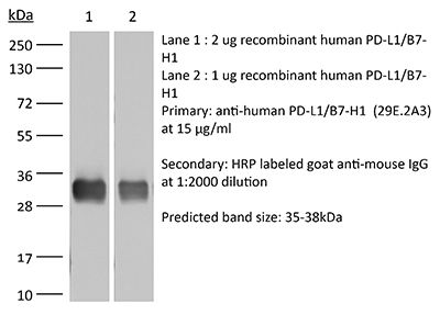
Specifications
| Isotype | Mouse IgG2b, κ |
|---|---|
| Recommended Isotype Control(s) | InVivoMAb mouse IgG2b isotype control, unknown specificity |
| Recommended Dilution Buffer | InVivoPure pH 7.0 Dilution Buffer |
| Conjugation | This product is unconjugated. Conjugation is available via our Antibody Conjugation Services. |
| Immunogen | Full length human PD-L1 |
| Reported Applications |
in vitro PD-L1 blockade Functional assays Immunohistochemistry (frozen) Flow cytometry in vitro Organoids/Organ-on-Chip |
| Formulation |
PBS, pH 7.0 Contains no stabilizers or preservatives |
| Endotoxin |
≤1EU/mg (≤0.001EU/μg) Determined by LAL assay |
| Purity |
≥95% Determined by SDS-PAGE |
| Sterility | 0.2 µm filtration |
| Production | Purified from cell culture supernatant in an animal-free facility |
| Purification | Protein G |
| RRID | AB_2687808 |
| Molecular Weight | 150 kDa |
| Storage | The antibody solution should be stored at the stock concentration at 4°C. Do not freeze. |
| Need a Custom Formulation? | See All Antibody Customization Options |
Application References
-
Broos, K., et al (2019). "Single Domain Antibody-Mediated Blockade of Programmed Death-Ligand 1 on Dendritic Cells Enhances CD8 T-cell Activation and Cytokine Production" Vaccines (Basel) 7(3).
PubMed
Dendritic cell [DC] vaccines can induce durable clinical responses, at least in a fraction of previously treated, late stage cancer patients. Several preclinical studies suggest that shielding programmed death-ligand 1 [PD-L1] on the DC surface may be an attractive strategy to extend such clinical benefits to a larger patient population. In this study, we evaluated the use of single domain antibody [sdAb] K2, a high affinity, antagonistic, PD-L1 specific sdAb, for its ability to enhance DC mediated T-cell activation and benchmarked it against the use of the monoclonal antibodies [mAbs], MIH1, 29E.2A3 and avelumab. Similar to mAbs, sdAb K2 enhanced antigen-specific T-cell receptor signaling in PD-1 positive (PD-1(pos)) reporter cells activated by DCs. We further showed that the activation and function of antigen-specific CD8 positive (CD8(pos)) T cells, activated by DCs, was enhanced by inclusion of sdAb K2, but not mAbs. The failure of mAbs to enhance T-cell activation might be explained by their low efficacy to bind PD-L1 on DCs when compared to binding of PD-L1 on non-immune cells, whereas sdAb K2 shows high binding to PD-L1 on immune as well as non-immune cells. These data provide a rationale for the inclusion of sdAb K2 in DC-based immunotherapy strategies.
-
Sivakumar, R., et al (2019). "Organotypic tumor slice cultures provide a versatile platform for immuno-oncology and drug discovery" Oncoimmunology 8(12): e1670019.
PubMed
Organotypic tumor slices represent a physiologically-relevant culture system for studying the tumor microenvironment. Systematic characterization of the tumor slice culture system will enable its effective application for translational research. Here, using flow cytometry-based immunophenotyping, we performed a comprehensive characterization of the immune cell composition in organotypic tumor slices prepared from four syngeneic mouse tumor models and a human liver tumor. We found that the immune cell compositions of organotypic tumor slices prepared on the same day as the tumor cores were harvested are similar. Differences were primarily observed in the lymphocyte population of a clinical hepatocellular carcinoma case. Viable populations of immune cells persisted in the tumor slices for 7 days. Despite some changes in the immune cell populations, we showed the utility of mouse tumor slices for assessing responses to immune-modulatory agents. Further, we demonstrated the ability to use patient-derived xenograft tumor slices for assessing responses to targeted and cytotoxic drugs. Overall, tumor slices provide a broadly useful platform for studying the tumor microenvironment and evaluating the preclinical efficacy of cancer therapeutics.
-
Porichis, F., et al (2014). "Differential impact of PD-1 and/or interleukin-10 blockade on HIV-1-specific CD4 T cell and antigen-presenting cell functions" J Virol 88(5): 2508-2518.
PubMed
Antigen persistence in chronic infections and cancer upregulates inhibitory networks, such as the PD-1 and interleukin-10 (IL-10) pathways, that impair immunity and lead to disease progression. These pathways are attractive targets for immunotherapy, as demonstrated by recent clinical trials of PD-1/PD-L1 blockade in cancer patients. However, in HIV-1 infection not all subjects respond to inhibition of either pathway and the mechanistic interactions between these two networks remain to be better defined. Here we demonstrate that in vitro blockade of PD-L1 and/or IL-10Ralpha results in markedly different profiles of HIV-1-specific CD4 T cell restoration. Whereas PD-L1 blockade leads to balanced increase in gamma interferon (IFN-gamma), IL-2, and IL-13 secretion, IL-10Ralpha blockade preferentially restores IFN-gamma production. In viremic subjects, combined PD-L1/IL-10Ralpha blockade results in a striking 10-fold increase in IFN-gamma secretion by HIV-1-specific CD4 T cells that is not observed in subjects with spontaneous (elite controllers) or therapy-induced control of viral replication. In contrast to the dramatic increase in IFN-gamma production, concurrent blockade has a marginal additive effect on IL-2 production, IL-13 secretion, and HIV-1-specific CD4 T cell proliferation. IFN-gamma produced by Thelper cells upregulates PD-L1, HLA I/II, and IL-12 expression by monocytes. The effect of combined blockade on IFN-gamma was dependent on reciprocal reinforcement through IL-12. These studies provide crucial information on the different immunoregulatory qualities of PD-1 and IL-10 in progressive disease and link exhausted virus-specific CD4 T cells and monocytes in the regulation of IFN-gamma and IL-12 secretion. IMPORTANCE: Infection with HIV results in most people in uncontrolled viral replication and progressive weakening of the body defenses. In the absence of antiviral therapy, this process results in clinical disease, or AIDS. An important reason why HIV continues to multiply is that a population of white blood cells called CD4 T cells that targets the virus fails to work properly. At least part of this impairment is under the control of inhibitory mechanisms that can be blocked to improve the function of these CD4 T cells. In this report, we show that blocking one or two of the molecules involved, called PD-1 and IL-10, has different effects on the individual functions of these cells and that one is strongly improved. We investigate how these effects are caused by interactions between CD4 T cells and antigen-presenting cells. These observations can have implications for new therapeutic approaches in HIV infection.
-
Hegde, S., et al (2011). "Human NKT cells direct the differentiation of myeloid APCs that regulate T cell responses via expression of programmed cell death ligands" J Autoimmun 37(1): 28-38.
PubMed
NKT cells are innate lymphocytes that can recognize self or foreign lipids presented by CD1d molecules. NKT cells have been shown to inhibit the development of autoimmunity in murine model systems, however, the pathways by which they foster immune tolerance remain poorly understood. Here we show that autoreactive human NKT cells stimulate monocytes to differentiate into myeloid APCs that have a regulatory phenotype characterized by poor conjugate formation with T cells. The NKT cell instructed myeloid APCs show elevated expression of the inhibitory ligand PD-L2, and blocking PD-L1 and PD-L2 during interactions of the APCs with T cells results in improved cluster formation and significantly increased T cell proliferative responses. The elevated expression of PD-L molecules on NKT-instructed APCs appears to result from exposure to extracellular ATP that is produced during NKT-monocyte interactions, and blocking purinergic signaling during monocyte differentiation results in APCs that form clusters with T cells and stimulate their proliferation. Finally, we show that human monocytes and NKT cells that are injected into immunodeficient mice co-localize together in spleen and liver, and after 3 days in vivo in the presence of NKT cells a fraction of the myeloid cells have upregulated markers associated with differentiation into professional APCs. These results suggest that autoreactive human NKT cells may promote tolerance by inducing the differentiation of regulatory myeloid APCs that limit T cell proliferation through expression of PD-L molecules.
Product Citations
-
Single Domain Antibody-Mediated Blockade of Programmed Death-Ligand 1 on Dendritic Cells Enhances CD8 T-cell Activation and Cytokine Production.
In Vaccines on 7 August 2019 by Broos, K., Lecocq, Q., et al.
PubMed
Dendritic cell [DC] vaccines can induce durable clinical responses, at least in a fraction of previously treated, late stage cancer patients. Several preclinical studies suggest that shielding programmed death-ligand 1 [PD-L1] on the DC surface may be an attractive strategy to extend such clinical benefits to a larger patient population. In this study, we evaluated the use of single domain antibody [sdAb] K2, a high affinity, antagonistic, PD-L1 specific sdAb, for its ability to enhance DC mediated T-cell activation and benchmarked it against the use of the monoclonal antibodies [mAbs], MIH1, 29E.2A3 and avelumab. Similar to mAbs, sdAb K2 enhanced antigen-specific T-cell receptor signaling in PD-1 positive (PD-1pos) reporter cells activated by DCs. We further showed that the activation and function of antigen-specific CD8 positive (CD8pos) T cells, activated by DCs, was enhanced by inclusion of sdAb K2, but not mAbs. The failure of mAbs to enhance T-cell activation might be explained by their low efficacy to bind PD-L1 on DCs when compared to binding of PD-L1 on non-immune cells, whereas sdAb K2 shows high binding to PD-L1 on immune as well as non-immune cells. These data provide a rationale for the inclusion of sdAb K2 in DC-based immunotherapy strategies.
-
Design of a bispecific peptide-nanozyme conjugate for cancer immunotherapy.
In Cell Rep Med on 17 February 2026 by Chen, D., Xu, R., et al.
PubMed
Despite advances in cancer immunotherapy, clinical efficacy remains constrained by immunosuppressive tumor microenvironment (TME), including PD-L1-mediated T cell dysfunction and CXCL8-driven myeloid cell recruitment. To address this, a bispecific peptide-nanozyme conjugate (BsPNEC) is engineered. Leveraging iterative structure-guided optimization, we first develop q6w, a proteolysis-resistant D-peptide targeting CXCR1/2, and conjugate it to a PD-L1-blocking peptide to generate a bispecific peptide qGA. To augment the therapeutic efficacy, qGA is conjugated to Fe3O4 nanozymes with peroxidase-mimetic activity. The Fe3O4 nanozymes catalytically decompose H2O2 into reactive oxygen species (ROS), thus activating the cGAS-STING pathway to potentiate CD8+ T cell infiltration and activation in anti-PD-1-resistant tumor model. The BsPNEC platform integrates tumor-targeted delivery, magnetic resonance imaging (MRI) contrast capabilities, and robust inhibition of tumor growth. Our findings present a synergistic immunotherapeutic strategy that simultaneously skews immunosuppressive TME and amplifies T cell immune response.
-
Fluorescence Lifetime Imaging Enables In Vivo Quantification of PD-L1 Expression and Intertumoral Heterogeneity.
In Cancer Res on 1 February 2025 by Pal, R., Murali, K., et al.
PubMed
Patient selection for cancer immunotherapy requires precise, quantitative readouts of biomarker expression in intact tumors that can be reliably compared across multiple subjects over time. The current clinical standard biomarker for assessing immunotherapy response is PD-L1 expression, typically quantified using IHC. This method, however, only provides snapshots of PD-L1 expression status in microscopic regions of ex vivo specimens. Although various targeted probes have been investigated for in vivo imaging of PD-L1, nonspecific probe accumulation within the tumor microenvironment has hindered accurate quantification, limiting the utility for preclinical and clinical studies. Here, we demonstrated that in vivo time-domain fluorescence imaging of an anti-PD-L1 antibody tagged with the near-infrared fluorophore IRDye 800CW (αPDL1-800) can yield quantitative estimates of baseline tumor PD-L1 heterogeneity across untreated mice, as well as variations in PD-L1 expression in mice undergoing clinically relevant anti-PD-1 treatment. The fluorescence lifetime (FLT) of PD-L1-bound αPDL1-800 was significantly longer than the FLT of nonspecifically accumulated αPDL1-800 in the tumor microenvironment. This FLT contrast allowed quantification of PD-L1 expression across mice both in superficial breast tumors using planar FLT imaging and in deep-seated liver tumors (>5 mm depth) using the asymptotic time-domain algorithm for fluorescence tomography. These findings suggest that FLT imaging can accelerate the preclinical investigation and clinical translation of new immunotherapy treatments by enabling robust quantification of receptor expression across subjects. Significance: Fluorescence lifetime imaging can quantify PD-L1 expression across multiple mice undergoing anti-PD-1 treatment, providing a critically needed noninvasive imaging method to quantify immunotherapy targets in vivo.
-
Development of a peptide-based tumor-activated checkpoint inhibitor for cancer immunotherapy.
In Acta Biomater on 24 January 2025 by Zhao, Z., Fetse, J., et al.
PubMed
Antibody-based checkpoint inhibitors have achieved great success in cancer immunotherapy, but their uncontrollable immune-related adverse events remain a major challenge. In this study, we developed a tumor-activated nanoparticle that is specifically active in tumors but not in normal tissues. We discovered a short anti-PD-L1 peptide that blocks the PD-1/PD-L1 interaction. The peptide was modified with a PEG chain through a novel matrix metalloproteinase-2 (MMP-2)-specific cleavage linker. The modified TR3 peptide self-assembles into a micelle-like nanoparticle (TR3-M-NP), which remains inactive and unable to block the PD-1/PD-L1 interaction in its native form. However, upon cleavage by MMP-2 in tumors, it releases the active peptide. The TR3-M-NP5k nanoparticle was specifically activated in tumors through enzyme-mediated cleavage, leading to the inhibition of tumor growth and extended survival compared to control groups. In summary, TR3-M-NP shows great potential as a tumor-responsive immunotherapy agent with reduced toxicities. STATEMENT OF SIGNIFICANCE: In this study, we developed a bioactive peptide-based checkpoint inhibitor that is active only in tumors and not in normal tissues, thereby potentially avoiding immune-related adverse effects. We discovered a short anti-PD-L1 peptide, TR3, that blocks the PD-1/PD-L1 interaction. We chemically modified the TR3 peptide to self-assemble into a micelle-like nanoparticle (TR3-M-NP), which itself cannot block the PD-1/PD-L1 interaction but releases the active TR3 peptide in tumors upon cleavage by MMP-2. In contrast, the nanoparticle is randomly degraded in normal tissues into peptides fragments that cannot block the PD-1/PD-L1 interaction. Upon intraperitoneal injection, TR3-M-NP5k was activated specifically in tumors through enzyme cleavage, leading to the inhibition of tumor growth and extended survival compared to the control groups. In summary, TR3-M-NP holds significant promise as a tumor-responsive immunotherapy agent with reduced toxicities. The bioactive platform has the potential to be used for other types of checkpoint inhibitor.