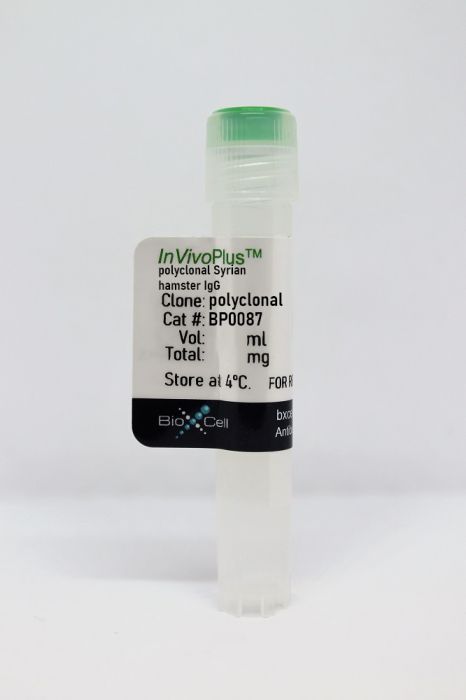
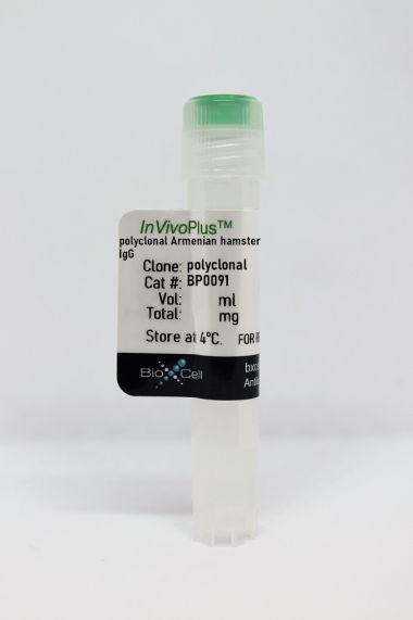
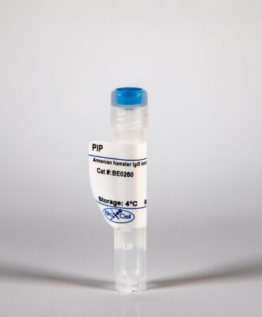
InVivoPlus polyclonal Syrian hamster IgG
Product Details
The polyclonal Syrian hamster IgG is purified from Syrian hamster serum. It is ideal for use as a non-reactive control IgG for Syrian hamster antibodies in most in vivo and in vitro applications.Specifications
| Isotype | Syrian hamster IgG |
|---|---|
| Recommended Dilution Buffer | InVivoPure pH 7.0 Dilution Buffer |
| Conjugation | This product is unconjugated. Conjugation is available via our Antibody Conjugation Services. |
| Formulation |
PBS, pH 7.0 Contains no stabilizers or preservatives |
| Aggregation* |
<5% Determined by SEC |
| Purity |
>95% Determined by SDS-PAGE |
| Sterility | 0.2 µm filtration |
| Purification | Protein G |
| RRID | AB_1107782 |
| Molecular Weight | 150 kDa |
| Murine Pathogen Tests* |
Ectromelia/Mousepox Virus: Negative Hantavirus: Negative K Virus: Negative Lactate Dehydrogenase-Elevating Virus: Negative Lymphocytic Choriomeningitis virus: Negative Mouse Adenovirus: Negative Mouse Cytomegalovirus: Negative Mouse Hepatitis Virus: Negative Mouse Minute Virus: Negative Mouse Norovirus: Negative Mouse Parvovirus: Negative Mouse Rotavirus: Negative Mycoplasma Pulmonis: Negative Pneumonia Virus of Mice: Negative Polyoma Virus: Negative Reovirus Screen: Negative Sendai Virus: Negative Theiler’s Murine Encephalomyelitis: Negative |
| Storage | The antibody solution should be stored at the stock concentration at 4°C. Do not freeze. |
Additional Formats
Recommended Products
Bradley, T., et al. (2020). "Immune checkpoint modulation enhances HIV-1 antibody induction" Nat Commun 11(1): 948. PubMed
Eliciting protective titers of HIV-1 broadly neutralizing antibodies (bnAbs) is a goal of HIV-1 vaccine development, but current vaccine strategies have yet to induce bnAbs in humans. Many bnAbs isolated from HIV-1-infected individuals are encoded by immunoglobulin gene rearrangments with infrequent naive B cell precursors and with unusual genetic features that may be subject to host regulatory control. Here, we administer antibodies targeting immune cell regulatory receptors CTLA-4, PD-1 or OX40 along with HIV envelope (Env) vaccines to rhesus macaques and bnAb immunoglobulin knock-in (KI) mice expressing diverse precursors of CD4 binding site HIV-1 bnAbs. CTLA-4 blockade augments HIV-1 Env antibody responses in macaques, and in a bnAb-precursor mouse model, CTLA-4 blocking or OX40 agonist antibodies increase germinal center B and T follicular helper cells and plasma neutralizing antibodies. Thus, modulation of CTLA-4 or OX40 immune checkpoints during vaccination can promote germinal center activity and enhance HIV-1 Env antibody responses.
Klepsch, V., et al. (2020). "Targeting the orphan nuclear receptor NR2F6 in T cells primes tumors for immune checkpoint therapy" Cell Commun Signal 18(1): 8. PubMed
BACKGROUND: NR2F6 has been proposed as an alternative cancer immune checkpoint in the effector T cell compartment. However, a realistic assessment of the in vivo therapeutic potential of NR2F6 requires acute depletion. METHODS: Employing primary T cells isolated from Cas9-transgenic mice for electroporation of chemically synthesized sgRNA, we established a CRISPR/Cas9-mediated acute knockout protocol of Nr2f6 in primary mouse T cells. RESULTS: Analyzing these Nr2f6(CRISPR/Cas9 knockout) T cells, we reproducibly observed a hyper-reactive effector phenotype upon CD3/CD28 stimulation in vitro, highly reminiscent to Nr2f6(-/-) T cells. Importantly, CRISPR/Cas9-mediated Nr2f6 ablation prior to adoptive cell therapy (ACT) of autologous polyclonal T cells into wild-type tumor-bearing recipient mice in combination with PD-L1 or CTLA-4 tumor immune checkpoint blockade significantly delayed MC38 tumor progression and induced superior survival, thus further validating a T cell-inhibitory function of NR2F6 during tumor progression. CONCLUSIONS: These findings indicate that Nr2f6(CRISPR/Cas9 knockout) T cells are comparable to germline Nr2f6(-/-) T cells, a result providing an independent confirmation of the immune checkpoint function of lymphatic NR2F6. Taken together, CRISPR/Cas9-mediated acute Nr2f6 gene ablation in primary mouse T cells prior to ACT appeared feasible for potentiating established PD-L1 and CTLA-4 blockade therapies, thereby pioneering NR2F6 inhibition as a sensitizing target for augmented tumor regression. Video abstract.
Wang, Q., et al. (2019). "Single-cell profiling guided combinatorial immunotherapy for fast-evolving CDK4/6 inhibitor-resistant HER2-positive breast cancer" Nat Commun 10(1): 3817. PubMed
Acquired resistance to targeted cancer therapy is a significant clinical challenge. In parallel with clinical trials combining CDK4/6 inhibitors to treat HER2+ breast cancer, we sought to prospectively model tumor evolution in response to this regimen in vivo and identify a clinically actionable strategy to combat drug resistance. Despite a promising initial response, acquired resistance emerges rapidly to the combination of anti-HER2/neu antibody and CDK4/6 inhibitor Palbociclib. Using high-throughput single-cell profiling over the course of treatments, we reveal a distinct immunosuppressive immature myeloid cell (IMC) population to infiltrate the resistant tumors. Guided by single-cell transcriptome analysis, we demonstrate that combination of IMC-targeting tyrosine kinase inhibitor cabozantinib and immune checkpoint blockade enhances anti-tumor immunity, and overcomes the resistance. Furthermore, sequential combinatorial immunotherapy enables a sustained control of the fast-evolving CDK4/6 inhibitor-resistant tumors. Our study demonstrates a translational framework for treating rapidly evolving tumors through preclinical modeling and single-cell analyses.
Su, W., et al. (2019). "The Polycomb Repressor Complex 1 Drives Double-Negative Prostate Cancer Metastasis by Coordinating Stemness and Immune Suppression" Cancer Cell 36(2): 139-155.e110. PubMed
The mechanisms that enable immune evasion at metastatic sites are poorly understood. We show that the Polycomb Repressor Complex 1 (PRC1) drives colonization of the bones and visceral organs in double-negative prostate cancer (DNPC). In vivo genetic screening identifies CCL2 as the top prometastatic gene induced by PRC1. CCL2 governs self-renewal and induces the recruitment of M2-like tumor-associated macrophages and regulatory T cells, thus coordinating metastasis initiation with immune suppression and neoangiogenesis. A catalytic inhibitor of PRC1 cooperates with immune checkpoint therapy to reverse these processes and suppress metastasis in genetically engineered mouse transplantation models of DNPC. These results reveal that PRC1 coordinates stemness with immune evasion and neoangiogenesis and point to the potential clinical utility of targeting PRC1 in DNPC.
Binnewies, M., et al. (2019). "Unleashing Type-2 Dendritic Cells to Drive Protective Antitumor CD4(+) T Cell Immunity" Cell 177(3): 556-571.e516. PubMed
Differentiation of proinflammatory CD4(+) conventional T cells (T(conv)) is critical for productive antitumor responses yet their elicitation remains poorly understood. We comprehensively characterized myeloid cells in tumor draining lymph nodes (tdLN) of mice and identified two subsets of conventional type-2 dendritic cells (cDC2) that traffic from tumor to tdLN and present tumor-derived antigens to CD4(+) T(conv), but then fail to support antitumor CD4(+) T(conv) differentiation. Regulatory T cell (T(reg)) depletion enhanced their capacity to elicit strong CD4(+) T(conv) responses and ensuing antitumor protection. Analogous cDC2 populations were identified in patients, and as in mice, their abundance relative to T(reg) predicts protective ICOS(+) PD-1(lo) CD4(+) T(conv) phenotypes and survival. Further, in melanoma patients with low T(reg) abundance, intratumoral cDC2 density alone correlates with abundant CD4(+) T(conv) and with responsiveness to anti-PD-1 therapy. Together, this highlights a pathway that restrains cDC2 and whose reversal enhances CD4(+) T(conv) abundance and controls tumor growth.
Lymphatic platelet thrombosis limits bone repair by precluding lymphatic transporting DAMPs
Preprint on Research Square on 14 November 2023 by Wang, Y., Zheng, Y., et al.
PubMed
Lymphatic vessels (LVs) interdigitated with blood vessels, travel and form an extensive transport network in the musculoskeletal system. Blood vessels in bone regulate osteogenesis and hematopoiesis, however, whether LVs in bone affect fracture healing is unclear. Here, by near infrared indocyanine green lymphatic imaging (NIR-ICG), we examined lymphatic draining function at the tibial fracture sites and found lymphatic drainage insufficiency (LDI) occurred as early as two weeks after fracture. Sufficient lymphatic drainage facilitates fracture healing. In addition, we identified that lymphatic platelet thrombosis (LPT) blocks the draining lymphoid sinus and LVs, caused LDI and then inhibited fracture healing, which can be rescued by a pharmacological approach. Moreover, unblocked lymphatic drainage decreased neutrophils and increased M2-like macrophages of hematoma niche to support osteoblast (OB) survival and bone marrow-derived mesenchymal stem cell (BMSC) proliferation via transporting damage-associated molecular patterns (DAMPs). These findings demonstrate that LPT limits bone regeneration by blocking lymphatic drainage from transporting DAMPs. Together, these findings represent a novel way forward in the treatment of bone repair.
- Cancer Research,
- Genetics
Defining the spatial distribution of extracellular adenosine revealed a myeloid-dependent immunosuppressive microenvironment in pancreatic ductal adenocarcinoma.
In Journal for Immunotherapy of Cancer on 1 August 2023 by Graziano, V., Dannhorn, A., et al.
PubMed
The prognosis for patients with pancreatic ductal adenocarcinoma (PDAC) remains extremely poor. It has been suggested that the adenosine pathway contributes to the ability of PDAC to evade the immune system and hence, its resistance to immuno-oncology therapies (IOT), by generating extracellular adenosine (eAdo). Using genetically engineered allograft models of PDAC in syngeneic mice with defined and different immune infiltration and response to IOT and autochthonous tumors in KPC mice we investigated the impact of the adenosine pathway on the PDAC tumor microenvironment (TME). Flow cytometry and imaging mass cytometry (IMC) were used to characterize the subpopulation frequency and spatial distribution of tumor-infiltrating immune cells. Mass spectrometry imaging (MSI) was used to visualize adenosine compartmentalization in the PDAC tumors. RNA sequencing was used to evaluate the influence of the adenosine pathway on the shaping of the immune milieu and correlate our findings to published data sets in human PDAC. We demonstrated high expression of adenosine pathway components in tumor-infiltrating immune cells (particularly myeloid populations) in the murine models. MSI demonstrated that extracellular adenosine distribution is heterogeneous in tumors, with high concentrations in peri-necrotic, hypoxic regions, associated with rich myeloid infiltration, demonstrated using IMC. Protumorigenic M2 macrophages express high levels of the Adora2a receptor; particularly in the IOT resistant model. Blocking the in vivo formation and function of eAdo (Adoi), using a combination of anti-CD73 antibody and an Adora2a inhibitor slowed tumor growth and reduced metastatic burden. Additionally, blocking the adenosine pathway improved the efficacy of combinations of cytotoxic agents or immunotherapy. Adoi remodeled the TME, by reducing the infiltration of M2 macrophages and regulatory T cells. RNA sequencing analysis showed that genes related to immune modulation, hypoxia and tumor stroma were downregulated following Adoi and a specific adenosine signature derived from this is associated with a poorer prognosis in patients with PDAC. The formation of eAdo promotes the development of the immunosuppressive TME in PDAC, contributing to its resistance to conventional and novel therapies. Therefore, inhibition of the adenosine pathway may represent a strategy to modulate the PDAC immune milieu and improve therapy response in patients with PDAC. © Author(s) (or their employer(s)) 2023. Re-use permitted under CC BY. Published by BMJ.
- Cancer Research,
- Immunology and Microbiology
Tumor Treating Fields (TTFields) Concomitant with Immune Checkpoint Inhibitors Are Therapeutically Effective in Non-Small Cell Lung Cancer (NSCLC) In Vivo Model.
In International Journal of Molecular Sciences on 15 November 2022 by Barsheshet, Y., Voloshin, T., et al.
PubMed
Tumor Treating Fields (TTFields) are electric fields that exert physical forces to disrupt cellular processes critical for cancer cell viability and tumor progression. TTFields induce anti-mitotic effects through the disruption of the mitotic spindle and abnormal chromosome segregation, which trigger several forms of cell death, including immunogenic cell death (ICD). The efficacy of TTFields concomitant with anti-programmed death-1 (anti-PD-1) treatment was previously shown in vivo and is currently under clinical investigation. Here, the potential of TTFields concomitant with anti- PD-1/anti-cytotoxic T-lymphocyte-associated protein 4 (anti-CTLA-4) or anti-programmed death-ligand 1 (anti-PD-L1) immune checkpoint inhibitors (ICI) to improve therapeutic efficacy was examined in lung tumor-bearing mice. Increased circulating levels of high mobility group box 1 protein (HMGB1) and elevated intratumoral levels of phosphorylated eukaryotic translation initiation factor 2α (p-eIF2α) were found in the TTFields-treated mice, indicative of ICD induction. The concomitant application of TTFields and ICI led to a significant decrease in tumor volume as compared to all other groups. In addition, significant increases in the number of tumor-infiltrating immune cells, specifically cytotoxic T-cells, were observed in the TTFields plus anti-PD-1/anti-CTLA-4 or anti-PD-L1 groups. Correspondingly, cytotoxic T-cells isolated from these tumors showed higher levels of IFN-γ production. Collectively, these results suggest that TTFields have an immunoactivating role that may be leveraged for concomitant treatment with ICI to achieve better tumor control by enhancing antitumor immunity.
- Immunology and Microbiology,
- Neuroscience
CD8+ T cells induce interferon-responsive oligodendrocytes and microglia in white matter aging.
In Nature Neuroscience on 1 November 2022 by Kaya, T., Mattugini, N., et al.
PubMed
A hallmark of nervous system aging is a decline of white matter volume and function, but the underlying mechanisms leading to white matter pathology are unknown. In the present study, we found age-related alterations of oligodendrocyte cell state with a reduction in total oligodendrocyte density in aging murine white matter. Using single-cell RNA-sequencing, we identified interferon (IFN)-responsive oligodendrocytes, which localize in proximity to CD8+ T cells in aging white matter. Absence of functional lymphocytes decreased the number of IFN-responsive oligodendrocytes and rescued oligodendrocyte loss, whereas T-cell checkpoint inhibition worsened the aging response. In addition, we identified a subpopulation of lymphocyte-dependent, IFN-responsive microglia in the vicinity of the CD8+ T cells in aging white matter. In summary, we provide evidence that CD8+ T-cell-induced, IFN-responsive oligodendrocytes and microglia are important modifiers of white matter aging. © 2022. The Author(s).
- Cancer Research,
- Genetics
The heterogeneous distribution of extracellular adenosine reveals a myeloid-dependent axis, shaping the immunosuppressive microenvironment in pancreatic ductal adenocarcinoma
Preprint on BioRxiv : the Preprint Server for Biology on 25 May 2022 by Graziano, V., Dannhorn, A., et al.
PubMed
The prognosis for patients with pancreatic ductal adenocarcinoma (PDAC) remains extremely poor. It has been suggested that the adenosine pathway contributes to the ability of PDAC to evade the immune system and its resistance to immunotherapies (Immuno-Oncology Therapy, IOT), by generating extracellular adenosine (eAdo). Using syngeneic genetically engineered mouse allograft models of PDAC with differential immune infiltration and response to IOT, we showed enrichment of the adenosine pathway in tumour-infiltrating immune cells (in particular, myeloid populations). Extracellular adenosine distribution is heterogeneous in tumours, with high concentrations in hypoxic margins that surround necrotic areas, associated with a rich myeloid infiltration. Pro-tumorigenic M2 macrophages express high levels of the Adora2a receptor; particularly in the IOT resistant model. Blocking the in vivo formation and function of eAdo (Adoi), using a combination of anti-CD73 antibody and an Adora2a inhibitor slowed tumour growth and reduced metastatic burden. In addition, blocking the adenosine pathway improved the efficacy of combinations of cytotoxic agents or immunotherapy. Finally, Adoi remodelled the tumour microenvironment (TME), as evidenced by reduced infiltration of M2 macrophages and Tregs. RNAseq analysis showed that genes related to immune modulation, hypoxia and tumour stroma were downregulated following Adoi and a specific adenosine signature derived from this is associated with a poorer prognosis in PDAC patients. The formation of eAdo appears to promote the development of the immunosuppressive TME in PDAC, contributing to its resistance to conventional and novel therapies. Therefore, inhibition of the adenosine pathway may represent a strategy to modulate the stroma and improve therapy response in patients with PDAC.
- In Vivo,
- Mus musculus (House mouse),
- Cancer Research,
- Immunology and Microbiology
Loss of Rnf43 Accelerates Kras-Mediated Neoplasia and Remodels the Tumor Immune Microenvironment in Pancreatic Adenocarcinoma.
In Gastroenterology on 1 April 2022 by Hosein, A. N., Dangol, G., et al.
PubMed
RNF43 is an E3 ubiquitin ligase that is recurrently mutated in pancreatic ductal adenocarcinoma (PDAC) and precursor cystic neoplasms of the pancreas. The impact of RNF43 mutations on PDAC is poorly understood and autochthonous models have not been characterized sufficiently. In this study, we describe a genetically engineered mouse model (GEMM) of PDAC with conditional expression of oncogenic Kras and deletion of the catalytic domain of Rnf43 in exocrine cells. We generated Ptf1a-Cre;LSL-KrasG12D;Rnf43flox/flox (KRC) and Ptf1a-Cre; LSL-KrasG12D (KC) mice and animal survival was assessed. KRC mice were sacrificed at 2 months, 4 months, and at moribund status followed by analysis of pancreata by single-cell RNA sequencing. Comparative analyses between moribund KRC and a moribund Kras/Tp53-driven PDAC GEMM (KPC) was performed. Cell lines were isolated from KRC and KC tumors and interrogated by cytokine array analyses, ATAC sequencing, and in vitro drug assays. KRC GEMMs were also treated with an anti-CTLA4 neutralizing antibody with treatment response measured by magnetic response imaging. We demonstrate that KRC mice display a marked increase in incidence of high-grade cystic lesions of the pancreas and PDAC compared with KC. Importantly, KRC mice have a significantly decreased survival compared with KC mice. Using single-cell RNA sequencing, we demonstrated that KRC tumor progression is accompanied by a decrease in macrophages, as well as an increase in T and B lymphocytes, with evidence of increased immune checkpoint molecule expression and affinity maturation, respectively. This was in stark contrast to the tumor immune microenvironment observed in the KPC PDAC GEMM. Furthermore, expression of the chemokine CXCL5 was found to be specifically decreased in KRC cancer cells by means of epigenetic regulation and emerged as a putative candidate for mediating the unique KRC immune landscape. The KRC GEMM establishes RNF43 as a bona fide tumor suppressor gene in PDAC. This GEMM features a markedly different immune microenvironment compared with previously reported PDAC GEMMs and puts forth a rationale for an immunotherapy approach in this subset of PDAC cases. Copyright © 2022 AGA Institute. Published by Elsevier Inc. All rights reserved.
- Cancer Research
The immunosuppressive role of Edn3 overexpression in the melanoma microenvironment.
In Pigment Cell Melanoma Research on 1 November 2021 by Freitas, J. T., López, J., et al.
PubMed
Endothelins are cytokines expressed in the microenvironment of several tumors. To identify which stromal cells in the melanoma microenvironment respond to endothelin, we injected murine melanoma cell lines B16F10, YUMM1.7, and YUMMER1.7 in a transgenic mouse that overexpresses endothelin 3 (Edn3) under the control of the keratin 5 promoter in the skin (K5-Edn3). All cell lines developed larger tumors in K5-Edn3 mice than in control animals. In YUMM1.7 tumors, the Edn3 receptor, endothelin receptor B (Ednrb), was expressed in several stromal cell types including immune cells. This result was validated by the identification of Ednrb-positive stromal cells in human melanoma from previously published RNA-seq data. Regulatory T cells (Tregs) and dendritic cell numbers were significantly higher in K5-Edn3 tumors when compared to control tumors. Edn3 increased Treg proliferation in vitro and the expression of FOXP3. YUMM1.7-GFP tumors in K5-Edn3 mice were sensitive to immune checkpoint inhibitor (anti-CTLA-4) as well as to Ednrb blockage (BQ-788). Our results indicate that Ednrb signaling has an important role in the melanoma microenvironment where it mediates immunosuppression resulting in escape from tumor immunity. © 2021 John Wiley Sons A/S. Published by John Wiley Sons Ltd.
- Mus musculus (House mouse),
- Cancer Research,
- Immunology and Microbiology
Loss of i>Rnf43/i> accelerates i>Kras/i>-mediated neoplasia and remodels the tumor immune microenvironment in pancreatic adenocarcinoma
Preprint on BioRxiv : the Preprint Server for Biology on 30 May 2021 by Hosein, A. N., Dangol, G., et al.
PubMed
RNF43 is an E3 ubiquitin ligase that is recurrently mutated in pancreatic ductal adenocarcinoma (PDAC) and precursor cystic neoplasms of the pancreas. The impact of RNF43 mutations on PDAC is poorly understood and autochthonous models have not been sufficiently characterized. In this study we describe a genetically engineered mouse model (GEMM) of PDAC with conditional expression of oncogenic Kras and deletion of the catalytic domain of Rnf43 (KRC) in exocrine cells. We demonstrate that Rnf43 loss results in an increased incidence of high-grade cystic lesions of the pancreas and PDAC. Importantly, KRC mice have a significantly decreased survival compared to mice containing only an oncogenic Kras mutation. By use of single cell RNA sequencing we demonstrated that KRC tumor progression is accompanied by a decrease in macrophages, as well as an increase in T and B lymphocytes with evidence of increased immune checkpoint molecule expression and affinity maturation, respectively. This was in stark contrast to the tumor immune microenvironment observed in the Kras / Tp53 driven PDAC GEMM. Furthermore, expression of the chemokine, CXCL5, was found to be specifically decreased in KRC cancer cells by means of epigenetic regulation and emerged as a putative candidate for mediating the unique KRC immune landscape. This GEMM establishes RNF43 as a bona fide tumor suppressor gene in PDAC and puts forth a rationale for an immunotherapy approach in this subset of PDAC cases.
- Control,
- In Vivo,
- Mus musculus (House mouse),
- Cancer Research,
- Immunology and Microbiology
Blocking Antibodies Targeting the CD39/CD73 Immunosuppressive Pathway Unleash Immune Responses in Combination Cancer Therapies.
In Cell Reports on 21 May 2019 by Perrot, I., Michaud, H. A., et al.
PubMed
Immune checkpoint inhibitors have revolutionized cancer treatment. However, many cancers are resistant to ICIs, and the targeting of additional inhibitory signals is crucial for limiting tumor evasion. The production of adenosine via the sequential activity of CD39 and CD73 ectoenzymes participates to the generation of an immunosuppressive tumor microenvironment. In order to disrupt the adenosine pathway, we generated two antibodies, IPH5201 and IPH5301, targeting human membrane-associated and soluble forms of CD39 and CD73, respectively, and efficiently blocking the hydrolysis of immunogenic ATP into immunosuppressive adenosine. These antibodies promoted antitumor immunity by stimulating dendritic cells and macrophages and by restoring the activation of T cells isolated from cancer patients. In a human CD39 knockin mouse preclinical model, IPH5201 increased the anti-tumor activity of the ATP-inducing chemotherapeutic drug oxaliplatin. These results support the use of anti-CD39 and anti-CD73 monoclonal antibodies and their combination with immune checkpoint inhibitors and chemotherapies in cancer.Copyright © 2019 The Author(s). Published by Elsevier Inc. All rights reserved.

