InVivoMAb anti-human PD-1 (CD279)
Product Details
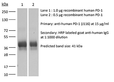
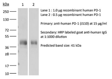
The J116 monoclonal antibody reacts with human PD-1 (programmed death-1) also known as CD279. PD-1 is a 50-55 kDa cell surface receptor encoded by the Pdcd1 gene that belongs to the CD28 family of the Ig superfamily. PD-1 is transiently expressed on CD4 and CD8 thymocytes as well as activated T and B lymphocytes and myeloid cells. PD-1 expression declines after successful elimination of antigen. Additionally, Pdcd1 mRNA is expressed in developing B lymphocytes during the pro-B-cell stage. PD-1’s structure includes a ITIM (immunoreceptor tyrosine-based inhibitory motif) suggesting that PD-1 negatively regulates TCR signals. PD-1 signals via binding its two ligands, PD-L1 and PD-L2 both members of the B7 family. Upon ligand binding, PD-1 signaling inhibits T-cell activation, leading to reduced proliferation, cytokine production, and T cell death. Additionally, PD-1 is known to play key roles in peripheral tolerance and prevention of autoimmune disease in mice as PD-1 knockout animals show dilated cardiomyopathy, splenomegaly, and loss of peripheral tolerance. Induced PD-L1 expression is common in many tumors including squamous cell carcinoma, colon adenocarcinoma, and breast adenocarcinoma. PD-L1 overexpression results in increased resistance of tumor cells to CD8 T cell mediated lysis. In mouse models of melanoma, tumor growth can be transiently arrested via treatment with antibodies which block the interaction between PD-L1 and its receptor PD-1. For these reasons anti-PD-1 mediated immunotherapies are currently being explored as cancer treatments. Binding of the J116 antibody is reported to inhibit PD-1 signal transduction, however, it is not reported to block PD-L1 binding.Specifications
| Isotype | Mouse IgG1, κ |
|---|---|
| Recommended Isotype Control(s) | InVivoMAb mouse IgG1 isotype control, unknown specificity |
| Recommended Dilution Buffer | InVivoPure pH 7.0 Dilution Buffer |
| Conjugation | This product is unconjugated. Conjugation is available via our Antibody Conjugation Services. |
| Immunogen | Not available or unknown |
| Reported Applications |
in vitro PD-1 neutralization in vivo PD-1 blockade in humanized mice |
| Formulation |
PBS, pH 7.0 Contains no stabilizers or preservatives |
| Endotoxin |
<2EU/mg (<0.002EU/μg) Determined by LAL gel clotting assay |
| Purity |
>95% Determined by SDS-PAGE |
| Sterility | 0.2 µm filtration |
| Production | Purified from cell culture supernatant in an animal-free facility |
| Purification | Protein G |
| RRID | AB_10950318 |
| Molecular Weight | 150 kDa |
| Storage | The antibody solution should be stored at the stock concentration at 4°C. Do not freeze. |
Recommended Products
in vitro PD-1 neutralization
Tkachev, V., et al. (2015). "Programmed death-1 controls T cell survival by regulating oxidative metabolism" J Immunol 194(12): 5789-5800. PubMed
The coinhibitory receptor programmed death-1 (PD-1) maintains immune homeostasis by negatively regulating T cell function and survival. Blockade of PD-1 increases the severity of graft-versus-host disease (GVHD), but the interplay between PD-1 inhibition and T cell metabolism is not well studied. We found that both murine and human alloreactive T cells concomitantly upregulated PD-1 expression and increased levels of reactive oxygen species (ROS) following allogeneic bone marrow transplantation. This PD-1(Hi)ROS(Hi) phenotype was specific to alloreactive T cells and was not observed in syngeneic T cells during homeostatic proliferation. Blockade of PD-1 signaling decreased both mitochondrial H2O2 and total cellular ROS levels, and PD-1-driven increases in ROS were dependent upon the oxidation of fatty acids, because treatment with etomoxir nullified changes in ROS levels following PD-1 blockade. Downstream of PD-1, elevated ROS levels impaired T cell survival in a process reversed by antioxidants. Furthermore, PD-1-driven changes in ROS were fundamental to establishing a cell’s susceptibility to subsequent metabolic inhibition, because blockade of PD-1 decreased the efficacy of later F1F0-ATP synthase modulation. These data indicate that PD-1 facilitates apoptosis in alloreactive T cells by increasing ROS in a process dependent upon the oxidation of fat. In addition, blockade of PD-1 undermines the potential for subsequent metabolic inhibition, an important consideration given the increasing use of anti-PD-1 therapies in the clinic.
in vivo PD-1 blockade in humanized mice
Tsukahara, T., et al. (2015). "The Tol2 transposon system mediates the genetic engineering of T-cells with CD19-specific chimeric antigen receptors for B-cell malignancies" Gene Ther 22(2): 209-215. PubMed
Engineered T-cell therapy using a CD19-specific chimeric antigen receptor (CD19-CAR) is a promising strategy for the treatment of advanced B-cell malignancies. Gene transfer of CARs to T-cells has widely relied on retroviral vectors, but transposon-based gene transfer has recently emerged as a suitable nonviral method to mediate stable transgene expression. The advantages of transposon vectors compared with viral vectors include their simplicity and cost-effectiveness. We used the Tol2 transposon system to stably transfer CD19-CAR into human T-cells. Normal human peripheral blood lymphocytes were co-nucleofected with the Tol2 transposon donor plasmid carrying CD19-CAR and the transposase expression plasmid and were selectively propagated on NIH3T3 cells expressing human CD19. Expanded CD3(+) T-cells with stable and high-level transgene expression (~95%) produced interferon-gamma upon stimulation with CD19 and specifically lysed Raji cells, a CD19(+) human B-cell lymphoma cell line. Adoptive transfer of these T-cells suppressed tumor progression in Raji tumor-bearing Rag2(-/-)gammac(-/-) immunodeficient mice compared with control mice. These results demonstrate that the Tol2 transposon system could be used to express CD19-CAR in genetically engineered T-cells for the treatment of refractory B-cell malignancies.
in vivo PD-1 blockade in humanized mice
Wang, C., et al. (2013). "Rapamycin-treated human endothelial cells preferentially activate allogeneic regulatory T cells" J Clin Invest 123(4): 1677-1693. PubMed
Human graft endothelial cells (ECs) can act as antigen-presenting cells to initiate allograft rejection by host memory T cells. Rapamycin, an mTOR inhibitor used clinically to suppress T cell responses, also acts on DCs, rendering them tolerogenic. Here, we report the effects of rapamycin on EC alloimmunogenicity. Compared with mock-treated cells, rapamycin-pretreated human ECs (rapa-ECs) stimulated less proliferation and cytokine secretion from allogeneic CD4+ memory cells, an effect mimicked by shRNA knockdown of mTOR or raptor in ECs. The effects of rapamycin persisted for several days and were linked to upregulation of the inhibitory molecules PD-L1 and PD-L2 on rapa-ECs. Additionally, rapa-ECs produced lower levels of the inflammatory cytokine IL-6. CD4+ memory cells activated by allogeneic rapa-ECs became hyporesponsive to restimulation in an alloantigen-specific manner and contained higher percentages of suppressive CD4+CD25(hi)CD127(lo)FoxP3+ cells that did not produce effector cytokines. In a human-mouse chimeric model of allograft rejection, rapamycin pretreatment of human arterial allografts increased graft EC expression of PD-L1 and PD-L2 and reduced subsequent infiltration of allogeneic effector T cells into the artery intima and intimal expansion. Preoperative conditioning of allograft ECs with rapamycin could potentially reduce immune-mediated rejection.
in vitro PD-1 neutralization
Singh, A., et al. (2012). "Foxp3+ regulatory T cells among tuberculosis patients: impact on prognosis and restoration of antigen specific IFN-gamma producing T cells" PLoS One 7(9): e44728. PubMed
CD4(+)CD25(+)Foxp3(+) regulatory T cells (Treg) and programmed death-1 (PD-1) molecules have emerged as pivotal players in immune suppression of chronic diseases. However, their impact on the disease severity, therapeutic response and restoration of immune response in human tuberculosis remains unclear. Here, we describe the possible role of Treg cells, their M. tuberculosis driven expansion and contribution of PD-1 pathway to the suppressive function of Treg cells among pulmonary tuberculosis (PTB) patients. Multicolor flow cytometry, cell culture, cells sorting and ELISA were employed to execute the study. Our results showed significant increase in frequency of antigen-reactive Treg cells, which gradually declined during successful therapy and paralleled with decline of M. tuberculosis-specific IL-10 along with elevation of IFN-gamma production, and raising the IFN-gamma/IL-4 ratio. Interestingly, persistence of Treg cells tightly correlated with MDR tuberculosis. Also, we show that blocking PD-1/PD-L1 pathway abrogates Treg-mediated suppression, suggesting that the PD-1/PD-L1 pathway is required for Treg-mediated suppression of the antigen-specific T cells. Treg cells possibly play a role in dampening the effector immune response and abrogating PD-1 pathway on Treg cells significantly rescued protective T cell response, suggesting its importance in immune restoration among tuberculosis patients.
in vitro PD-1 neutralization
Rosignoli, G., et al. (2009). "Programmed death (PD)-1 molecule and its ligand PD-L1 distribution among memory CD4 and CD8 T cell subsets in human immunodeficiency virus-1-infected individuals" Clin Exp Immunol 157(1): 90-97. PubMed
Human immunodeficiency virus (HIV)-1 causes T cell anergy and affects T cell maturation. Various mechanisms are responsible for impaired anti-HIV-1-specific responses: programmed death (PD)-1 molecule and its ligand PD-L1 are negative regulators of T cell activity and their expression is increased during HIV-1 infection. This study examines correlations between T cell maturation, expression of PD-1 and PD-L1, and the effects of their blockade. Peripheral blood mononuclear cells (PBMC) from 24 HIV-1(+) and 17 uninfected individuals were phenotyped for PD-1 and PD-L1 expression on CD4(+) and CD8(+) T cell subsets. The effect of PD-1 and PD-L1 blockade on proliferation and interferon (IFN)-gamma production was tested on eight HIV-1(+) patients. Naive (CCR7(+)CD45RA(+)) CD8(+) T cells were reduced in HIV-1 aviraemic (P = 0.0065) and viraemic patients (P = 0.0130); CD8 T effector memory subsets [CCR7(-)CD45RA(-)(T(EM))] were increased in HIV-1(+) aviraemic (P = 0.0122) and viraemic (P = 0.0023) individuals versus controls. PD-1 expression was increased in CD4 naive (P = 0.0496), central memory [CCR7(+)CD45RA(-) (T(CM)); P = 0.0116], T(EM) (P = 0.0037) and CD8 naive T cells (P = 0.0133) of aviraemic HIV-1(+) versus controls. PD-L1 was increased in CD4 T(EMRA) (CCR7(-)CD45RA(+), P = 0.0119), CD8 T(EM) (P = 0.0494) and CD8 T(EMRA) (P = 0.0282) of aviraemic HIV-1(+)versus controls. PD-1 blockade increased HIV-1-specific proliferative responses in one of eight patients, whereas PD-L1 blockade restored responses in four of eight patients, but did not increase IFN-gamma-production. Alteration of T cell subsets, accompanied by increased PD-1 and PD-L1 expression in HIV-1 infection contributes to anergy and impaired anti-HIV-1-specific responses which are not rescued when PD-1 is blocked, in contrast to when PD-L1 is blocked, due possibly to an ability to bind to receptors other than PD-1.
in vitro PD-1 neutralization
urado, J. O., et al. (2008). "Programmed death (PD)-1:PD-ligand 1/PD-ligand 2 pathway inhibits T cell effector functions during human tuberculosis" J Immunol 181(1): 116-125. PubMed
Protective immunity against Mycobacterium tuberculosis requires the generation of cell-mediated immunity. We investigated the expression and role of programmed death 1 (PD-1) and its ligands, molecules known to modulate T cell activation, in the regulation of IFN-gamma production and lytic degranulation during human tuberculosis. We demonstrated that specific Ag-stimulation increased CD3+PD-1+ lymphocytes in peripheral blood and pleural fluid from tuberculosis patients in direct correlation with IFN-gamma production from these individuals. Moreover, M. tuberculosis-induced IFN-gamma participated in the up-regulation of PD-1 expression. Blockage of PD-1 or PD-1 and its ligands (PD-Ls: PD-L1, PD-L2) enhanced the specific degranulation of CD8+ T cells and the percentage of specific IFN-gamma-producing lymphocytes against the pathogen, demonstrating that the PD-1:PD-Ls pathway inhibits T cell effector functions during active M. tuberculosis infection. Furthermore, the simultaneous blockage of the inhibitory receptor PD-1 together with the activation of the costimulatory protein signaling lymphocytic activation molecule led to the promotion of protective IFN-gamma responses to M. tuberculosis, even in patients with weak cell-mediated immunity against the bacteria. Together, we demonstrated that PD-1 interferes with T cell effector functions against M. tuberculosis, suggesting that PD-1 has a key regulatory role during the immune response of the host to the pathogen.
- Immunology and Microbiology,
Genomic Profiling of Radiation-Induced Sarcomas Reveals the Immunologic Characteristics and Its Response to Immune Checkpoint Blockade.
In Clinical Cancer Research on 1 August 2023 by Hong, D. C., Yang, J., et al.
PubMed
Radiation-induced sarcomas (RIS) have a poor prognosis and lack effective treatments. Its genome and tumor microenvironment are not well characterized and need further exploration. Here, we performed whole-exome sequencing (WES) and mRNA sequencing (mRNA-seq) on patients with RIS and primary sarcomas (WES samples 46 vs. 48, mRNA-seq samples 16 vs. 8, mainly in head and neck), investigated the antitumor effect of programmed cell death protein 1 (PD-1) blockade in RIS patient-derived xenograft models, and analyzed clinical data of patients with RIS treated with chemotherapy alone or combined with an anti-PD-1 antibody. Compared with primary sarcomas, RIS manifested different patterns of copy-number variations, a significantly higher number of predicted strong MHC-binding neoantigens, and significantly increased immune cell infiltration. Clinical data showed that the combinatorial use of chemotherapy and PD-1 blockade achieved a higher objective response rate (36.67% vs. 8.00%; P = 0.003), longer overall survival (31.9 months vs. 14.8 months; P = 0.014), and longer progression-free survival (4.7 months vs. 9.5 months; P = 0.032) in patients with RIS compared with single chemotherapy. Elevated genomic instability and higher immune cell infiltrations were found in RIS than in primary sarcomas. Moreover, higher efficacy of chemotherapy plus PD-1 blockade was observed in animal experiments and clinical practice. This evidence indicated the promising application of immune checkpoint inhibitors in the treatment of RIS. ©2023 The Authors; Published by the American Association for Cancer Research.
- Biochemistry and Molecular biology,
- Cell Biology,
- Immunology and Microbiology
Low-density lipoprotein balances T cell metabolism and enhances response to anti-PD-1 blockade in a HCT116 spheroid model.
In Frontiers in Oncology on 14 February 2023 by Babl, N., Hofbauer, J., et al.
PubMed
The discovery of immune checkpoints and the development of their specific inhibitors was acclaimed as a major breakthrough in cancer therapy. However, only a limited patient cohort shows sufficient response to therapy. Hence, there is a need for identifying new checkpoints and predictive biomarkers with the objective of overcoming immune escape and resistance to treatment. Having been associated with both, treatment response and failure, LDL seems to be a double-edged sword in anti-PD1 immunotherapy. Being embedded into complex metabolic conditions, the impact of LDL on distinct immune cells has not been sufficiently addressed. Revealing the effects of LDL on T cell performance in tumor immunity may enable individual treatment adjustments in order to enhance the response to routinely administered immunotherapies in different patient populations. The object of this work was to investigate the effect of LDL on T cell activation and tumor immunity in-vitro. Experiments were performed with different LDL dosages (LDLlow = 50 μg/ml and LDLhigh = 200 μg/ml) referring to medium control. T cell phenotype, cytokines and metabolism were analyzed. The functional relevance of our findings was studied in a HCT116 spheroid model in the context of anti-PD-1 blockade. The key points of our findings showed that LDLhigh skewed the CD4+ T cell subset into a central memory-like phenotype, enhanced the expression of the co-stimulatory marker CD154 (CD40L) and significantly reduced secretion of IL-10. The exhaustion markers PD-1 and LAG-3 were downregulated on both T cell subsets and phenotypical changes were associated with a balanced T cell metabolism, in particular with a significant decrease of reactive oxygen species (ROS). T cell transfer into a HCT116 spheroid model resulted in a significant reduction of the spheroid viability in presence of an anti-PD-1 antibody combined with LDLhigh. Further research needs to be conducted to fully understand the impact of LDL on T cells in tumor immunity and moreover, to also unravel LDL effects on other lymphocytes and myeloid cells for improving anti-PD-1 immunotherapy. The reason for improved response might be a resilient, less exhausted phenotype with balanced ROS levels. Copyright © 2023 Babl, Hofbauer, Matos, Voll, Menevse, Rechenmacher, Mair, Beckhove, Herr, Siska, Renner, Kreutz and Schnell.
- Cancer Research,
- Immunology and Microbiology
Preclinical Platform Using a Triple-negative Breast Cancer Syngeneic Murine Model to Evaluate Immune Checkpoint Inhibitors.
In Anticancer Research on 1 January 2023 by Katuwal, N. B., Park, N., et al.
PubMed
To evaluate the feasibility of syngeneic mouse models of breast cancer by analyzing the efficacy of immune checkpoint inhibitors (ICIs) and potential predictive biomarkers. To establish the murine triple-negative breast cancer (TNBC) models, JC, 4T1, EMT6, and E0771 cells were subcutaneously implanted into female syngeneic mice. When the tumor reached 50-100 mm3, each mouse model was divided into a treatment (using a murine PD-1 antibody) and a no-treatment control group. The treatment group was further divided into the responder and non-responder groups. Potential predictive biomarkers were evaluated by analyzing serum cytokines, peripheral blood T cells and tumor infiltrating immune cells. The EMT6 model showed the highest tumor response rate (54%, 6/11) of the syngeneic models: 4T1 (45%, 5/11), JC (40%, 4/10), or E0771 (23%, 3/13). Early changes in tumor size at 7 days post-PD-1 inhibitor treatment predicted the final efficacy of the PD-1 inhibitor. Peripheral blood CD8+ and CD4+ T cells with or without Ki67 expression at 7 days post-PD-1 inhibitor treatment were higher in the finally designated responder group than in the non-responder group. At the time of sacrifice, analyses of tumor infiltrating lymphocytes consistently supported these results. We also demonstrated that retro-orbital blood sampling procedures (baseline, 7 days post-treatment, time of sacrifice) were safe for serum cytokine analyses, suggesting that our preclinical platform may be used for biomarker research using serum cytokines. Our syngeneic mouse model of TNBC is a feasible preclinical platform to evaluate ICI efficacy combined with other drugs and predictive biomarkers in the screening process of immune-oncology drug development. Copyright © 2023 International Institute of Anticancer Research (Dr. George J. Delinasios), All rights reserved.
- Cancer Research,
- Immunology and Microbiology
FGFR4-Targeted Chimeric Antigen Receptors Combined with Anti-Myeloid Polypharmacy Effectively Treat Orthotopic Rhabdomyosarcoma.
In Molecular Cancer Therapeutics on 7 October 2022 by Sullivan, P. M., Kumar, R., et al.
PubMed
Rhabdomyosarcoma (RMS) is the most common soft tissue cancer in children. Treatment outcomes, particularly for relapsed/refractory or metastatic disease, have not improved in decades. The current lack of novel therapies and low tumor mutational burden suggest that chimeric antigen receptor (CAR) T-cell therapy could be a promising approach to treating RMS. Previous work identified FGF receptor 4 (FGFR4, CD334) as being specifically upregulated in RMS, making it a candidate target for CAR T cells. We tested the feasibility of an FGFR4-targeted CAR for treating RMS using an NSG mouse with RH30 orthotopic (intramuscular) tumors. The first barrier we noted was that RMS tumors produce a collagen-rich stroma, replete with immunosuppressive myeloid cells, when T-cell therapy is initiated. This stromal response is not seen in tumor-only xenografts. When scFV-based binders were selected from phage display, CARs targeting FGFR4 were not effective until our screening approach was refined to identify binders to the membrane-proximal domain of FGFR4. Having improved the CAR, we devised a pharmacologic strategy to augment CAR T-cell activity by inhibiting the myeloid component of the T-cell-induced tumor stroma. The combined treatment of mice with anti-myeloid polypharmacy (targeting CSF1R, IDO1, iNOS, TGFbeta, PDL1, MIF, and myeloid misdifferentiation) allowed FGFR4 CAR T cells to successfully clear orthotopic RMS tumors, demonstrating that RMS tumors, even with very low copy-number targets, can be targeted by CAR T cells upon reversal of an immunosuppressive microenvironment. ©2022 The Authors; Published by the American Association for Cancer Research.
- Immunology and Microbiology
Targeting cathepsin B by cycloastragenol enhances antitumor immunity of CD8 T cells via inhibiting MHC-I degradation.
In Journal for Immunotherapy of Cancer on 1 October 2022 by Deng, G., Zhou, L., et al.
PubMed
The loss of tumor antigens and depletion of CD8 T cells caused by the PD-1/PD-L1 pathway are important factors for tumor immune escape. In recent years, there has been increasing research on traditional Chinese medicine in tumor treatment. Cycloastragenol (CAG), an effective active molecule in Astragalus membranaceus, has been found to have antiviral, anti-aging, anti-inflammatory, and other functions. However, its antitumor effect and mechanism are not clear. The antitumor effect of CAG was investigated in MC38 and CT26 mouse transplanted tumor models. The antitumor effect of CAG was further analyzed via single-cell multiomics sequencing. Target responsive accessibility profiling technology was used to find the target protein of CAG. Subsequently, the antitumor mechanism of CAG was explored using confocal microscopy, coimmunoprecipitation and transfection of mutant plasmids. Finally, the combined antitumor effect of CAG and PD-1 antibodies in mice or organoids were investigated. We found that CAG effectively inhibited tumor growth in vivo. Our single-cell multiomics atlas demonstrated that CAG promoted the presentation of tumor cell-surface antigens and was characterized by the enhanced killing function of CD8+ T cells. Mechanistically, CAG bound to its target protein cathepsin B, which then inhibited the lysosomal degradation of major histocompatibility complex I (MHC-I) and promoted the aggregation of MHC-I to the cell membrane, boosting the presentation of the tumor antigen. Meanwhile, the combination of CAG with PD-1 antibody effectively enhanced the tumor killing ability of CD8+ T cells in xenograft mice and colorectal cancer organoids. Our data reported for the first time that cathepsin B downregulation confers antitumor immunity and explicates the antitumor mechanism of natural product CAG. © Author(s) (or their employer(s)) 2022. Re-use permitted under CC BY-NC. No commercial re-use. See rights and permissions. Published by BMJ.
- Cancer Research,
- Immunology and Microbiology
Breast cancer cell-derived extracellular vesicles promote CD8+ T cell exhaustion via TGF-β type II receptor signaling.
In Nature Communications on 1 August 2022 by Xie, F., Zhou, X., et al.
PubMed
Cancer immunotherapies have shown clinical success in various types of tumors but the patient response rate is low, particularly in breast cancer. Here we report that malignant breast cancer cells can transfer active TGF-β type II receptor (TβRII) via tumor-derived extracellular vesicles (TEV) and thereby stimulate TGF-β signaling in recipient cells. Up-take of extracellular vesicle-TβRII (EV-TβRII) in low-grade tumor cells initiates epithelial-to-mesenchymal transition (EMT), thus reinforcing cancer stemness and increasing metastasis in intracardial xenograft and orthotopic transplantation models. EV-TβRII delivered as cargo to CD8+ T cells induces the activation of SMAD3 which we demonstrated to associate and cooperate with TCF1 transcription factor to impose CD8+ T cell exhaustion, resulting in failure of immunotherapy. The levels of TβRII+ circulating extracellular vesicles (crEV) appears to correlate with tumor burden, metastasis and patient survival, thereby serve as a non-invasive screening tool to detect malignant breast tumor stages. Thus, our findings not only identify a possible mechanism by which breast cancer cells can promote T cell exhaustion and dampen host anti-tumor immunity, but may also identify a target for immune therapy against the most devastating breast tumors. © 2022. The Author(s).
- Biochemistry and Molecular biology,
- Cancer Research,
- Cell Biology
IFNα Potentiates Anti-PD-1 Efficacy by Remodeling Glucose Metabolism in the Hepatocellular Carcinoma Microenvironment.
In Cancer Discovery on 6 July 2022 by Hu, B., Yu, M., et al.
PubMed
The overall response rate for anti-PD-1 therapy remains modest in hepatocellular carcinoma (HCC). We found that a combination of IFNα and anti-PD-1-based immunotherapy resulted in enhanced antitumor activity in patients with unresectable HCC. In both immunocompetent orthotopic and spontaneous HCC models, IFNα therapy synergized with anti-PD-1 and the combination treatment led to significant enrichment of cytotoxic CD27+CD8+ T cells. Mechanistically, IFNα suppressed HIF1α signaling by inhibiting FosB transcription in HCC cells, resulting in reduced glucose consumption capacity and consequentially establishing a high-glucose microenvironment that fostered transcription of the T-cell costimulatory molecule Cd27 via mTOR-FOXM1 signaling in infiltrating CD8+ T cells. Together, these data reveal that IFNα reprograms glucose metabolism within the HCC tumor microenvironment, thereby liberating T-cell cytotoxic capacities and potentiating the PD-1 blockade-induced immune response. Our findings suggest that IFNα and anti-PD-1 cotreatment is an effective novel combination strategy for patients with HCC. Our study supports a role of tumor glucose metabolism in IFNα-mediated antitumor immunity in HCC, and tumor-infiltrating CD27+CD8+ T cells may be a promising biomarker for stratifying patients for anti-PD-1 therapy. See related commentary by Kao et al., p. 1615. This article is highlighted in the In This Issue feature, p. 1599. ©2022 American Association for Cancer Research.
- In Vivo,
- Mus musculus (House mouse),
- Cancer Research,
- Immunology and Microbiology
Blocking TIGIT/CD155 signalling reverses CD8+ T cell exhaustion and enhances the antitumor activity in cervical cancer.
In Journal of Translational Medicine on 21 June 2022 by Liu, L., Wang, A., et al.
PubMed
TIGIT/CD155 has attracted widespread attention as a new immune checkpoint and a potential target for cancer immunotherapy. In our study, we evaluated the role of TIGIT/CD155 checkpoints in the progression of cervical cancer. The expression of CD155 and TIGIT in cervical cancer tissues was detected using flow cytometry, immunohistochemistry (IHC) and gene expression profiling. In vivo and in vitro experiments have proven that blocking TIGIT/CD155 restores the ability of CD8+ T cells to produce cytokines. Changes in the NF-κB and ERK pathways were detected using western blotting (WB) after blocking TIGIT/CD155 signalling. TIGIT expression was elevated in patients with cervical cancer. High TIGIT expression in CD8+ T lymphocytes from patients with cervical cancer promotes the exhaustion of CD8+ T lymphocytes. In addition, CD155 is expressed at high levels in cervical cancer tissues and is negatively correlated with the level of infiltrating CD8+ T cells. We found that TIGIT, upon binding to CD155 and being phosphorylated, inhibited NF-κB and ERK activation by recruiting SHIP-1, resulting in the downregulation of cytokine production. Blocking TIGIT in activated CD8+ T cells attenuates the inhibitory effect of SHIP-1 on CD8+ T cells and enhances the activation of NF-κB and ERK. In vivo and in vitro experiments have proven that blocking TIGIT/CD155 restores the ability of CD8+ T cells to produce cytokines. Injecting the blocking antibody TIGIT in vivo inhibits tumour growth and enhances CD8+ T lymphocyte function. Treatment with a combination of TIGIT and PD-1 inhibitors further increases the efficacy of the TIGIT blocking antibody. Our research shows that TIGIT/CD155 is a potential therapeutic target for cervical cancer. © 2022. The Author(s).
- Cancer Research,
- Immunology and Microbiology
Blocking TIGIT/CD155 Signal Reverses CD8+T Cell Exhaustion and Enhances the Anti-Tumor Ability of Cervical Cancer
Preprint on Research Square on 28 February 2022 by Liu, L., Wang, A., et al.
PubMed
h4>Objective: /h4> : TIGIT/CD155 has attracted widespread attention as a new immune checkpoint and a potential target for cancer immunotherapy. In our study, we evaluated the role of TIGIT/CD155 checkpoints in the progression of cervical cancer.Methods. Detect the expression of CD155 and TIGIT in cervical cancer tissues by flow cytometry, immunohistochemistry and gene expression profiling. In vivo and in vitro experiments have proved that blocking TIGIT/CD155 restores the ability of CD8+T cells to produce cytokines. h4>Methods: /h4>. :Detect the expression of CD155 and TIGIT in cervical cancer tissues by flow cytometry, immunohistochemistry (IHC) and gene expression profiling. In vivo and in vitro experiments have proved that blocking TIGIT/CD155 restores the ability of CD8+T cells to produce cytokines.Changes in NF-κB and ERK pathways detected by western blot (WB) after blocking TIGIT/CD155 signaling h4>Results: /h4> :We found that the expression of TIGIT is elevated in patients with cervical cancer. The high expression of TIGIT in CD8 + T lymphocytes of cervical cancer patients promotes the failure of CD8 + T lymphocytes. In addition, CD155 is highly expressed in cervical cancer tissues and negatively correlated with the infiltration level of CD8 + T cells. We showed that phosphorylated TIGIT binds to CD155 to inhibit NF-κB and ERK activation by recruiting SHIP-1, leading to down-regulation of cytokine production. Inactivated CD8 + T cells, after blocking TIGIT, the inhibitory effect of SHIP-1 on CD8 + T cells is weakened, and the activation of NF-κB and ERK is enhanced. In vivo and in vitro experiments have proved that blocking TIGIT/CD155 restores the ability of CD8 + T cells to produce cytokines. Injecting blocking antibody TIGIT in vivo could inhibit tumor growth and promote CD8 + T lymphocyte function. Combining blocking TIGIT and PD-1 further increased the effect of the blocking antibody TIGIT. h4>Conclusions: /h4>: Our research shows that TIGIT/CD155 is a potential therapeutic target for cervical cancer.
- Cancer Research,
- Immunology and Microbiology
T cells drive negative feedback mechanisms in cancer associated fibroblasts, promoting expression of co-inhibitory ligands, CD73 and IL-27 in non-small cell lung cancer.
In Oncoimmunology on 23 July 2021 by O'Connor, R. A., Chauhan, V., et al.
PubMed
The success of immune checkpoint therapy shows tumor-reactive T cells can eliminate cancer cells but are restrained by immunosuppression within the tumor micro-environment (TME). Cancer associated fibroblasts (CAFs) are the dominant stromal cell in the TME and co-localize with T cells in non-small cell lung cancer. We demonstrate the bidirectional nature of CAF/T cell interactions; T cells promote expression of co-inhibitory ligands, MHC molecules and CD73 on CAFs, increasing their production of IL-6 and eliciting production of IL-27. In turn CAFs upregulate co-inhibitory receptors on T cells including the ectonucleotidase CD39 promoting development of an exhausted but highly cytotoxic phenotype. Our results highlight the bidirectional interaction between T cells and CAFs in promoting components of the immunosuppressive CD39, CD73 adenosine pathway and demonstrate IL-27 production can be induced in CAF by activated T cells. © 2021 The Author(s). Published with license by Taylor Francis Group, LLC.
- Cancer Research,
- Immunology and Microbiology
Integrin αvβ6-TGFβ-SOX4 Pathway Drives Immune Evasion in Triple-Negative Breast Cancer.
In Cancer Cell on 11 January 2021 by Bagati, A., Kumar, S., et al.
PubMed
Cancer immunotherapy shows limited efficacy against many solid tumors that originate from epithelial tissues, including triple-negative breast cancer (TNBC). We identify the SOX4 transcription factor as an important resistance mechanism to T cell-mediated cytotoxicity for TNBC cells. Mechanistic studies demonstrate that inactivation of SOX4 in tumor cells increases the expression of genes in a number of innate and adaptive immune pathways important for protective tumor immunity. Expression of SOX4 is regulated by the integrin αvβ6 receptor on the surface of tumor cells, which activates TGFβ from a latent precursor. An integrin αvβ6/8-blocking monoclonal antibody (mAb) inhibits SOX4 expression and sensitizes TNBC cells to cytotoxic T cells. This integrin mAb induces a substantial survival benefit in highly metastatic murine TNBC models poorly responsive to PD-1 blockade. Targeting of the integrin αvβ6-TGFβ-SOX4 pathway therefore provides therapeutic opportunities for TNBC and other highly aggressive human cancers of epithelial origin. Copyright © 2020 Elsevier Inc. All rights reserved.
- Homo sapiens (Human)
Preclinical platform for long-term evaluation of immuno-oncology drugs using hCD34+ humanized mouse model.
In Journal for Immunotherapy of Cancer on 1 November 2020 by Park, N., Pandey, K., et al.
PubMed
Well-characterized preclinical models are essential for immune-oncology research. We investigated the feasibility of our humanized mouse model for evaluating the long-term efficacy of immunotherapy and biomarkers. Humanized mice were generated by injecting human fetal cord blood-derived CD34+ hematopoietic stem cells to NOD-scid IL2rγnull (NSG) mice myeloablated with irradiation or busulfan. The humanization success was defined as a 25% or higher ratio of human CD45+ cells to mice peripheral blood mononuclear cells. Busulfan was ultimately selected as the appropriate myeloablative method because it provided a higher success rate of humanization (approximately 80%) and longer survival time (45 weeks). We proved the development of functional T cells by demonstrating the anticancer effect of the programmed cell death-1 (PD-1) inhibitor in our humanized mice but not in non-humanized NSG mice. After confirming the long-lasting humanization state (45 weeks), we further investigated the response durability of the PD-1 inhibitor and biomarkers in our humanized mice. Early increase in serum tumor necrosis factor α levels, late increase in serum interleukin 6 levels and increase in tumor-infiltrating CD8+ T lymphocytes correlated more with a durable response over 60 days than with a non-durable response. Our CD34+ humanized mouse model is the first in vivo platform for testing the long-term efficacy of anticancer immunotherapies and biomarkers, given that none of the preclinical models has ever been evaluated for such a long duration. © Author(s) (or their employer(s)) 2020. Re-use permitted under CC BY-NC. No commercial re-use. See rights and permissions. Published by BMJ.
- Immunology and Microbiology
In situ immunization of a TLR9 agonist virus-like particle enhances anti-PD1 therapy.
In Journal for Immunotherapy of Cancer on 1 October 2020 by Cheng, Y., Lemke-Miltner, C. D., et al.
PubMed
CMP-001 is a novel Toll-like receptor-9 agonist that consists of an unmethylated CpG-A motif-rich G10 oligodeoxynucleotide (ODN) encapsulated in virus-like particles. In situ vaccination of CMP-001 is believed to activate local tumor-associated plasmacytoid dendritic cells (pDCs) leading to type I interferon secretion and tumor antigen presentation to T cells and systemic antitumor T cell responses. This study is designed to investigate if CMP-001 would enhance head and neck squamous cell carcinoma (HNSCC) tumor response to anti-programmed cell death protein-1 (anti-PD-1) therapy in a human papilloma virus-positive (HPV+) tumor mouse model. Immune cell activation in response to CMP-001±anti-Qβ was performed using co-cultures of peripheral blood mononuclear cells and HPV+/HPV- HNSCC cells and then analyzed by flow cytometry. In situ vaccination with CMP-001 alone and in combination with anti-PD-1 was investigated in C57BL/6 mice-bearing mEERL HNSCC tumors and analyzed for anti-Qβ development, antitumor response, survival and immune cell recruitment. The role of antitumor immune response due to CMP-001+anti-PD-1 treatment was investigated by the depletion of natural killer (NK), CD4+ T, and CD8+ T cells. Results showed that the activity of CMP-001 on immune cell (pDCs, monocytes, CD4+/CD8+ T cells and NK cells) activation depends on the presence of anti-Qβ. A 2-week 'priming' period after subcutaneous administration of CMP-001 was required for robust anti-Qβ development in mice. In situ vaccination of CMP-001 was superior to unencapsulated G10 CpG-A ODN at suppressing both injected and uninjected (distant) tumors. In situ vaccination of CMP-001 in combination with anti-PD-1 therapy induced durable tumor regression at injected and distant tumors and significantly prolonged mouse survival compared with anti-PD-1 therapy alone. The antitumor effect of CMP-001+anti-PD-1 was accompanied by increased interferon gamma (IFNγ)+ CD4+/CD8+ T cells compared with control-treated mice. The therapeutic and abscopal effect of CMP-001+ anti-PD-1 therapy was completely abrogated by CD8+ T cell depletion. These results demonstrate that in situ vaccination with CMP-001 can induce both local and abscopal antitumor immune responses. Additionally, the antitumor efficacy of CMP-001 combined with α-PD-1 therapy warrants further study as a novel immunotherapeutic strategy for the treatment of HNSCC. © Author(s) (or their employer(s)) 2020. Re-use permitted under CC BY-NC. No commercial re-use. See rights and permissions. Published by BMJ.
- Cancer Research,
- Cell Biology,
- Immunology and Microbiology
Mitochondrial dysregulation and glycolytic insufficiency functionally impair CD8 T cells infiltrating human renal cell carcinoma.
In JCI Insight on 15 June 2017 by Siska, P. J., Beckermann, K. E., et al.
PubMed
Cancer cells can inhibit effector T cells (Teff) through both immunomodulatory receptors and the impact of cancer metabolism on the tumor microenvironment. Indeed, Teff require high rates of glucose metabolism, and consumption of essential nutrients or generation of waste products by tumor cells may impede essential T cell metabolic pathways. Clear cell renal cell carcinoma (ccRCC) is characterized by loss of the tumor suppressor von Hippel-Lindau (VHL) and altered cancer cell metabolism. Here, we assessed how ccRCC influences the metabolism and activation of primary patient ccRCC tumor infiltrating lymphocytes (TIL). CD8 TIL were abundant in ccRCC, but they were phenotypically distinct and both functionally and metabolically impaired. ccRCC CD8 TIL were unable to efficiently uptake glucose or perform glycolysis and had small, fragmented mitochondria that were hyperpolarized and generated large amounts of ROS. Elevated ROS was associated with downregulated mitochondrial SOD2. CD8 T cells with hyperpolarized mitochondria were also visible in the blood of ccRCC patients. Importantly, provision of pyruvate to bypass glycolytic defects or scavengers to neutralize mitochondrial ROS could partially restore TIL activation. Thus, strategies to improve metabolic function of ccRCC CD8 TIL may promote the immune response to ccRCC.

