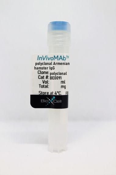
InVivoMAb hamster IgG f(ab')2 fragments
Product Details
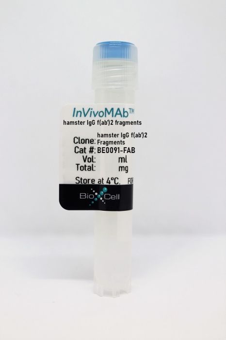
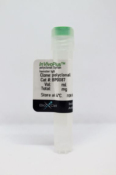
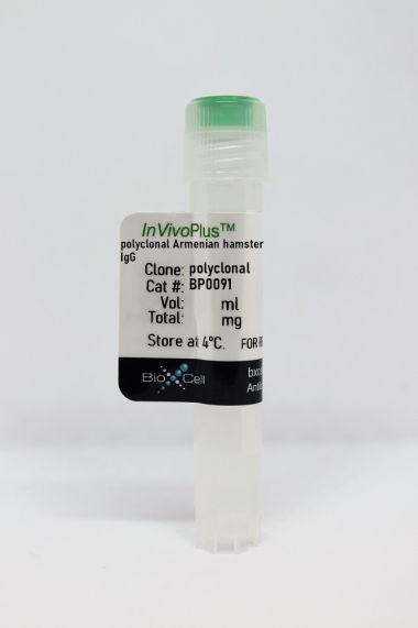
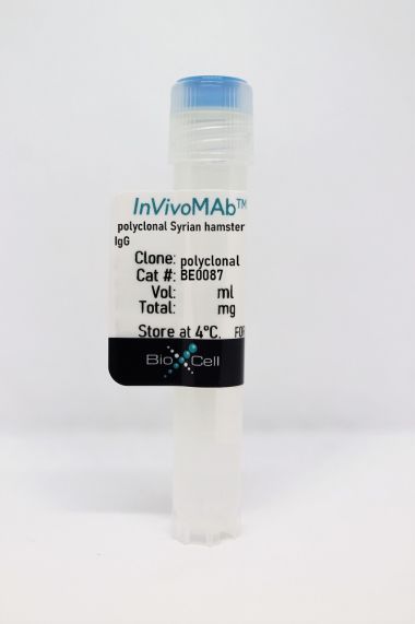
The hamster IgG f(ab’)2 fragments are the f(ab’)2 fragments of polyclonal hamster IgG. The majority of the Fc fragment has been removed via pepsin digestion. This product is commonly used as a non-reactive control for the anti-mouse CD3ε F(ab’)2 fragment BE0001-1FAB.Specifications
| Isotype | Polyclonal |
|---|---|
| Recommended Dilution Buffer | InVivoPure pH 7.0 Dilution Buffer |
| Formulation |
PBS, pH 7.0 Contains no stabilizers or preservatives |
| Endotoxin |
<2EU/mg (<0.002EU/μg) Determined by LAL gel clotting assay |
| Purity |
>95% Determined by SDS-PAGE |
| Sterility | 0.2 µm filtration |
| Production | Pepsin Digest |
| Purification | Protein A |
| RRID | AB_2687680 |
| Storage | The antibody solution should be stored at the stock concentration at 4°C. Do not freeze. |
Recommended Products
Wilhelmson, A. S., et al. (2018). "Testosterone Protects Against Atherosclerosis in Male Mice by Targeting Thymic Epithelial Cells-Brief Report" Arterioscler Thromb Vasc Biol 38(7): 1519-1527. PubMed
OBJECTIVE: Androgen deprivation therapy has been associated with increased cardiovascular risk in men. Experimental studies support that testosterone protects against atherosclerosis, but the target cell remains unclear. T cells are important modulators of atherosclerosis, and deficiency of testosterone or its receptor, the AR (androgen receptor), induces a prominent increase in thymus size. Here, we tested the hypothesis that atherosclerosis induced by testosterone deficiency in male mice is T-cell dependent. Further, given the important role of the thymic epithelium for T-cell homeostasis and development, we hypothesized that depletion of the AR in thymic epithelial cells will result in increased atherosclerosis. APPROACH AND RESULTS: Prepubertal castration of male atherosclerosis-prone apoE(-/-) mice increased atherosclerotic lesion area. Depletion of T cells using an anti-CD3 antibody abolished castration-induced atherogenesis, demonstrating a role of T cells. Male mice with depletion of the AR specifically in epithelial cells (E-ARKO [epithelial cell-specific AR knockout] mice) showed increased thymus weight, comparable with that of castrated mice. E-ARKO mice on an apoE(-/-) background displayed significantly increased atherosclerosis and increased infiltration of T cells in the vascular adventitia, supporting a T-cell-driven mechanism. Consistent with a role of the thymus, E-ARKO apoE(-/-) males subjected to prepubertal thymectomy showed no atherosclerosis phenotype. CONCLUSIONS: We show that atherogenesis induced by testosterone/AR deficiency is thymus- and T-cell dependent in male mice and that the thymic epithelial cell is a likely target cell for the antiatherogenic actions of testosterone. These insights may pave the way for new therapeutic strategies for safer endocrine treatment of prostate cancer.
- Immunology and Microbiology,
- Cell Biology,
- Biochemistry and Molecular biology
PHGDH-mediated endothelial metabolism drives glioblastoma resistance to chimeric antigen receptor T cell immunotherapy.
In Cell Metabolism on 7 March 2023 by Zhang, D., Li, A. M., et al.
PubMed
The efficacy of immunotherapy is limited by the paucity of T cells delivered and infiltrated into the tumors through aberrant tumor vasculature. Here, we report that phosphoglycerate dehydrogenase (PHGDH)-mediated endothelial cell (EC) metabolism fuels the formation of a hypoxic and immune-hostile vascular microenvironment, driving glioblastoma (GBM) resistance to chimeric antigen receptor (CAR)-T cell immunotherapy. Our metabolome and transcriptome analyses of human and mouse GBM tumors identify that PHGDH expression and serine metabolism are preferentially altered in tumor ECs. Tumor microenvironmental cues induce ATF4-mediated PHGDH expression in ECs, triggering a redox-dependent mechanism that regulates endothelial glycolysis and leads to EC overgrowth. Genetic PHGDH ablation in ECs prunes over-sprouting vasculature, abrogates intratumoral hypoxia, and improves T cell infiltration into the tumors. PHGDH inhibition activates anti-tumor T cell immunity and sensitizes GBM to CAR T therapy. Thus, reprogramming endothelial metabolism by targeting PHGDH may offer a unique opportunity to improve T cell-based immunotherapy. Copyright © 2023 Elsevier Inc. All rights reserved.
- In Vivo,
- Mus musculus (House mouse),
- Cell Biology
Endothelial Caspase-8 prevents fatal necroptotic hemorrhage caused by commensal bacteria.
In Cell Death and Differentiation on 1 January 2023 by Bader, S. M., Preston, S. P., et al.
PubMed
Caspase-8 transduces signals from death receptor ligands, such as tumor necrosis factor, to drive potent responses including inflammation, cell proliferation or cell death. This is a developmentally essential function because in utero deletion of endothelial Caspase-8 causes systemic circulatory collapse during embryogenesis. Whether endothelial Caspase-8 is also required for cardiovascular patency during adulthood was unknown. To address this question, we used an inducible Cre recombinase system to delete endothelial Casp8 in 6-week-old conditionally gene-targeted mice. Extensive whole body vascular gene targeting was confirmed, yet the dominant phenotype was fatal hemorrhagic lesions exclusively within the small intestine. The emergence of these intestinal lesions was not a maladaptive immune response to endothelial Caspase-8-deficiency, but instead relied upon aberrant Toll-like receptor sensing of microbial commensals and tumor necrosis factor receptor signaling. This lethal phenotype was prevented in compound mutant mice that lacked the necroptotic cell death effector, MLKL. Thus, distinct from its systemic role during embryogenesis, our data show that dysregulated microbial- and death receptor-signaling uniquely culminate in the adult mouse small intestine to unleash MLKL-dependent necroptotic hemorrhage after loss of endothelial Caspase-8. These data support a critical role for Caspase-8 in preserving gut vascular integrity in the face of microbial commensals. © 2022. The Author(s).
- In Vivo,
- Mus musculus (House mouse),
- Endocrinology and Physiology,
- Immunology and Microbiology
T cells mediate cell non-autonomous arterial ageing in mice.
In The Journal of Physiology on 1 August 2021 by Trott, D. W., Machin, D. R., et al.
PubMed
Increased large artery stiffness and impaired endothelium-dependent dilatation occur with advanced age. We sought to determine whether T cells mechanistically contribute to age-related arterial dysfunction. We found that old mice exhibited greater proinflammatory T cell accumulation around both the aorta and mesenteric arteries. Pharmacologic depletion or genetic deletion of T cells in old mice resulted in ameliorated large artery stiffness and greater endothelium-dependent dilatation compared with mice with T cells intact. Ageing of the arteries is characterized by increased large artery stiffness and impaired endothelium-dependent dilatation. T cells contribute to hypertension in acute rodent models but whether they contribute to chronic age-related arterial dysfunction is unknown. To determine whether T cells directly mediate age-related arterial dysfunction, we examined large elastic artery and resistance artery function in young (4-6 months) and old (22-24 months) wild-type mice treated with anti-CD3 F(ab'2) fragments to deplete T cells (150 μg, i.p. every 7 days for 28 days) or isotype control fragments. Old mice exhibited greater numbers of T cells in both aorta and mesenteric vasculature when compared with young mice. Old mice treated with anti-CD3 fragments exhibited depletion of T cells in blood, spleen, aorta and mesenteric vasculature. Old mice also exhibited greater numbers of aortic and mesenteric IFN-γ and TNF-α-producing T cells when compared with young mice. Old control mice exhibited greater large artery stiffness and impaired resistance artery endothelium-dependent dilatation in comparison with young mice. In old mice, large artery stiffness was ameliorated with anti-CD3 treatment. Anti-CD3-treated old mice also exhibited greater endothelium-dependent dilatation than age-matched controls. We also examined arterial function in young and old Rag-1-/- mice, which lack lymphocytes. Rag-1-/- mice exhibited blunted increases in large artery stiffness with age compared with wild-type mice. Old Rag-1-/- mice also exhibited greater endothelium-dependent dilatation compared with old wild-type mice. Collectively, these results demonstrate that T cells play an important role in age-related arterial dysfunction. © 2021 The Authors. The Journal of Physiology © 2021 The Physiological Society.
- In Vivo,
- Mus musculus (House mouse),
- Immunology and Microbiology
Immunostimulatory bacterial antigen-armed oncolytic measles virotherapy significantly increases the potency of anti-PD1 checkpoint therapy.
In The Journal of Clinical Investigation on 1 July 2021 by Panagioti, E., Kurokawa, C., et al.
PubMed
Clinical immunotherapy approaches are lacking efficacy in the treatment of glioblastoma (GBM). In this study, we sought to reverse local and systemic GBM-induced immunosuppression using the Helicobacter pylori neutrophil-activating protein (NAP), a potent TLR2 agonist, as an immunostimulatory transgene expressed in an oncolytic measles virus (MV) platform, retargeted to allow viral entry through the urokinase-type plasminogen activator receptor (uPAR). While single-agent murine anti-PD1 treatment or repeat in situ immunization with MV-s-NAP-uPA provided modest survival benefit in MV-resistant syngeneic GBM models, the combination treatment led to synergy with a cure rate of 80% in mice bearing intracranial GL261 tumors and 72% in mice with CT-2A tumors. Combination NAP-immunovirotherapy induced massive influx of lymphoid cells in mouse brain, with CD8+ T cell predominance; therapeutic efficacy was CD8+ T cell dependent. Inhibition of the IFN response pathway using the JAK1/JAK2 inhibitor ruxolitinib decreased PD-L1 expression on myeloid-derived suppressor cells in the brain and further potentiated the therapeutic effect of MV-s-NAP-uPA and anti-PD1. Our findings support the notion that MV strains armed with bacterial immunostimulatory antigens represent an effective strategy to overcome the limited efficacy of immune checkpoint inhibitor-based therapies in GBM, creating a promising translational strategy for this lethal brain tumor.
- FC/FACS,
- Mus musculus (House mouse),
- Cancer Research
Malignant cell-specific CXCL14 promotes tumor lymphocyte infiltration in oral cavity squamous cell carcinoma.
In Journal for Immunotherapy of Cancer on 1 September 2020 by Parikh, A., Shin, J., et al.
PubMed
To explore lymphocyte infiltration as a potential mechanism behind CXCL14-mediated tumor growth suppression in oral cavity squamous cell carcinoma (OSCC). We analyzed single cell RNA-sequencing (scRNA-seq) data from OSCC to identify expression changes among malignant cells in lymph nodes (LN) versus primary tumors. CXCL14 expression in murine OSCC cell lines was quantified using qRT-PCR. Short hairpin RNA knockdown of CXCL14 was performed in mouse oral cavity (MOC)1 cells, and CXCL14 overexpression was performed in MOC2 cells. Cells in each condition were injected into C57BL/6 mice with and without T cell depletion, and tumor volume was measured. At 30 days, tumors were dissociated and analyzed by flow cytometry for CD45+CD3+ T cells. CXCL14 expression was correlated with gene expression signatures of tumor infiltrating lymphocytes (TIL) in scRNA-seq data, as well as TCGA tumors. scRNA-seq revealed CXCL14 as the most significantly downregulated gene among malignant cells in LNs relative to primary tumor, supporting a role in preventing invasion and/or metastasis. In a murine immunocompetent model, CXCL14 expression was higher in indolent MOC1 cells than in more aggressive MOC2 cells. Tumor growth in vivo was significantly increased by CXCL14 knockdown in MOC1 cells relative to control, with a corresponding decrease in TIL. In MOC2 cells, tumor growth was significantly reduced by CXCL14 overexpression relative to control and TIL were increased. Both effects were lost with T cell depletion. In a human tumor scRNA-seq cohort, we found that only malignant cell CXCL14, but not non-malignant cell or fibroblast CXCL14, was associated with TIL. Bulk CXCL14 from the TCGA cohort had no association with TIL. Higher CXCL14 expression by tumor cells is associated with reduced tumor growth and increased TIL, supporting immune-mediated suppression of tumor growth in OSCC. Given that CXCL14 is downregulated in LN metastases compared with primary tumors, our data raise the possibility that CXCL14-mediated immune infiltration may discourage invasion and metastasis. In human scRNA-seq data, only malignant cell-specific CXCL14 was associated with TIL, suggesting a critical context-dependent effect of CXCL14 expression. © Author(s) (or their employer(s)) 2020. Re-use permitted under CC BY-NC. No commercial re-use. See rights and permissions. Published by BMJ.
- In Vivo,
- Immu-depl,
- Mus musculus (House mouse),
- Cardiovascular biology,
- Endocrinology and Physiology
Testosterone Protects Against Atherosclerosis in Male Mice by Targeting Thymic Epithelial Cells-Brief Report.
In Arteriosclerosis, Thrombosis, and Vascular Biology on 1 July 2018 by Wilhelmson, A. S., Lantero Rodriguez, M., et al.
PubMed
Androgen deprivation therapy has been associated with increased cardiovascular risk in men. Experimental studies support that testosterone protects against atherosclerosis, but the target cell remains unclear. T cells are important modulators of atherosclerosis, and deficiency of testosterone or its receptor, the AR (androgen receptor), induces a prominent increase in thymus size. Here, we tested the hypothesis that atherosclerosis induced by testosterone deficiency in male mice is T-cell dependent. Further, given the important role of the thymic epithelium for T-cell homeostasis and development, we hypothesized that depletion of the AR in thymic epithelial cells will result in increased atherosclerosis. Prepubertal castration of male atherosclerosis-prone apoE-/- mice increased atherosclerotic lesion area. Depletion of T cells using an anti-CD3 antibody abolished castration-induced atherogenesis, demonstrating a role of T cells. Male mice with depletion of the AR specifically in epithelial cells (E-ARKO [epithelial cell-specific AR knockout] mice) showed increased thymus weight, comparable with that of castrated mice. E-ARKO mice on an apoE-/- background displayed significantly increased atherosclerosis and increased infiltration of T cells in the vascular adventitia, supporting a T-cell-driven mechanism. Consistent with a role of the thymus, E-ARKO apoE-/- males subjected to prepubertal thymectomy showed no atherosclerosis phenotype. We show that atherogenesis induced by testosterone/AR deficiency is thymus- and T-cell dependent in male mice and that the thymic epithelial cell is a likely target cell for the antiatherogenic actions of testosterone. These insights may pave the way for new therapeutic strategies for safer endocrine treatment of prostate cancer. © 2018 American Heart Association, Inc.

