

How the Tumor Microenvironment Drives Cancer Progression
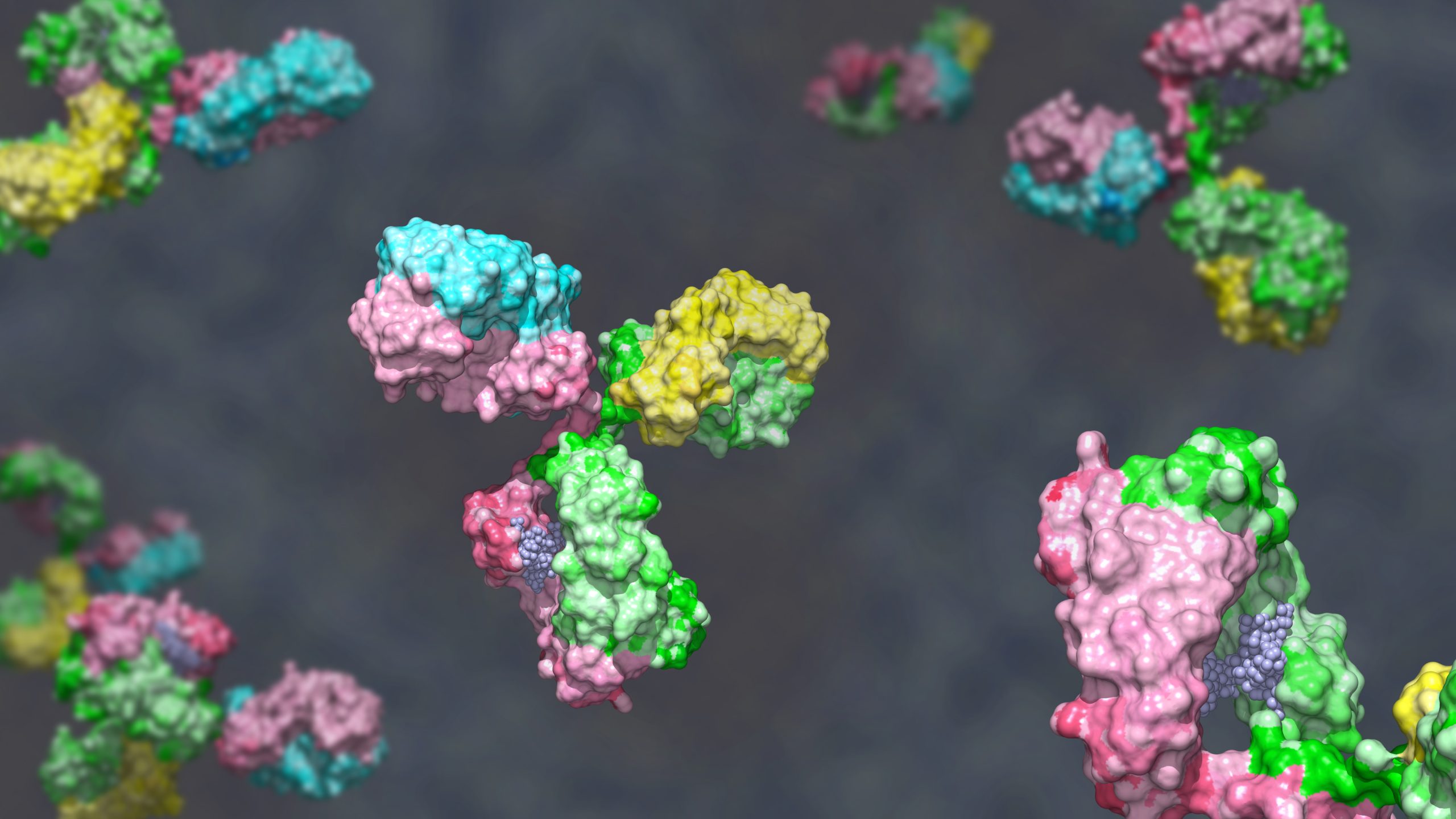
 Functional antibodies are key experimental tools for targeting the complex tumor microenvironment and developing new immunotherapies. The saying goes that no man is an island, and as it turns out, neither is cancer. Tumors are surrounded by a complex non-cancerous ecosystem containing immune, stromal, and vascular cells, diverse cytokines, and immune checkpoints.[1] Scientists have discovered that this tumor microenvironment (TME) plays a crucial role throughout cancer development, from initiation to metastasis.
Importantly, developing tumors can distort immune cell signaling to drive chronic, damaging inflammation, which promotes both tumor progression and resistance to therapy.[1][2]
The TME also supports tumoral immune evasion. Myeloid cells, for example, can secrete mediators such as IL-10 and TGF-β to inhibit T cells and natural killer cells, trigger inflammation via the release of IL-1β, TNF-α, and IL-6, and express PD-L1, a checkpoint ligand that prevents immune cells from identifying and eliminating cancer cells.[1]
The TME Presents Experimental Challenges and Scientific Opportunities
It is perhaps no surprise that preclinical cancer models that fail to consider the TME show limited success in therapeutic development. Subcutaneous cell line-derived xenograft (CDX) models, in which scientists inject human cancer cell lines below a mouse’s skin, are a case in point.[3] CDX models do not recapitulate the TME of the tumor’s organ of origin, and the US Food and Drug Administration has only approved 3–5 percent of the cancer drugs developed using these models. As a result, incorporating the complicated, dynamic TME into model design has become increasingly important for therapeutic discovery.
Taking the TME fully into account when developing immunotherapies and preclinical cancer models is no easy feat, but doing so may usher in new, paradigm-shifting discoveries.
One of the hurdles to studying the TME is the trade-off between complexity and controllability that is broadly inherent to cancer models.[3] In vivo models more faithfully recapitulate the TME but are difficult to manipulate, while simpler in vitro models can be subjected to minute experimental perturbations but do not fully capture the TME’s complexity and heterogeneity. Therefore, functional antibodies have become powerful tools that can precisely target the TME in both in vivo and non-animal models, enabling new immunotherapy insights and deeper scientific understanding of this intricate ecosystem.
Functional Antibodies and the Tumor Microenvironment
Bio X Cell’s ready-made functional antibodies are widely used by the research community to investigate and manipulate the TME, with thousands of peer-reviewed publications demonstrating their significance in cancer biology. The company’s cancer biology portfolio includes hundreds of functional antibodies that can deplete, modulate, or block the cells, cytokines, and immune checkpoints that shape the TME.
For example, researchers frequently use anti-CD25 to deplete regulatory T cells, anti-Gr-1 to target myeloid-derived suppressor cells, and anti-CSF1R to modulate macrophage activity. These tools allow precise interrogation of key immune populations within the TME.
In addition, researchers can block immunosuppressive cytokine pathways using antibodies such as anti-TGF-β or anti-IL-10R. They can also activate co-stimulatory signaling with agonists such as anti-4-1BB or anti-CD40.
Similarly, immune checkpoint inhibitors such as anti-mPD-1 and anti-mCTLA-4 remain central reagents in cancer research. This is illustrated by the work of Yumeng Wang and colleagues at Fudan University.[4] Wang's team investigated how dendritic cells in the TME promote immune tolerance in cervical cancer. They found that cervical cancer cells drive dendritic cells toward a low-immunogenicity, high-immunotolerance phenotype via sialoglycan/Siglec-10 signaling.
Blocking Siglec-10 enhanced dendritic cell-mediated tumor cell apoptosis in a patient-derived tumor fragment model. However, adding anti-mPD-1 antibody produced an even stronger immune activation effect, demonstrating the value of synergistic checkpoint blockade. These results help clarify mechanisms that may inform future cervical cancer research.
Studies of this kind rely on antibodies that demonstrate functional activity in living systems. Bio X Cell functional antibodies are purpose built for sensitive in vivo and advanced in vitro model system experiments due to their high purity, extremely low endotoxin levels, and preservative- and stabilizer-free formulations. With reliable, high-quality functional antibodies in hand, cancer researchers can continue to dissect the complexities of the tumor microenvironment.
References
- de Visser KE and Joyce JA. The evolving tumor microenvironment: From cancer initiation to metastatic outgrowth. Cancer Cell. 2023;41(3):374-403.
- Zhao H, et al. Inflammation and tumor progression: signaling pathways and targeted intervention. Signal Transduct Target Ther. 2021;6:263.
- Crouigneau R, et al. Mimicking and analyzing the tumor microenvironment. Cell Rep Methods. 2024;4(10):100866.
- Wang C, et al. Identification of Siglec-10 as a new dendritic cell checkpoint for cervical cancer immunotherapy. J Immunother Cancer. 2024;12:e009404.