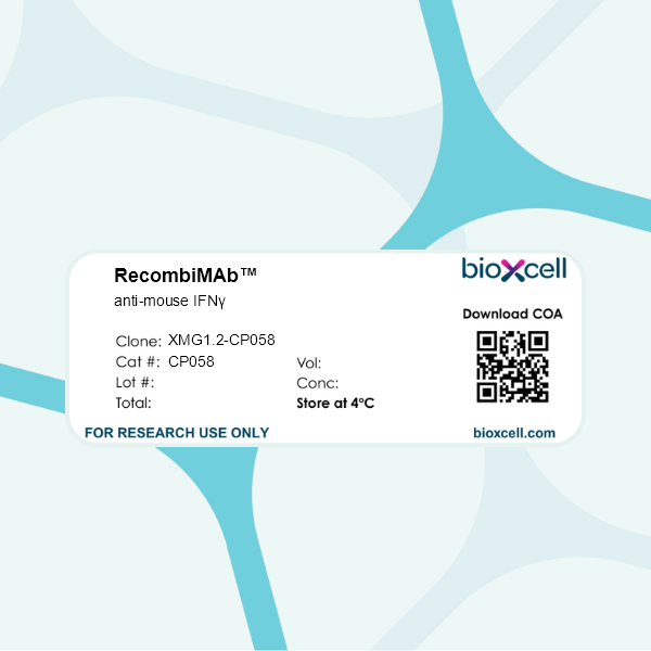
RecombiMAb anti-mouse IFNγ
(switched from rat IgG1, kappa)
Product Details
The XMG1.2-CP058 monoclonal antibody is a recombinant, chimeric version of the original XMG1.2 antibody. The variable domain sequences are identical but the constant region sequences have been switched from Rat IgG1, κ to mouse IgG1, κ for use in murine models. Species-matched chimeric antibodies exhibit regulated effector functions—including Fc receptor binding and complement activation—and cause less immunogenicity and formation of anti-drug antibodies (ADAs) than xenogenic antibodies in animal models. The highly controlled sequence and lack of genetic drift in recombinant antibodies provide more reliable and reproducible results over hybridoma derived antibodies.The XMG1.2 monoclonal antibody reacts with mouse IFNγ (interferon gamma) a 20 kDa soluble pleiotropic cytokine and the sole member of the type II class of interferons. IFNγ is primarily produced by activated lymphocytes including T, B, NK cells, and ILCs. IFNγ exerts immunoregulatory, anti-proliferative, anti-viral, and proinflammatory activities and plays an important role in activation, growth, and differentiation of T and B lymphocytes, macrophages, NK cells and other non-hematopoietic cell types. Additionally, IFNγ induces the production of cytokines, Fc receptor, and adhesion molecules and up-regulates MHC class I and II antigen expression by antigen presenting cells during an immune response. IFNγ has also been shown to modulate macrophage effector functions, influence isotype switching and induce the secretion of immunoglobulins by B cells. IFNγ signals through the IFN gamma receptor which exists as a heterodimer composed of CD119 (IFN gamma receptor 1) and AF-1 (IFN gamma receptor 2). The IFNγ receptor is expressed ubiquitously on almost all cell types except for mature erythrocytes. The XMG1.2 antibody is a neutralizing antibody.
Specifications
Isotype | Mouse IgG1 |
---|---|
Recommended Isotype Control(s) | RecombiMAb mouse IgG1 isotype control, anti-hen egg lysozyme |
Recommended Dilution Buffer | InVivoPure pH 7.0 Dilution Buffer |
Immunogen | Recombinant mouse IFNγ |
Reported Applications |
Flow Cytometry Western Blot ELISPOT in vitro IFNγ neutralization in vivo IFNγ neutralization *Reported for the original XMG1.2 antibody |
Formulation |
PBS, pH 7.0 Contains no stabilizers or preservatives |
Endotoxin |
<1EU/mg (<0.001EU/μg) Determined by LAL gel clotting assay |
Aggregation |
<5% Determined by SEC |
Purity |
>95% Determined by SDS-PAGE |
Sterility | 0.2 µm filtration |
Production | Purified from HEK293 cell supernatant in an animal-free facility |
Purification | Protein G |
Molecular Weight | 150 kDa |
Murine Pathogen Tests |
Ectromelia/Mousepox Virus: Negative Hantavirus: Negative K Virus: Negative Lactate Dehydrogenase-Elevating Virus: Negative Lymphocytic Choriomeningitis virus: Negative Mouse Adenovirus: Negative Mouse Cytomegalovirus: Negative Mouse Hepatitis Virus: Negative Mouse Minute Virus: Negative Mouse Norovirus: Negative Mouse Parvovirus: Negative Mouse Rotavirus: Negative Mycoplasma Pulmonis: Negative Pneumonia Virus of Mice: Negative Polyoma Virus: Negative Reovirus Screen: Negative Sendai Virus: Negative Theiler’s Murine Encephalomyelitis: Negative |
Storage | The antibody solution should be stored at the stock concentration at 4°C. Do not freeze. |
Additional Formats
Recommended Products
in vivo IFNγ neutralization
Elewaut A, Estivill G, Bayerl F, Castillon L, Novatchkova M, Pottendorfer E, Hoffmann-Haas L, Schönlein M, Nguyen TV, Lauss M, Andreatta F, Vulin M, Krecioch I, Bayerl J, Pedde AM, Fabre N, Holstein F, Cronin SM, Rieser S, Laniti DD, Barras D, Coukos G, Quek C, Bai X, Muñoz I Ordoño M, Wiesner T, Zuber J, Jönsson G, Böttcher JP, Vanharanta S, Obenauf AC. (2025). "Cancer cells impair monocyte-mediated T cell stimulation to evade immunity" Nature 637(8046):716-725. PubMed
The tumour microenvironment is programmed by cancer cells and substantially influences anti-tumour immune responses1,2. Within the tumour microenvironment, CD8+ T cells undergo full effector differentiation and acquire cytotoxic anti-tumour functions in specialized niches3-7. Although interactions with type 1 conventional dendritic cells have been implicated in this process3-5,8-10, the underlying cellular players and molecular mechanisms remain incompletely understood. Here we show that inflammatory monocytes can adopt a pivotal role in intratumoral T cell stimulation. These cells express Cxcl9, Cxcl10 and Il15, but in contrast to type 1 conventional dendritic cells, which cross-present antigens, inflammatory monocytes obtain and present peptide-major histocompatibility complex class I complexes from tumour cells through 'cross-dressing'. Hyperactivation of MAPK signalling in cancer cells hampers this process by coordinately blunting the production of type I interferon (IFN-I) cytokines and inducing the secretion of prostaglandin E2 (PGE2), which impairs the inflammatory monocyte state and intratumoral T cell stimulation. Enhancing IFN-I cytokine production and blocking PGE2 secretion restores this process and re-sensitizes tumours to T cell-mediated immunity. Together, our work uncovers a central role of inflammatory monocytes in intratumoral T cell stimulation, elucidates how oncogenic signalling disrupts T cell responses through counter-regulation of PGE2 and IFN-I, and proposes rational combination therapies to enhance immunotherapies.
in vivo IFNγ neutralization
Fumagalli V, Ravà M, Marotta D, Di Lucia P, Bono EB, Giustini L, De Leo F, Casalgrandi M, Monteleone E, Mouro V, Malpighi C, Perucchini C, Grillo M, De Palma S, Donnici L, Marchese S, Conti M, Muramatsu H, Perlman S, Pardi N, Kuka M, De Francesco R, Bianchi ME, Guidotti LG, Iannacone M. (2024). "Antibody-independent protection against heterologous SARS-CoV-2 challenge conferred by prior infection or vaccination" Nat Immunol 25(4):633-643. PubMed
Vaccines have reduced severe acute respiratory syndrome coronavirus 2 (SARS-CoV-2) morbidity and mortality, yet emerging variants challenge their effectiveness. The prevailing approach to updating vaccines targets the antibody response, operating under the presumption that it is the primary defense mechanism following vaccination or infection. This perspective, however, can overlook the role of T cells, particularly when antibody levels are low or absent. Here we show, through studies in mouse models lacking antibodies but maintaining functional B cells and lymphoid organs, that immunity conferred by prior infection or mRNA vaccination can protect against SARS-CoV-2 challenge independently of antibodies. Our findings, using three distinct models inclusive of a novel human/mouse ACE2 hybrid, highlight that CD8+ T cells are essential for combating severe infections, whereas CD4+ T cells contribute to managing milder cases, with interferon-γ having an important function in this antibody-independent defense. These findings highlight the importance of T cell responses in vaccine development, urging a broader perspective on protective immunity beyond just antibodies.
Huang CC, Sung HH, Li HC, Miaw SC, Kung JT, Chou MY, Wu-Hsieh BA. (2023). "A novel trivalent non-Fc anti-CD3 Collabody preferentially induces Th1 cell apoptosis in vitro and long-lasting remission in recent-onset diabetic NOD mice" Front Immunol . PubMed
Specific anti-CD3 treatment is deemed to be a promising therapy for allograft rejection and type 1 diabetes (T1D). Fc receptor (FcR) reduced-binding antibodies, by avoiding adverse effects of Fc and FcR interaction, have good therapeutic potential. We generated a trivalent anti-mouse-CD3 Collabody, h145CSA, by using a triplex-forming collagen-like peptide (Gly-Pro-Pro)10 to drive the trimerization of the Fab fragments. Exposure to h145CSA, but not its bivalent counterparts 145-2C11 and h145chIgGAA (FcR reduced-binding format), upregulates FasL expression on Th1 cells and causes Th1 cell apoptosis. Administration of h145CSA invokes minimal mitogenic effects in mice. The ability of multiple dosing of h145CSA to induce splenic CD4+ T-cell depletion is comparable to bivalent antibodies but is characterized by more rapid CD4+ T-cell recovery kinetics. h145CSA is more potent than h145chIgGAA in inducing long-lasting remission in recent-onset diabetic NOD mice. Its therapeutic effect is accompanied by a significantly lower percentage of CD4+IFNγ+ T cells and a higher Treg/Th1 ratio in pancreatic and mesenteric lymph nodes. The results of our study demonstrate that trivalent non-Fc anti-CD3 Collabody has the potential to be used in the treatment of T1D.
Flow Cytometry, in vivo IFNγ neutralization
Shibuya M, Tamiya S, Kawai A, Hirai T, Cragg MS, Yoshioka Y. (2021). "Synergistic effect of non-neutralizing antibodies and interferon-γ for cross-protection against influenza" iScience 24(10):103131. PubMed
Current influenza vaccines do not typically confer cross-protection against antigenically mismatched strains. To develop vaccines conferring broader cross-protection, recent evidence indicates the crucial role of both cross-reactive antibodies and viral-specific CD4+ T cells; however, the precise mechanism of cross-protection is unclear. Furthermore, adjuvants that can efficiently induce cross-protective CD4+ T cells have not been identified. Here we show that CpG oligodeoxynucleotides combined with aluminum salts work as adjuvants for influenza vaccine and confer strong cross-protection in mice. Both cross-reactive antibodies and viral-specific CD4+ T cells contributed to cross-protection synergistically, with each individually ineffective. Furthermore, we found that downregulated expression of Fcγ receptor IIb on alveolar macrophages due to IFN-γ secreted by viral-specific CD4+ T cells improves the activity of cross-reactive antibodies. Our findings inform the development of optimal adjuvants for vaccines and how influenza vaccines confer broader cross-protection.