InVivoMAb anti-mouse IFNγ

Catalog #BE0055
Product Citations:
278
Clone:
XMG1.2
Reactivities:
Mouse

$172.00 - $4,494.00

Choose an Option...
  • 100 mg - $4,494.00
  • 50 mg - $3,175.00
  • 25 mg - $2,109.00
  • 5 mg - $630.00
  • 1 mg - $172.00
  • Custom Amount (Quotes Only)
In stock
Only %1 left

Product Details

The XMG1.2 monoclonal antibody reacts with mouse IFNγ (interferon gamma) a 20 kDa soluble pleiotropic cytokine and the sole member of the type II class of interferons. IFNγ is primarily produced by activated lymphocytes including T, B, NK cells, and ILCs. IFNγ exerts immunoregulatory, anti-proliferative, anti-viral, and proinflammatory activities and plays an important role in activation, growth, and differentiation of T and B lymphocytes, macrophages, NK cells and other non-hematopoietic cell types. Additionally, IFNγ induces the production of cytokines, Fc receptor, and adhesion molecules and up-regulates MHC class I and II antigen expression by antigen presenting cells during an immune response. IFNγ has also been shown to modulate macrophage effector functions, influence isotype switching and induce the secretion of immunoglobulins by B cells. IFNγ signals through the IFN gamma receptor which exists as a heterodimer composed of CD119 (IFN gamma receptor 1) and AF-1 (IFN gamma receptor 2). The IFNγ receptor is expressed ubiquitously on almost all cell types with the exception of mature erythrocytes. The XMG1.2 antibody is a neutralizing antibody.

Specifications

Isotype Rat IgG1, κ
Recommended Isotype Control(s) InVivoMAb rat IgG1 isotype control, anti-horseradish peroxidase
Recommended Dilution Buffer InVivoPure pH 8.0T Dilution Buffer
Conjugation This product is unconjugated. Conjugation is available via our Antibody Conjugation Services.
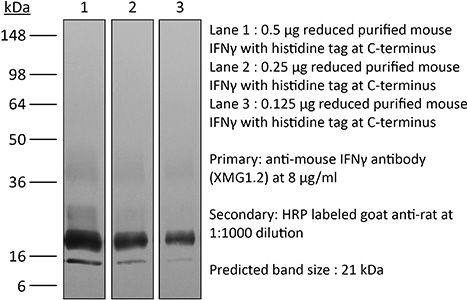
Immunogen Recombinant mouse IFNγ
Reported Applications in vivo IFNγ neutralization
in vitro IFNγ neutralization
ELISPOT
Flow cytometry
Western blot
Formulation PBS + 0.01% Tween, pH 8.0
Contains no stabilizers or preservatives
Endotoxin <2EU/mg (<0.002EU/μg)
Determined by LAL gel clotting assay
Purity >95%
Determined by SDS-PAGE
Sterility 0.2 µm filtration
Production Purified from cell culture supernatant in an animal-free facility
Purification Protein G
RRID AB_1107694
Molecular Weight 150 kDa
Storage The antibody solution should be stored at the stock concentration at 4°C. Do not freeze.

Additional Formats

in vivo IFNγ neutralization
Glasner, A., et al. (2018). "NKp46 Receptor-Mediated Interferon-gamma Production by Natural Killer Cells Increases Fibronectin 1 to Alter Tumor Architecture and Control Metastasis" Immunity 48(1): 107-119 e104. PubMed

Natural killer (NK) cells are innate lymphoid cells, and their presence within human tumors correlates with better prognosis. However, the mechanisms by which NK cells control tumors in vivo are unclear. Here, we used reflectance confocal microscopy (RCM) imaging in humans and in mice to visualize tumor architecture in vivo. We demonstrated that signaling via the NK cell receptor NKp46 (human) and Ncr1 (mouse) induced interferon-gamma (IFN-gamma) secretion from intratumoral NK cells. NKp46- and Ncr1-mediated IFN-gamma production led to the increased expression of the extracellular matrix protein fibronectin 1 (FN1) in the tumors, which altered primary tumor architecture and resulted in decreased metastases formation. Injection of IFN-gamma into tumor-bearing mice or transgenic overexpression of Ncr1 in NK cells in mice resulted in decreased metastasis formation. Thus, we have defined a mechanism of NK cell-mediated control of metastases in vivo that may help develop NK cell-dependent cancer therapies.

in vitro IFNγ neutralization
Clever, D., et al. (2016). "Oxygen Sensing by T Cells Establishes an Immunologically Tolerant Metastatic Niche" Cell 166(5): 1117-1131 e1114. PubMed

Cancer cells must evade immune responses at distant sites to establish metastases. The lung is a frequent site for metastasis. We hypothesized that lung-specific immunoregulatory mechanisms create an immunologically permissive environment for tumor colonization. We found that T-cell-intrinsic expression of the oxygen-sensing prolyl-hydroxylase (PHD) proteins is required to maintain local tolerance against innocuous antigens in the lung but powerfully licenses colonization by circulating tumor cells. PHD proteins limit pulmonary type helper (Th)-1 responses, promote CD4(+)-regulatory T (Treg) cell induction, and restrain CD8(+) T cell effector function. Tumor colonization is accompanied by PHD-protein-dependent induction of pulmonary Treg cells and suppression of IFN-gamma-dependent tumor clearance. T-cell-intrinsic deletion or pharmacological inhibition of PHD proteins limits tumor colonization of the lung and improves the efficacy of adoptive cell transfer immunotherapy. Collectively, PHD proteins function in T cells to coordinate distinct immunoregulatory programs within the lung that are permissive to cancer metastasis.

in vitro IFNγ neutralization, Flow Cytometry
Gu, A. D., et al. (2015). "A critical role for transcription factor Smad4 in T cell function that is independent of transforming growth factor beta receptor signaling" Immunity 42(1): 68-79. PubMed

Transforming growth factor-beta (TGF-beta) suppresses T cell function to maintain self-tolerance and to promote tumor immune evasion. Yet how Smad4, a transcription factor component of TGF-beta signaling, regulates T cell function remains unclear. Here we have demonstrated an essential role for Smad4 in promoting T cell function during autoimmunity and anti-tumor immunity. Smad4 deletion rescued the lethal autoimmunity resulting from transforming growth factor-beta receptor (TGF-betaR) deletion and compromised T-cell-mediated tumor rejection. Although Smad4 was dispensable for T cell generation, homeostasis, and effector function, it was essential for T cell proliferation after activation in vitro and in vivo. The transcription factor Myc was identified to mediate Smad4-controlled T cell proliferation. This study thus reveals a requirement of Smad4 for T-cell-mediated autoimmunity and tumor rejection, which is beyond the current paradigm. It highlights a TGF-betaR-independent role for Smad4 in promoting T cell function, autoimmunity, and anti-tumor immunity.

in vitro IFNγ neutralization, Flow Cytometry
Sell, S., et al. (2015). "Control of murine cytomegalovirus infection by gammadelta T cells" PLoS Pathog 11(2): e1004481. PubMed

Infections with cytomegalovirus (CMV) can cause severe disease in immunosuppressed patients and infected newborns. Innate as well as cellular and humoral adaptive immune effector functions contribute to the control of CMV in immunocompetent individuals. None of the innate or adaptive immune functions are essential for virus control, however. Expansion of gammadelta T cells has been observed during human CMV (HCMV) infection in the fetus and in transplant patients with HCMV reactivation but the protective function of gammadelta T cells under these conditions remains unclear. Here we show for murine CMV (MCMV) infections that mice that lack CD8 and CD4 alphabeta-T cells as well as B lymphocytes can control a MCMV infection that is lethal in RAG-1(-/-) mice lacking any T- and B-cells. gammadelta T cells, isolated from infected mice can kill MCMV infected target cells in vitro and, importantly, provide long-term protection in infected RAG-1(-/-) mice after adoptive transfer. gammadelta T cells in MCMV infected hosts undergo a prominent and long-lasting phenotypic change most compatible with the view that the majority of the gammadelta T cell population persists in an effector/memory state even after resolution of the acute phase of the infection. A clonotypically focused Vgamma1 and Vgamma2 repertoire was observed at later stages of the infection in the organs where MCMV persists. These findings add gammadelta T cells as yet another protective component to the anti-CMV immune response. Our data provide clear evidence that gammadelta T cells can provide an effective control mechanism of acute CMV infections, particularly when conventional adaptive immune mechanisms are insufficient or absent, like in transplant patient or in the developing immune system in utero. The findings have implications in the stem cell transplant setting, as antigen recognition by gammadelta T cells is not MHC-restricted and dual reactivity against CMV and tumors has been described.

in vitro IFNγ neutralization
Cao, A. T., et al. (2015). "Interleukin (IL)-21 promotes intestinal IgA response to microbiota" Mucosal Immunol 8(5): 1072-1082. PubMed

Commensal microbiota-specific T helper type 17 (Th17) cells are enriched in the intestines, which can convert into T follicular helper (Tfh) in Peyer’s patches, and are crucial for production of intestinal immunoglobulin A (IgA) against microbiota; however, the role of Th17 and Tfh cytokines in regulating the mucosal IgA response to enteric microbiota is still not completely known. In this study, we found that intestinal IgA was impaired in mice deficient in interleukin (IL)-17 or IL-21 signaling. IL-21, but not IL-17, is able to augment B-cell differentiation to IgA(+) cells as mediated by transforming growth factor beta1 (TGFbeta1) and accelerate IgA class switch recombination (CSR). IL-21 and retinoic acid (RA) induce IgA(+) B-cell development and IgA production and drives autocrine TGFbeta1 production to initiate IgA CSR. Repletion of T-cell-deficient TCRbetaxdelta(-/-) mice with Th17 cells specific for commensal bacterial antigen increased the levels of IgA(+) B cells and IgA production in the intestine, which was blocked by neutralizing IL-21. Thus IL-21 functions to strongly augment IgA production under intestinal environment. Furthermore, IL-21 promotes intestinal B-cell homing through alpha4beta7 expression, alone or with TGFbeta and RA. Together, IL-21 from microbiota-specific Th17 and/or Tfh cells contributes to robust intestinal IgA levels by enhancing IgA(+) CSR, IgA production and B-cell trafficking into the intestine.

in vitro IFNγ neutralization
Choi, Y. S., et al. (2015). "LEF-1 and TCF-1 orchestrate TFH differentiation by regulating differentiation circuits upstream of the transcriptional repressor Bcl6" Nat Immunol 16(9): 980-990. PubMed

Follicular helper T cells (TFH cells) are specialized effector CD4(+) T cells that help B cells develop germinal centers (GCs) and memory. However, the transcription factors that regulate the differentiation of TFH cells remain incompletely understood. Here we report that selective loss of Lef1 or Tcf7 (which encode the transcription factor LEF-1 or TCF-1, respectively) resulted in TFH cell defects, while deletion of both Lef1 and Tcf7 severely impaired the differentiation of TFH cells and the formation of GCs. Forced expression of LEF-1 enhanced TFH differentiation. LEF-1 and TCF-1 coordinated such differentiation by two general mechanisms. First, they established the responsiveness of naive CD4(+) T cells to TFH cell signals. Second, they promoted early TFH differentiation via the multipronged approach of sustaining expression of the cytokine receptors IL-6Ralpha and gp130, enhancing expression of the costimulatory receptor ICOS and promoting expression of the transcriptional repressor Bcl6.

in vivo IFNγ neutralization
Zander, R. A., et al. (2015). "PD-1 Co-inhibitory and OX40 Co-stimulatory Crosstalk Regulates Helper T Cell Differentiation and Anti-Plasmodium Humoral Immunity" Cell Host Microbe 17(5): 628-641. PubMed

The differentiation and protective capacity of Plasmodium-specific T cells are regulated by both positive and negative signals during malaria, but the molecular and cellular details remain poorly defined. Here we show that malaria patients and Plasmodium-infected rodents exhibit atypical expression of the co-stimulatory receptor OX40 on CD4 T cells and that therapeutic enhancement of OX40 signaling enhances helper CD4 T cell activity, humoral immunity, and parasite clearance in rodents. However, these beneficial effects of OX40 signaling are abrogated following coordinate blockade of PD-1 co-inhibitory pathways, which are also upregulated during malaria and associated with elevated parasitemia. Co-administration of biologics blocking PD-1 and promoting OX40 signaling induces excessive interferon-gamma that directly limits helper T cell-mediated support of humoral immunity and decreases parasite control. Our results show that targeting OX40 can enhance Plasmodium control and that crosstalk between co-inhibitory and co-stimulatory pathways in pathogen-specific CD4 T cells can impact pathogen clearance.

in vitro IFNγ neutralization, Flow Cytometry
Hou, L., et al. (2015). "The protease cathepsin L regulates Th17 cell differentiation" J Autoimmun. S 0896-8411(15): 30024-X. PubMed

Previously we reported that IL-17+ T cells, primarily IL-17+ gammadelta cells, are increased in mice lacking the protease inhibitor serpinB1 (serpinb1-/- mice). Here we show that serpinB1-deficient CD4 cells exhibit a cell-autonomous and selective deficiency in suppressing T helper 17 (Th17) cell differentiation. This suggested an opposing role for one or more protease in promoting Th17 differentiation. We found that several SerpinB1-inhibitable cysteine cathepsins are induced in Th17 cells, most prominently cathepsin L (catL); this was verified by peptidase assays, active site labeling and Western blots. Moreover, Th17 differentiation was suppressed by both broad cathepsin inhibitors and catL selective inhibitors. CatL is present in Th17 cells as single chain (SC)- and two-chain (TC)-forms. Inhibiting asparagine endopeptidase (AEP) blocked conversion of SC-catL to TC-catL and increased generation of serpinb1-/- Th17 cells, but not wild-type Th17 cells. These findings suggest that SC-catL is biologically active in promoting Th17 generation and is counter-regulated by serpinB1 and secondarily by AEP. Thus, in addition to regulation by cytokines and transcription factors, differentiation of CD4 cells to Th17 cells is actively regulated by a catL-serpinB1-AEP module. Targeting this protease regulatory module could be an approach to treating Th17 cell-driven autoimmune disorders.

in vitro IFNγ neutralization, Flow Cytometry
Rabenstein, H., et al. (2014). "Differential kinetics of antigen dependency of CD4+ and CD8+ T cells" J Immunol 192(8): 3507-3517. PubMed

Ag recognition via the TCR is necessary for the expansion of specific T cells that then contribute to adaptive immunity as effector and memory cells. Because CD4+ and CD8+ T cells differ in terms of their priming APCs and MHC ligands we compared their requirements of Ag persistence during their expansion phase side by side. Proliferation and effector differentiation of TCR transgenic and polyclonal mouse T cells were thus analyzed after transient and continuous TCR signals. Following equally strong stimulation, CD4+ T cell proliferation depended on prolonged Ag presence, whereas CD8+ T cells were able to divide and differentiate into effector cells despite discontinued Ag presentation. CD4+ T cell proliferation was neither affected by Th lineage or memory differentiation nor blocked by coinhibitory signals or missing inflammatory stimuli. Continued CD8+ T cell proliferation was truly independent of self-peptide/MHC-derived signals. The subset divergence was also illustrated by surprisingly broad transcriptional differences supporting a stronger propensity of CD8+ T cells to programmed expansion. These T cell data indicate an intrinsic difference between CD4+ and CD8+ T cells regarding the processing of TCR signals for proliferation. We also found that the presentation of a MHC class II-restricted peptide is more efficiently prolonged by dendritic cell activation in vivo than a class I bound one. In summary, our data demonstrate that CD4+ T cells require continuous stimulation for clonal expansion, whereas CD8+ T cells can divide following a much shorter TCR signal.

in vivo IFNγ neutralization, Flow Cytometry
Uddin, M. N., et al. (2014). "TNF-alpha-dependent hematopoiesis following Bcl11b deletion in T cells restricts metastatic melanoma" J Immunol 192(4): 1946-1953. PubMed

Using several tumor models, we demonstrate that mice deficient in Bcl11b in T cells, although having reduced numbers of T cells in the peripheral lymphoid organs, developed significantly less tumors compared with wild-type mice. Bcl11b(-/-) CD4(+) T cells, with elevated TNF-alpha levels, but not the Bcl11b(-/-) CD8(+) T cells, were required for the reduced tumor burden, as were NK1.1(+) cells, found in increased numbers in Bcl11b(F/F)/CD4-Cre mice. Among NK1.1(+) cells, the NK cell population was predominant in number and was the only population displaying elevated granzyme B levels and increased degranulation, although not increased proliferation. Although the number of myeloid-derived suppressor cells was increased in the lungs with metastatic tumors of Bcl11b(F/F)/CD4-Cre mice, their arginase-1 levels were severely reduced. The increase in NK cell and myeloid-derived suppressor cell numbers was associated with increased bone marrow and splenic hematopoiesis. Finally, the reduced tumor burden, increased numbers of NK cells in the lung, and increased hematopoiesis in Bcl11b(F/F)/CD4-Cre mice were all dependent on TNF-alpha. Moreover, TNF-alpha treatment of wild-type mice also reduced the tumor burden and increased hematopoiesis and the numbers and activity of NK cells in the lung. In vitro treatment with TNF-alpha of lineage-negative hematopoietic progenitors increased NK and myeloid differentiation, further supporting a role of TNF-alpha in promoting hematopoiesis. These studies reveal a novel role for TNF-alpha in the antitumor immune response, specifically in stimulating hematopoiesis and increasing the numbers and activity of NK cells.

in vitro IFNγ neutralization
Bertin, S., et al. (2014). "The ion channel TRPV1 regulates the activation and proinflammatory properties of CD4(+) T cells" Nat Immunol 15(11): 1055-1063. PubMed

TRPV1 is a Ca(2+)-permeable channel studied mostly as a pain receptor in sensory neurons. However, its role in other cell types is poorly understood. Here we found that TRPV1 was functionally expressed in CD4(+) T cells, where it acted as a non-store-operated Ca(2+) channel and contributed to T cell antigen receptor (TCR)-induced Ca(2+) influx, TCR signaling and T cell activation. In models of T cell-mediated colitis, TRPV1 promoted colitogenic T cell responses and intestinal inflammation. Furthermore, genetic and pharmacological inhibition of TRPV1 in human CD4(+) T cells recapitulated the phenotype of mouse Trpv1(-/-) CD4(+) T cells. Our findings suggest that inhibition of TRPV1 could represent a new therapeutic strategy for restraining proinflammatory T cell responses.

in vitro IFNγ neutralization, ELISPOT
Deng, L., et al. (2014). "Irradiation and anti-PD-L1 treatment synergistically promote antitumor immunity in mice" J Clin Invest 124(2): 687-695. PubMed

High-dose ionizing irradiation (IR) results in direct tumor cell death and augments tumor-specific immunity, which enhances tumor control both locally and distantly. Unfortunately, local relapses often occur following IR treatment, indicating that IR-induced responses are inadequate to maintain antitumor immunity. Therapeutic blockade of the T cell negative regulator programmed death-ligand 1 (PD-L1, also called B7-H1) can enhance T cell effector function when PD-L1 is expressed in chronically inflamed tissues and tumors. Here, we demonstrate that PD-L1 was upregulated in the tumor microenvironment after IR. Administration of anti-PD-L1 enhanced the efficacy of IR through a cytotoxic T cell-dependent mechanism. Concomitant with IR-mediated tumor regression, we observed that IR and anti-PD-L1 synergistically reduced the local accumulation of tumor-infiltrating myeloid-derived suppressor cells (MDSCs), which suppress T cells and alter the tumor immune microenvironment. Furthermore, activation of cytotoxic T cells with combination therapy mediated the reduction of MDSCs in tumors through the cytotoxic actions of TNF. Our data provide evidence for a close interaction between IR, T cells, and the PD-L1/PD-1 axis and establish a basis for the rational design of combination therapy with immune modulators and radiotherapy.

in vivo IFNγ neutralization, Flow Cytometry
Yu, X., et al. (2013). "A multifunctional chimeric chaperone serves as a novel immune modulator inducing therapeutic antitumor immunity" Cancer Res 73(7): 2093-2103. PubMed

Converting the immunosuppressive tumor environment into one that is favorable to the induction of antitumor immunity is indispensable for effective cancer immunotherapy. Here, we strategically incorporate a pathogen (i.e., flagellin)-derived, NF-kappaB-stimulating “danger” signal into the large stress protein or chaperone Grp170 (HYOU1/ORP150) that was previously shown to facilitate antigen crosspresentation. This engineered chimeric molecule (i.e., Flagrp170) is capable of transporting tumor antigens and concurrently inducing functional activation of dendritic cells (DC). Intratumoral administration of adenoviruses expressing Flagrp170 induces a superior antitumor response against B16 melanoma and its distant lung metastasis compared with unmodified Grp170 and flagellin. The enhanced tumor destruction is accompanied with significantly increased tumor infiltration by CD8(+) cells as well as elevation of IFN-gamma and interleukin (IL)-12 levels in the tumor sites. In situ Ad.Flagrp170 therapy provokes systemic activation of CTLs that recognize several antigens naturally expressing in melanoma (e.g., gp100/PMEL and TRP2/DCT). The mechanistic studies using CD11c-DTR transgenic mice and Batf3-deficient mice reveal that CD8alpha(+) DCs are required for the improved T-cell crosspriming. Antibody neutralization assays show that IL-12 and IFN-gamma are essential for the Flagrp170-elicited antitumor response, which also involves CD8(+) T cells and natural killer cells. The therapeutic efficacy of Flagrp170 and its immunostimulating activity are also confirmed in mouse prostate cancer and colon carcinoma. Together, targeting the tumor microenvironment with this chimeric chaperone is highly effective in mobilizing or restoring antitumor immunity, supporting the potential therapeutic use of this novel immunomodulator in the treatment of metastatic diseases.

in vivo IFNγ neutralization, Flow Cytometry
Simons, D. M., et al. (2013). "Autoreactive Th1 cells activate monocytes to support regional Th17 responses in inflammatory arthritis" J Immunol 190(7): 3134-3141. PubMed

We have examined mechanisms underlying the formation of pathologic Th17 cells using a transgenic mouse model in which autoreactive CD4(+) T cells recognize influenza virus hemagglutinin (HA) as a ubiquitously expressed self-Ag and induce inflammatory arthritis. The lymph nodes of arthritic mice contain elevated numbers of inflammatory monocytes (iMO) with an enhanced capacity to promote CD4(+) Th17 cell differentiation, and a regional inflammatory response develops in the paw-draining lymph nodes by an IL-17-dependent mechanism. The activation of these Th17-trophic iMO precedes arthritis development and occurs in the context of an autoreactive CD4(+) Th1 cell response. Adoptive transfer of HA-specific CD4(+) T cells into nonarthritic mice expressing HA as a self-Ag similarly led to the formation of Th1 cells and of iMO that could support Th17 cell formation, and, notably, the accumulation of these iMO in the lymph nodes was blocked by IFN-gamma neutralization. These studies show that autoreactive CD4(+) Th1 cells directed to a systemically distributed self-Ag can promote the development of a regional Th17 cell inflammatory response by driving the recruitment of Th17-trophic iMO to the lymph nodes.

in vivo IFNγ neutralization, Flow Cytometry
Kugler, D. G., et al. (2013). "CD4+ T cells are trigger and target of the glucocorticoid response that prevents lethal immunopathology in toxoplasma infection" J Exp Med 210(10): 1919-1927. PubMed

Synthetic glucocorticoids (GCs) are commonly used in the treatment of inflammatory diseases, but the role of endogenous GCs in the regulation of host-protective immune responses is poorly understood. Here we show that GCs are induced during acute Toxoplasma gondii infection and directly control the T cell response to the parasite. When infected with toxoplasma, mice that selectively lack GC receptor (GR) expression in T cells (GR(lck-Cre)) rapidly succumb to infection despite displaying parasite burdens indistinguishable from control animals and unaltered levels of the innate cytokines IL-12 and IL-27. Mortality in the GR(lck-Cre) mice was associated with immunopathology and hyperactive Th1 cell function as revealed by enhanced IFN-gamma and TNF production in vivo. Unexpectedly, these CD4(+) T lymphocytes also overexpressed IL-10. Importantly, CD4(+) T cell depletion in wild-type or GR(lck-Cre) mice led to ablation of the GC response to infection. Moreover, in toxoplasma-infected RAG(-/-) animals, adoptive transfer of CD4(+) T lymphocytes was required for GC induction. These findings establish a novel IL-10-independent immunomodulatory circuit in which CD4(+) T cells trigger a GC response that in turn dampens their own effector function. In the case of T. gondii infection, this self-regulatory pathway is critical for preventing collateral tissue damage and promoting host survival.

in vivo IFNγ neutralization, Flow Cytometry
Mohr, E., et al. (2010). "IFN-{gamma} produced by CD8 T cells induces T-bet-dependent and -independent class switching in B cells in responses to alum-precipitated protein vaccine" Proc Natl Acad Sci U S A 107(40): 17292-17297. PubMed

Alum-precipitated protein (alum protein) vaccines elicit long-lasting neutralizing antibody responses that prevent bacterial exotoxins and viruses from entering cells. Typically, these vaccines induce CD4 T cells to become T helper 2 (Th2) cells that induce Ig class switching to IgG1. We now report that CD8 T cells also respond to alum proteins, proliferating extensively and producing IFN-gamma, a key Th1 cytokine. These findings led us to question whether adoptive transfer of antigen-specific CD8 T cells alters the characteristic CD4 Th2 response to alum proteins and the switching pattern in responding B cells. To this end, WT mice given transgenic ovalbumin (OVA)-specific CD4 (OTII) or CD8 (OTI) T cells, or both, were immunized with alum-precipitated OVA. Cotransfer of antigen-specific CD8 T cells skewed switching patterns in responding B cells from IgG1 to IgG2a and IgG2b. Blocking with anti-IFN-gamma antibody largely inhibited this altered B-cell switching pattern. The transcription factor T-bet is required in B cells for IFN-gamma-dependent switching to IgG2a. By contrast, we show that this transcription factor is dispensable in B cells both for IFN-gamma-induced switching to IgG2b and for inhibition of switching to IgG1. Thus, T-bet dependence identifies distinct transcriptional pathways in B cells that regulate IFN-gamma-induced switching to different IgG isotypes.

in vivo IFNγ neutralization, Flow Cytometry
Quezada, S. A., et al. (2010). "Tumor-reactive CD4(+) T cells develop cytotoxic activity and eradicate large established melanoma after transfer into lymphopenic hosts" J Exp Med 207(3): 637-650. PubMed

Adoptive transfer of large numbers of tumor-reactive CD8(+) cytotoxic T lymphocytes (CTLs) expanded and differentiated in vitro has shown promising clinical activity against cancer. However, such protocols are complicated by extensive ex vivo manipulations of tumor-reactive cells and have largely focused on CD8(+) CTLs, with much less emphasis on the role and contribution of CD4(+) T cells. Using a mouse model of advanced melanoma, we found that transfer of small numbers of naive tumor-reactive CD4(+) T cells into lymphopenic recipients induces substantial T cell expansion, differentiation, and regression of large established tumors without the need for in vitro manipulation. Surprisingly, CD4(+) T cells developed cytotoxic activity, and tumor rejection was dependent on class II-restricted recognition of tumors by tumor-reactive CD4(+) T cells. Furthermore, blockade of the coinhibitory receptor CTL-associated antigen 4 (CTLA-4) on the transferred CD4(+) T cells resulted in greater expansion of effector T cells, diminished accumulation of tumor-reactive regulatory T cells, and superior antitumor activity capable of inducing regression of spontaneous mouse melanoma. These findings suggest a novel potential therapeutic role for cytotoxic CD4(+) T cells and CTLA-4 blockade in cancer immunotherapy, and demonstrate the potential advantages of differentiating tumor-reactive CD4(+) cells in vivo over current protocols favoring in vitro expansion and differentiation.

    • Immunology and Microbiology
    • ,
    IGF1R Promotes Th17/Treg Cell Development in Experimental Autoimmune Prostatitis.

    In Journal of Inflammation Research on 5 May 2025 by Guan, Y., Yue, S., et al.

    Chronic prostatitis is a common urological disorder in young and middle-aged men, characterized by frequent relapses and an unknown etiology. We investigated the potential function of insulin-like growth factor 1 (IGF1) -related ligands in chronic prostatitis in the current study. In this study, we established the chronic experimental autoimmune prostatitis mouse model H&E staining was used to assess immune cell infiltration in prostate tissue, while RT-qPCR and Western blot analyses were performed to validate gene and protein expression differences across groups, respectively. Immunofluorescence staining was utilized to determine the spatial distribution of key proteins. Flow cytometry was conducted to analyze the proportions of immune cell populations in different experimental groups. Adeno-associated virus (AAV) was employed to knock down Igflr, and ELISA was used to measure cytokine levels in the peripheral blood of mice. Statistical significance was defined as P < 0.05, and all tests were conducted as two-tailed. Data analysis was performed using R software (version 4.2.2). We successful established the EAP model and discovered that the expression of IGF1R, content of IGF1-related ligands, was highest in prostate tissue and CD4+ T cell subset. Furthermore, protein expression levels of IGF1R were also validated that upregulated in mouse prostate tissue. Colocalization of immunofluorescence suggested that IGF1R protein is highly expressed on CD4+ T cells. Stimulation with desIGF1, a truncated analogue of IGF1, resulted in the significantly increased prostate inflammation and pain scores observed in the EAP+desIGF1 group mouse compared to other groups In vitro study further suggested that desIGF1 could increase the proportion of Th17 cells while decreasing the proportion of Treg cells. In the EAP+AAV-shIgf1r group, the knock down function of igf1r led to the alleviative prostate inflammation and response frequency of pain behavior test. We found that calcium ion associated pathways are active in EAP by bioinformatics, and further validated that PKC-β protein with significantly increased expression noted in the EAP+desIGF1 group, and decreased in the EAP+AAV-shIgf1r group. We also found that the proportion of Th17 cells increased after activation of PKC- β by flow cytometry. These findings support that PKC-β associated pathways mediated by IGF1/IGF1R axis may impact Th17 cell differentiation and exacerbating prostate inflammation in EAP mouse, providing new molecular targets for the clinical therapeutic strategy. © 2025 Guan et al.

    • Immunology and Microbiology
    Inhibition of Sphingosine-1-Phosphate Receptor 2 (S1P2) Attenuates Imiquimod-Induced Psoriasis-Like Skin Inflammation in BALB/c Mice.

    In Biomolecules Therapeutics on 1 May 2025 by Lee, J. H. & Im, D. S.

    Serum and epidermal levels of sphingosine 1-phosphate (S1P) are higher in patients with psoriasis than healthy subjects. Although roles of type 1 S1P receptor, S1P1, in the development of psoriasis has intensively been investigated, roles of S1P2 have not been elucidated. We aim to investigate whether blockage of S1P2 reduce imiquimod-induced psoriasis-like dermatitis using an S1P2 antagonist, JTE-013, in combination with S1pr2 wild-type (WT) and knock-out (KO) BALB/c mice. Imiquimod induced increase of erythematous papules and plaques with silver scaling, whereas administration of JTE-013 significantly suppressed those increases in S1pr2 WT mice. Deficiency of S1pr2 gene reduced the imiquimod-induced symptoms. Imiquimod increased mRNA expression levels of pro-inflammatory Th1/Th17 cytokines, whereas JTE-013 significantly suppressed those increases in S1pr2 WT mice. Deficiency of S1pr2 gene also suppressed the imiquimod-induced pro-inflammatory cytokine expression. Imiquimod induced enlargement of lymph nodes and spleens, whereas JTE-013 suppressed them in S1pr2 WT mice. Imiquimod induced increase of pro-inflammatory Th1/Th17 cytokine levels and Th17 cell numbers in lymph nodes and spleens, whereas JTE-013 suppressed them in S1pr2 WT mice. In summary, the present results suggest that blockage of S1P2 could suppress the characteristics of psoriasis-form dermatitis and be a therapeutic strategy.

    • Immunology and Microbiology
    OCA-B promotes pathogenic maturation of stem-like CD4+ T cells and autoimmune demyelination.

    In The Journal of Clinical Investigation on 29 April 2025 by Hughes, E. P., Syage, A. R., et al.

    Stem-like T cells selectively contribute to autoimmunity, but the activities that promote their pathogenicity are incompletely understood. Here, we identify the transcription coregulator OCA-B as a driver of the pathogenic maturation of stem-like CD4+ T cell to promote autoimmune demyelination. Using two human multiple sclerosis (MS) datasets, we show that POU2AF1, the gene encoding OCA-B, is elevated in CD4+ T cells from MS patients. We show that T cell-intrinsic OCA-B loss protects mice from experimental autoimmune encephalomyelitis (EAE) while preserving responses to viral CNS infection. In EAE models driven by antigen reencounter, OCA-B deletion nearly eliminates CNS infiltration, proinflammatory cytokine production and clinical disease. OCA-B-expressing CD4+ T cells of mice primed with autoantigen express an encephalitogenic gene program and preferentially confer disease. In a relapsing-remitting EAE model, OCA-B loss protects mice specifically at relapse. During remission, OCA-B promotes the expression of Tcf7, Slamf6, and Sell in proliferating CNS T cell populations. At relapse timepoints, OCA-B loss results in both the accumulation of an immunomodulatory CD4+ T cell population expressing Ccr9 and Bach2, and loss of pro-inflammatory gene expression from Th17 cells. These results identify OCA-B as a driver of pathogenic CD4+ T cells.

    Targeted labeling and depletion of alveolar macrophages using VeDTR mouse technology.

    In IScience on 21 March 2025 by Nakayama, Y., Sasai, M., et al.

    Alveolar macrophages (AMs) are essential for maintaining lung homeostasis. However, their roles in respiratory infections have been controversial because the methods of depleting them have often suffered from poor cell selectivity. To resolve this problem, we here used VeDTR technology to generate a transgenic mouse line in which AMs can be specifically depleted using diphtheria toxin. When various respiratory infections were examined using this system, we found that AMs prevented the proliferation of Mycobacterium abscessus. This result differed from previous findings using clodronate liposomes to deplete macrophages. We also revealed that the disappearance of AMs contributes to the reduction of bacterial load in the lungs and that AMs are indispensable for GM-CSF-mediated defense against M. abscessus infection. Taken together, the development of an AM-specific depletion system has provided an opportunity to study the roles of AMs in various respiratory infections from a different perspective. © 2025 The Author(s).

    Single-cell and chromatin accessibility profiling reveals regulatory programs of pathogenic Th2 cells in allergic asthma.

    In Nature Communications on 15 March 2025 by Khan, M., Alteneder, M., et al.

    Lung pathogenic T helper type 2 (pTh2) cells are important in mediating allergic asthma, but fundamental questions remain regarding their heterogeneity and epigenetic regulation. Here we investigate immune regulation in allergic asthma by single-cell RNA sequencing in mice challenged with house dust mite, in the presence and absence of histone deacetylase 1 (HDAC1) function. Our analyses indicate two distinct highly proinflammatory subsets of lung pTh2 cells and pinpoint thymic stromal lymphopoietin (TSLP) and Tumour Necrosis Factor Receptor Superfamily (TNFRSF) members as important drivers to generate pTh2 cells in vitro. Using our in vitro model, we uncover how signalling via TSLP and a TNFRSF member shapes chromatin accessibility at the type 2 cytokine gene loci by modulating HDAC1 repressive function. In summary, we have generated insights into pTh2 cell biology and establish an in vitro model for investigating pTh2 cells that proves useful for discovering molecular mechanisms involved in pTh2-mediated allergic asthma. © 2025. The Author(s).

    • Immunology and Microbiology
    RGS2 is an innate immune checkpoint for suppressing Gαq-mediated IFNγ generation and lung injury.

    In IScience on 21 February 2025 by Joshi, J. C., Joshi, B., et al.

    Interferon gamma (IFNγ), a type II interferon, augments tissue inflammation following infections, leading to lethal acute lung injury (ALI), yet the mechanisms controlling IFNγ generation in the lungs remain elusive. Here, we identified regulator of G protein signaling 2 (RGS2) as a gatekeeper of the lung's IFNγ levels during infections. Deletion of RGS2 sustained an increase in IFNγ levels in macrophages, leading to unresolvable inflammatory lung injury. This response was not seen in RGS2 null chimeric mice receiving wild-type (WT) bone marrow or the RGS2 gene in alveolar macrophages (AMs) or IFNγ-blocking antibody. RGS2 functioned by suppressing Gαq-mediated IFNγ generation and AM inflammatory signaling. Thus, the inhibition of Gαq blocked IFNγ generation in AMs and rewired AM transcriptomes from an inflammatory to a reparative phenotype in RGS2 null mice, pointing to the RGS2-Gαq axis as a potential target for suppressing inflammatory injury. © 2025 The Author(s).

    • Mus musculus (House mouse)
    • ,
    • Cancer Research
    • ,
    • Immunology and Microbiology
    Endothelial STING-JAK1 interaction promotes tumor vasculature normalization and antitumor immunity.

    In The Journal of Clinical Investigation on 16 January 2025 by Zhang, H., Wang, Z., et al.

    Stimulator of interferon genes (STING) agonists have been developed and tested in clinical trials for their antitumor activity. However, the specific cell population(s) responsible for such STING activation-induced antitumor immunity have not been completely understood. In this study, we demonstrated that endothelial STING expression was critical for STING agonist-induced antitumor activity. STING activation in endothelium promoted vessel normalization and CD8+ T cell infiltration - which required type I IFN (IFN-I) signaling- but not IFN-γ or CD4+ T cells. Rather than an upstream adaptor for inducing IFN-I signaling, STING acted downstream of interferon-α/β receptor (IFNAR) in endothelium for the JAK1-STAT signaling activation. Mechanistically, IFN-I stimulation induced JAK1-STING interaction and promoted JAK1 phosphorylation, which involved STING palmitoylation at the Cysteine 91 site but not its C-terminal tail (CTT) domain. Endothelial STING and JAK1 expression was significantly associated with immune cell infiltration in patients with cancer, and STING palmitoylation level correlated positively with CD8+ T cell infiltration around STING-positive blood vessels in tumor tissues from patients with melanoma. In summary, our findings uncover a previously unrecognized function of STING in regulating JAK1/STAT activation downstream of IFN-I stimulation and provide a new insight for future design and clinical application of STING agonists for cancer therapy.

    • Mus musculus (House mouse)
    A20’s Linear Ubiquitin Binding Motif Restrains Pathogenic Activation of TH17/22 cells and IL-22 Driven Enteritis

    Preprint on BioRxiv : the Preprint Server for Biology on 2 January 2025 by Bowman, C. J., Stibor, D., et al.

    A20, encoded by the TNFAIP3 gene, is a protein linked to Crohn’s disease and celiac disease in humans. We now find that mice expressing point mutations in A20’s M1 ubiquitin binding motif (ZF7) spontaneously develop proximate enteritis that requires both luminal microbes and T cells. Cellular and transcriptomic profiling reveal expansion of TH17/22 cells and aberrant expression of IL-17A and IL-22 in intestinal lamina propria of A20 ZF7 mice. While deletion of IL-17A from A20 ZF7/ZF7 mice exacerbates enteritis, deletion of IL-22 abrogates intestinal epithelial cell hyperproliferation, barrier dysfunction, and alarmin expression. A20 ZF7/ZF7 TH17/22 cells autonomously express more RORψt and IL-22 after differentiation in vitro . ATAC sequencing identified an enhancer region upstream of the Il22 gene in A20 ZF7/ZF7 T cells, and this enhancer demonstrated increased activating histone acetylation coupled with exaggerated Il22 transcription. Finally, CRISPR/Cas9-mediated ablation of A20 ZF7 in human T cells increases RORψt expression and IL22 transcription. These studies link A20’s M1 ubiquitin binding function with RORψt expression, epigenetic activation of TH17/22 cells, and IL-22 driven enteritis.

    • Immunology and Microbiology
    T cell-derived IFN-γ Suppresses T Follicular Helper Cell Differentiation and Antibody Responses

    Preprint on BioRxiv : the Preprint Server for Biology on 1 January 2025 by Sala, E., Nelli, M., et al.

    CD4 + T cells play a critical role in antiviral humoral and cellular immune responses. We have previously reported that subcutaneous lymphocytic choriomeningitis virus (s.c. LCMV) infection is characterized by a stark compartmentalization of CD4 + T cells, leading to strong T H 1 polarization but virtually absent T follicular helper (T FH ) cells, a key driver of humoral immunity. Here, we investigated the mechanisms responsible for this impaired T FH differentiation. We found that T-bet + cells induced by s.c. LCMV infection encompass a T H 1 subset expressing Granzyme-B (GzmB) and a Tcf-1 + subset that retains the potential for T FH differentiation without expressing mature T FH markers. Interestingly, IFN-γ blockade enables full differentiation of Tcf-1 + cells into T FH , formation of germinal centers and increased antibody production. Of note, the suppression of T FH cells by IFN-γ is not directly mediated through CD4 + T cells but rather involves another cell type, likely dendritic cells (DCs). Our study provides novel insights into the mechanisms directing early CD4 + T cell polarization and affecting humoral responses to viruses, laying a foundation for the development of effective vaccine strategies.

    • Mus musculus (House mouse)
    • ,
    • Immunology and Microbiology
    Dynamics of tissue repair regulatory T cells and damage in acute Trypanosoma cruzi infection.

    In PLoS Pathogens on 1 January 2025 by Boccardo, S., Rodriguez, C., et al.

    Tissue-repair regulatory T cells (trTregs) comprise a specialized cell subset essential for tissue homeostasis and repair. While well-studied in sterile injury models, their role in infection-induced tissue damage and antimicrobial immunity is less understood. We investigated trTreg dynamics during acute Trypanosoma cruzi infection, marked by extensive tissue damage and strong CD8+ immunity. Unlike sterile injury models, trTregs significantly declined in secondary lymphoid organs and non-lymphoid target tissues during infection, correlating with systemic and local tissue damage, and downregulation of function-associated genes in skeletal muscle. This decline was linked to decreased systemic IL-33 levels, a key trTreg growth factor, and promoted by the Th1 cytokine IFN-γ. Early recombinant IL-33 treatment increased trTregs, type 2 innate lymphoid cells, and parasite-specific CD8+ cells at specific time points after infection, leading to reduced tissue damage, lower parasite burden, and improved disease outcome. Our findings not only provide novel insights into trTregs during infection but also highlight the potential of optimizing immune balance by modulating trTreg responses to promote tissue repair while maintaining effective pathogen control during infection-induced injury. Copyright: © 2025 Boccardo et al. This is an open access article distributed under the terms of the Creative Commons Attribution License, which permits unrestricted use, distribution, and reproduction in any medium, provided the original author and source are credited.

    • Mus musculus (House mouse)
    • ,
    • Neuroscience
    Cathelicidin antimicrobial peptide expression in neutrophils and neurons antagonistically modulates neuroinflammation.

    In The Journal of Clinical Investigation on 10 December 2024 by Verma, S. C., Énée, É., et al.

    Multiple sclerosis (MS) is an autoimmune disease that affects the CNS, the pathophysiology of which remains unclear and for which there is no definitive cure. Antimicrobial peptides (AMPs) are immunomodulatory molecules expressed in various tissues, including the CNS. Here, we investigated whether the cathelicidin-related AMP (CRAMP) modulated the development of experimental autoimmune encephalomyelitis (EAE), a mouse model of MS. We showed that, at an early stage, CNS-recruited neutrophils produced neutrophil extracellular traps (NETs) rich in CRAMP that were required for EAE initiation. NET-associated CRAMP stimulated IL-6 production by dendritic cells via the cGAS/STING pathway, thereby promoting encephalitogenic Th17 response. However, at a later disease stage, neurons also expressed CRAMP that reduced EAE severity. Camp knockdown in neurons led to disease exacerbation, while local injection of CRAMP1-39 at the peak of EAE promoted disease remission. In vitro, CRAMP1-39 regulated the activation of microglia and astrocytes through the formyl peptide receptor (FPR) 2. Finally, administration of butyrate, a gut microbiota-derived metabolite, stimulated the expression of neural CRAMP via the free fatty acids receptors 2/3 (FFAR2/3), and prevented EAE. This study shows that CRAMP produced by different cell types has opposing effects on neuroinflammation, offering therapeutic opportunities for MS and other neuroinflammatory disorders.

    • Biochemistry and Molecular biology
    • ,
    • Cell Biology
    • ,
    • Immunology and Microbiology
    Neurotrophic factor Neuritin modulates T cell electrical and metabolic state for the balance of tolerance and immunity.

    In eLife on 20 November 2024 by Yu, H., Nishio, H., et al.

    The adaptive T cell response is accompanied by continuous rewiring of the T cell's electric and metabolic state. Ion channels and nutrient transporters integrate bioelectric and biochemical signals from the environment, setting cellular electric and metabolic states. Divergent electric and metabolic states contribute to T cell immunity or tolerance. Here, we report in mice that neuritin (Nrn1) contributes to tolerance development by modulating regulatory and effector T cell function. Nrn1 expression in regulatory T cells promotes its expansion and suppression function, while expression in the T effector cell dampens its inflammatory response. Nrn1 deficiency in mice causes dysregulation of ion channel and nutrient transporter expression in Treg and effector T cells, resulting in divergent metabolic outcomes and impacting autoimmune disease progression and recovery. These findings identify a novel immune function of the neurotrophic factor Nrn1 in regulating the T cell metabolic state in a cell context-dependent manner and modulating the outcome of an immune response. © 2024, Yu et al.

    • Cell Biology
    • ,
    • Immunology and Microbiology
    Probiotics and their metabolite spermidine enhance IFN-γ+CD4+ T cell immunity to inhibit hepatitis B virus.

    In Cell Reports Medicine on 19 November 2024 by Wang, T., Fan, Y., et al.

    The therapeutic potential of commensal microbes and their metabolites is promising in the functional cure of chronic hepatitis B virus (HBV) infection, which is defined as hepatitis B surface antigen (HBsAg) loss. Here, using both specific-pathogen-free and germ-free mice, we report that probiotics significantly promote the decline of HBsAg and inhibit HBV replication by enhancing intestinal homeostasis and provoking intrahepatic interferon (IFN)-γ+CD4+ T cell immune response. Depletion of CD4+ T cells or blockage of IFN-γ abolishes probiotics-mediated HBV inhibition. Specifically, probiotics-derived spermidine accumulates in the gut and transports to the liver, where it exhibits a similar anti-HBV effect. Mechanistically, spermidine enhances IFN-γ+CD4+ T cell immunity by autophagy. Strikingly, administration of probiotics in HBV patients reveals a preliminary trend to accelerate the decline of serum HBsAg. In conclusion, probiotics and their derived spermidine promote HBV clearance via autophagy-enhanced IFN-γ+CD4+ T cell immunity, highlighting the therapeutic potential of probiotics and spermidine for the functional cure of HBV patients. Copyright © 2024 The Author(s). Published by Elsevier Inc. All rights reserved.

    • In Vivo
    • ,
    • Mus musculus (House mouse)
    • ,
    • Cancer Research
    Decoy-resistant IL-18 reshapes the tumor microenvironment and enhances rejection by anti-CTLA-4 in renal cell carcinoma.

    In JCI Insight on 19 November 2024 by Schoenfeld, D. A., Djureinovic, D., et al.

    The cytokine IL-18 has immunostimulatory effects but is negatively regulated by a secreted binding protein, IL-18BP, that limits IL-18's anticancer efficacy. A decoy-resistant form of IL-18 (DR-18) that avoids sequestration by IL-18BP while maintaining its immunostimulatory potential has recently been developed. Here, we investigated the therapeutic potential of DR-18 in renal cell carcinoma (RCC). Using pantumor transcriptomic data, we found that clear cell RCC had among the highest expression of IL-18 receptor subunits and IL18BP of tumor types in the database. In samples from patients with RCC treated with immune checkpoint inhibitors, IL-18BP protein expression increased in the tumor microenvironment and in circulation within plasma in nonresponding patients, and it decreased in the majority of responding patients. We used immunocompetent RCC murine models to assess the efficacy of DR-18 in combination with single- and dual-agent anti-PD-1 and anti-CTLA-4. In contrast to preclinical models of other tumor types, in RCC models, DR-18 enhanced the activity of anti-CTLA-4 but not anti-PD-1 treatment. This activity correlated with intratumoral enrichment and clonal expansion of effector CD8+ T cells, decreased Treg levels, and enrichment of proinflammatory antitumor myeloid cell populations. Our findings support further clinical investigation of the combination of DR-18 and anti-CTLA-4 in RCC.

    • Mus musculus (House mouse)
    • ,
    • Immunology and Microbiology
    Stress from environmental change drives clearance of a persistent enteric virus

    Preprint on BioRxiv : the Preprint Server for Biology on 7 November 2024 by Herrmann, C., Zaldana, K., et al.

    Persistent viral infections are associated with long-term health issues and prolonged transmission. How external perturbations after initial exposure affect the duration of infection is unclear. We discovered that murine astrovirus, an enteric RNA virus, persists indefinitely when mice remain unperturbed but is cleared rapidly after cage change. Besides eliminating the external viral reservoir, cage change also induced a transcriptional defense response in the intestinal epithelium. We further identified that displacing infected animals initially caused a temporary period of immune suppression through the stress hormone corticosterone, which was followed by an immune rebound characterized by an increase in CD8 T cells responsible for the epithelial antiviral responses. Our findings show how viral persistence can be disrupted by preventing re-exposure and activating immunity upon stress recovery, indicating that external factors can be manipulated to shorten the duration of a viral infection.

    • Immunology and Microbiology
    • ,
    • Neuroscience
    MrgprA3 neurons drive cutaneous immunity against helminths through selective control of myeloid-derived IL-33.

    In Nature Immunology on 1 November 2024 by Inclan-Rico, J. M., Napuri, C. M., et al.

    Skin uses interdependent cellular networks for barrier integrity and host immunity, but most underlying mechanisms remain obscure. Herein, we demonstrate that the human parasitic helminth Schistosoma mansoni inhibited pruritus evoked by itch-sensing afferents bearing the Mas-related G-protein-coupled receptor A3 (MrgprA3) in mice. MrgprA3 neurons controlled interleukin (IL)-17+ γδ T cell expansion, epidermal hyperplasia and host resistance against S. mansoni through shaping cytokine expression in cutaneous antigen-presenting cells. MrgprA3 neuron activation downregulated IL-33 but induced IL-1β and tumor necrosis factor in macrophages and type 2 conventional dendritic cells partially through the neuropeptide calcitonin gene-related peptide. Macrophages exposed to MrgprA3-derived secretions or bearing cell-intrinsic IL-33 deletion showed increased chromatin accessibility at multiple inflammatory cytokine loci, promoting IL-17/IL-23-dependent changes to the epidermis and anti-helminth resistance. This study reveals a previously unrecognized intercellular communication mechanism wherein itch-inducing MrgprA3 neurons initiate host immunity against skin-invasive parasites by directing cytokine expression patterns in myeloid antigen-presenting cell subsets. © 2024. The Author(s), under exclusive licence to Springer Nature America, Inc.

    • Cancer Research
    • ,
    • Immunology and Microbiology
    Tumor cell-intrinsic Piezo2 drives radioresistance by impairing CD8+ T cell stemness maintenance.

    In The Journal of Experimental Medicine on 7 October 2024 by Miao, N., Cao, D., et al.

    Changes in mechanosensitive ion channels following radiation have seldom been linked to therapeutic sensitivity or specific factors involved in antitumor immunity. Here, in this study, we found that the mechanical force sensor, Piezo2, was significantly upregulated in tumor cells after radiation, and Piezo2 knockout in tumor cells enhanced tumor growth suppression by radiotherapy. Specifically, loss of Piezo2 in tumor cells induced their IL-15 expression via unleashing JAK2/STAT1/IRF-1 axis after radiation. This increase in IL-15 activates IL-15Rα on tumor-infiltrating CD8+ T cells, thereby leading to their augmented effector and stem cell-like properties, along with reduced terminal exhausted feature. Importantly, Piezo2 expression was negatively correlated with CD8 infiltration, as well as with radiosensitivity of patients with rectum adenocarcinoma receiving radiotherapy treatment. Together, our findings reveal that tumor cell-intrinsic Piezo2 induces radioresistance by dampening the IRF-1/IL-15 axis, thus leading to impaired CD8+ T cell-dependent antitumor responses, providing insights into the further development of combination strategies to treat radioresistant cancers. © 2024 Miao et al.

    • Biochemistry and Molecular biology
    TRAF3 regulates STAT6 activation and T-helper cell differentiation by modulating the phosphatase PTP1B.

    In The Journal of Biological Chemistry on 1 October 2024 by Arkee, T., Hornick, E. L., et al.

    The adaptor protein tumor necrosis factor receptor-associated factor 3 (TRAF3) is a multifaceted regulator of lymphocyte biology that plays key roles in modulation of the molecular signals required for T-cell activation and function. TRAF3 regulates signals mediated by the T-cell receptor (TCR), costimulatory molecules, and cytokine receptors, which each drive activation of the serine/threonine kinase Akt. The impact of TRAF3 upon TCR-CD28-mediated activation of Akt, and thus on the diverse cellular processes regulated by Akt, including CD4 T-cell fate decisions, remains poorly understood. We show here that TRAF3 deficiency led to impaired Akt activation and thus to impaired in vitro skewing of CD4 T cells into the TH1 and TH2 fates. We investigated the role of TRAF3 in regulation of signaling pathways that drive TH1 and TH2 differentiation and found that TRAF3 enhanced activation of signal transducer and activator of transcription 6 (STAT6), thus promoting skewing toward the TH2 fate. TRAF3 promoted STAT6 activation by regulating recruitment of the inhibitory molecule protein tyrosine phosphatase 1B to the IL-4R signaling complex, in a manner that required integration of TCR-CD28- and IL-4R-mediated signals. This work reveals a new mechanism for TRAF3-mediated regulation of STAT6 activation in CD4 T cells and adds to our understanding of the diverse roles played by TRAF3 as an important regulator of T-cell biology. Copyright © 2024 The Authors. Published by Elsevier Inc. All rights reserved.

    Macrophages Promote Atherosclerosis Development by Inhibiting CD8T Cell Apoptosis.

    In Mediators of Inflammation on 30 September 2024 by Xu, X., Wu, Y., et al.

    Atherosclerosis is an inflammatory cardiovascular disease. However, whether the association of immune cells in plaques promotes the progression of this disease has not yet been completely elucidated. Thus, this study aimed to investigate the relationship between C1q+ macrophages and CD8T cells through scRNA-seq data reanalysis, quantitative real-time PCR, and flow cytometry. Chromatin immunoprecipitation-quantitative polymerase chain reaction, western blot, and antibody-blocking experiments were performed to investigate the role of macrophage-CD8T interaction in atherosclerosis. An atherosclerotic mouse model was developed to confirm our findings. Mechanistically, Spi1 expression induced by granulocyte-macrophage colony-stimulating factor promoted C1q expression in the macrophages. Moreover, C1q+ macrophages suppressed CD8T cell apoptosis by upregulating Slc7a7 expression to enhance the L-arginine uptake of CD8T cells. CD8T-derived interferon-γ promoted macrophage activation to induce atherosclerosis. Blockade of the C1q-C1qbp axis attenuated atherosclerosis. In conclusion, macrophages interacting with CD8T promote atherosclerosis development via the C1q-C1qbp axis. Copyright © 2024 Xiaoming Xu et al.

    • Mus musculus (House mouse)
    • ,
    • Immunology and Microbiology
    HIF-2α-dependent induction of miR-29a restrains TH1 activity during T cell dependent colitis.

    In Nature Communications on 14 September 2024 by Czopik, A. K., McNamee, E. N., et al.

    Metabolic imbalance leading to inflammatory hypoxia and stabilization of hypoxia-inducible transcription factors (HIFs) is a hallmark of inflammatory bowel diseases. We hypothesize that HIF could be stabilized in CD4+ T cells during intestinal inflammation and alter the functional responses of T cells via regulation of microRNAs. Our assays reveal markedly increased T cell-intrinsic hypoxia and stabilization of HIF protein during experimental colitis. microRNA screen in primary CD4+ T cells points us towards miR-29a and our subsequent studies identify a selective role for HIF-2α in CD4-cell-intrinsic induction of miR-29a during hypoxia. Mice with T cell-intrinsic HIF-2α deletion display elevated T-bet (target of miR-29a) levels and exacerbated intestinal inflammation. Mice with miR-29a deficiency in T cells show enhanced intestinal inflammation. T cell-intrinsic overexpression of HIF-2α or delivery of miR-29a mimetic dampen TH1-driven colitis. In this work, we show a previously unrecognized function for hypoxia-dependent induction of miR-29a in attenuating TH1-mediated inflammation. © 2024. The Author(s).

1 2 3 4 5 6 7 8 9 10 11 12 13