RecombiMAb anti-mouse CD71 (TfR1)
(switched from rat IgG2a, kappa)
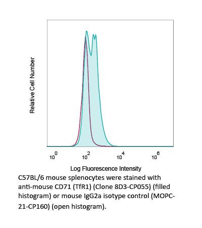
Product Details
The CP055 monoclonal antibody is a recombinant, chimeric version of the original 8D3 antibody. The variable domain sequences are identical to the original 8D3 but the constant region sequences have been switched from rat IgG2a to mouse IgG2a, kappa for use in murine models. Species-matched chimeric antibodies exhibit regulated effector functionsāincluding Fc receptor binding and complement activationāand result in less immunogenicity and formation of anti-drug antibodies (ADAs) than xenogenic antibodies in animal models. The anti-tumor and cytotoxic activities of anti-TfR1 antibodies have been demonstrated to require Fc effector function to elicit antibody-dependent cell-mediated cytotoxicity and antibody-dependent cell-mediated phagocytosis. The 8D3 monoclonal antibody reacts with native, soluble and denatured forms of murine CD71, also known as transferrin receptor protein 1 (TfR1). CD71 is a type II homodimeric transmembrane glycoprotein expressed on the surface of proliferating cells, reticulocytes, and erythroid precursors. CD71 plays a role in the control of cellular proliferation and is required for iron import from transferrin into cells by endocytosis. CD71 is overexpressed on many different types of cancer cells and expression level correlates with advanced stage and/or poorer prognosis in several cancers, including solid cancers. Elevated levels of CD71 expression on malignant cells, together with its extracellular accessibility, ability to internalize, and central role in cancer cell pathology make this receptor an attractive target for antibody-mediated therapy. In addition, cells of the vascular endothelium of brain capillaries that compose the blood-brain barrier (BBB) also express high levels of CD71 allowing for receptor-mediated transcytosis of large biomolecules into the brain. Chimeric anti-CD71 clone 8D3 has been used as a BBB transporter in mice and is suitable for studying CD71 expression and iron uptake into different tissues.Specifications
Isotype | Mouse IgG2a, Īŗ |
---|---|
Recommended Isotype Control(s) | RecombiMAb mouse IgG2a isotype control, unknown specificity |
Recommended Dilution Buffer | InVivoPure pH 7.0 Dilution Buffer |
Immunogen | Mouse transformed endothelioma cell line t.end1 |
Reported Applications |
Targeted drug delivery to the brain Immunohistochemistry Flow Cytometry Western Blot In vivo depletion of CD71+ cells In vivo anti-tumor activity *Reported for the original 8D3 antibody. For information on in vivo applications, please contact technicalservice@bioxcell.com |
Formulation |
PBS, pH 7.0 Contains no stabilizers or preservatives |
Endotoxin |
<1EU/mg (<0.001EU/μg) Determined by LAL gel clotting assay |
Aggregation |
<5% Determined by SEC |
Purity |
>95% Determined by SDS-PAGE |
Sterility | 0.2 µm filtration |
Production | Purified from HEK293 cell supernatant in an animal-free facility |
Purification | Protein G |
Molecular Weight | 150 kDa |
Murine Pathogen Tests |
Ectromelia/Mousepox Virus: Negative Hantavirus: Negative K Virus: Negative Lactate Dehydrogenase-Elevating Virus: Negative Lymphocytic Choriomeningitis virus: Negative Mouse Adenovirus: Negative Mouse Cytomegalovirus: Negative Mouse Hepatitis Virus: Negative Mouse Minute Virus: Negative Mouse Norovirus: Negative Mouse Parvovirus: Negative Mouse Rotavirus: Negative Mycoplasma Pulmonis: Negative Pneumonia Virus of Mice: Negative Polyoma Virus: Negative Reovirus Screen: Negative Sendai Virus: Negative Theilerās Murine Encephalomyelitis: Negative |
Storage | The antibody solution should be stored at the stock concentration at 4°C. Do not freeze. |
Additional Formats
Recommended Products
in vivo anti-tumor activity
Candelaria PV, Leoh LS, Penichet ML, Daniels-Wells TR. (2021). "Antibodies Targeting the Transferrin Receptor 1 (TfR1) as Direct Anti-cancer Agents" Front Immunol . PubMed
The transferrin receptor 1 (TfR1), also known as cluster of differentiation 71 (CD71), is a type II transmembrane glycoprotein that binds transferrin (Tf) and performs a critical role in cellular iron uptake through the interaction with iron-bound Tf. Iron is required for multiple cellular processes and is essential for DNA synthesis and, thus, cellular proliferation. Due to its central role in cancer cell pathology, malignant cells often overexpress TfR1 and this increased expression can be associated with poor prognosis in different types of cancer. The elevated levels of TfR1 expression on malignant cells, together with its extracellular accessibility, ability to internalize, and central role in cancer cell pathology make this receptor an attractive target for antibody-mediated therapy. The TfR1 can be targeted by antibodies for cancer therapy in two distinct ways: (1) indirectly through the use of antibodies conjugated to anti-cancer agents that are internalized by receptor-mediated endocytosis or (2) directly through the use of antibodies that disrupt the function of the receptor and/or induce Fc effector functions, such as antibody-dependent cell-mediated cytotoxicity (ADCC), antibody-dependent cell-mediated phagocytosis (ADCP), or complement-dependent cytotoxicity (CDC). Although TfR1 has been used extensively as a target for antibody-mediated cancer therapy over the years, interest continues to increase for both targeting the receptor for delivery purposes and for its use as direct anti-cancer agents. This review focuses on the developments in the use of antibodies targeting TfR1 as direct anti-tumor agents.
in vivo anti-tumor activity
Daniels-Wells TR, Candelaria PV, Leoh LS, Nava M, MartĆnez-Maza O, Penichet ML. (2020). "An IgG1 Version of the Anti-transferrin Receptor 1 Antibody ch128.1 Shows Significant Antitumor Activity Against Different Xenograft Models of Multiple Myeloma: A Brief Communication" J Immunother 43(2):48-52. PubMed
The transferrin receptor 1 (TfR1) is a meaningful target for antibody-based cancer therapy given its overexpression on malignant cells and its central role in cancer pathology. We previously developed a mouse/human chimeric IgG3 targeting human TfR1 (ch128.1), which exhibits significant antitumor activity against multiple myeloma (MM) in xenograft models of SCID-Beige mice bearing disseminated ARH-77 or KMS-11 tumors. This activity is observed in early and late disease stages of disseminated KMS-11 tumors and, in this model, the mechanism of antitumor activity is Fc-mediated, involving macrophages. As human IgG1 is the isotype of choice for therapeutic antibodies targeting malignant cells and has several advantages compared with IgG3, including established manufacturability, we now developed an IgG1 version of ch128.1. A single dose of ch128.1/IgG1 shows significant antitumor activity, not only against early and late stages of disseminated KMS-11 tumors (Asian origin) but also against these stages of disseminated disease following injection of human MM cells MM.1S (African American origin) or its variant that is resistant to dexamethasone MM.1R. Treatment with the Fc mutant version of ch128.1/IgG1 (L234A/L235A/P329S) with impaired effector functions fails to confer protection against MM.1S and MM.1R tumors, indicating a crucial role of the Fc fragment in the antitumor activity, similar to its IgG3 counterpart. In fact, we found that ch128.1/IgG1, but not the mutant, elicits antibody-dependent cell-mediated cytotoxicity and antibody-dependent cell-mediated phagocytosis in the presence of murine bone marrow-derived macrophages. Our results suggest that ch128.1/IgG1 is a promising therapeutic against human B-cell malignancies such as MM.
transport across the BBB
Cabezón I, Manich G, MartĆn-Venegas R, Camins A, PelegrĆ C, Vilaplana J. (2015). "Trafficking of Gold Nanoparticles Coated with the 8D3 Anti-Transferrin Receptor Antibody at the Mouse Blood-Brain Barrier" Mol Pharm 12(11):4137-45. PubMed
Receptor-mediated transcytosis has been widely studied as a possible strategy to transport neurotherapeutics across the blood-brain barrier (BBB). Monoclonal antibodies directed against the transferrin receptor (TfR) have been proposed as potential carrier candidates. A better understanding of the mechanisms involved in their cellular uptake and intracellular trafficking is required and could critically contribute to the improvement of delivery methods. Accordingly, we studied here the trafficking of gold nanoparticles (AuNPs) coated with the 8D3 anti-transferrin receptor antibody at the mouse BBB. 8D3-AuNPs were intravenously administered to mice and allowed to recirculate for a range of times, from 10 min to 24 h, before brain extraction and analysis by transmission electron microscope techniques. Our results indicated a TfR-mediated and clathrin-dependent internalization process by which 8D3-AuNPs internalize individually in vesicles. These vesicles then follow at least two different routes. On one hand, most vesicles enter intracellular processes of vesicular fusion and rearrangement in which the AuNPs end up accumulating in late endosomes, multivesicular bodies or lysosomes, which present a high AuNP content. On the other hand, a small percentage of the vesicles follow a different route in which they fuse with the abluminal membrane and open to the basal membrane. In these cases, the 8D3-AuNPs remain attached to the abluminal membrane, which suggests an endosomal escape, but not dissociation from TfR. Altogether, although receptor-mediated transport continues to be one of the most promising strategies to overcome the BBB, different optimization approaches need to be developed for efficient drug delivery.
transport across the BBB
Boado RJ, Zhang Y, Wang Y, Pardridge WM. (2009). "Engineering and expression of a chimeric transferrin receptor monoclonal antibody for blood-brain barrier delivery in the mouse" Biotechnol Bioeng 102(4):1251-8. PubMed
Protein therapeutics may be delivered across the blood-brain barrier (BBB) by genetic fusion to a BBB molecular Trojan horse. The latter is an endogenous peptide or a peptidomimetic monoclonal antibody (MAb) against a BBB receptor, such as the insulin receptor or the transferrin receptor (TfR). Fusion proteins have been engineered with the MAb against the human insulin receptor (HIR). However, the HIRMAb is not active against the rodent insulin receptor, and cannot be used for drug delivery across the mouse BBB. The rat 8D3 MAb against the mouse TfR is active as a drug delivery system in the mouse, and the present studies describe the cloning and sequencing of the variable region of the heavy chain (VH) and light chain (VL) of the rat 8D3 TfRMAb. The VH and VL were fused to the constant region of mouse IgG1 heavy chain and mouse kappa light chain, respectively, to produce a new chimeric TfRMAb. The chimeric TfRMAb was expressed in COS cells following dual transfection with the heavy and light chain expression plasmids, and was purified by protein G affinity chromatography. The affinity of the chimeric TfRMAb for the murine TfR was equal to the 8D3 MAb using a radio-receptor assay and mouse fibroblasts. The chimeric TfRMAb was radio-labeled and injected into mice for a pharmacokinetics study of the clearance of the chimeric TfRMAb. The chimeric TfRMAb was rapidly taken up by mouse brain in vivo at a level comparable to the rat 8D3 MAb. In summary, these studies describe the genetic engineering, expression, and validation of a chimeric TfRMAb with high activity for the mouse TfR, which can be used in future engineering of therapeutic fusion proteins for BBB drug delivery in the mouse.