


High performance fluorescently conjugated antibodies designed for flow cytometry and beyond help researchers improve data reliability and streamline experimental design.
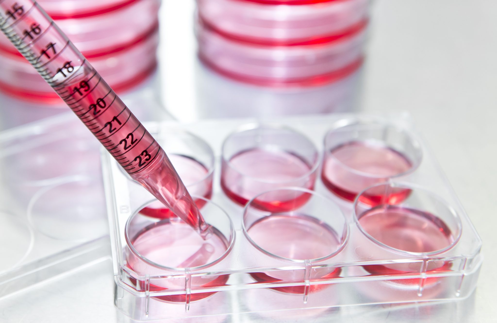
 Flow cytometry is a powerful technology used to analyze single cells or particles in liquid suspension. During the assay, cells or particles are processed in the lab using fluorescently labeled antibodies or dyes and are then analyzed by a laser-equipped instrument called a flow cytometer. The readouts provide scientists with information on parameters such as size, granularity, and emitted fluorescence intensity, enabling discrimination and study of various characteristics. This analysis allows for the investigation of multiple parameters simultaneously, facilitating the definition of complex phenotypes and functional states within samples. Flow cytometry has broad applications, including diagnostics, monitoring disease progression, and studying infections in humans, animals, food, and plants. <sup>1 2</sup>
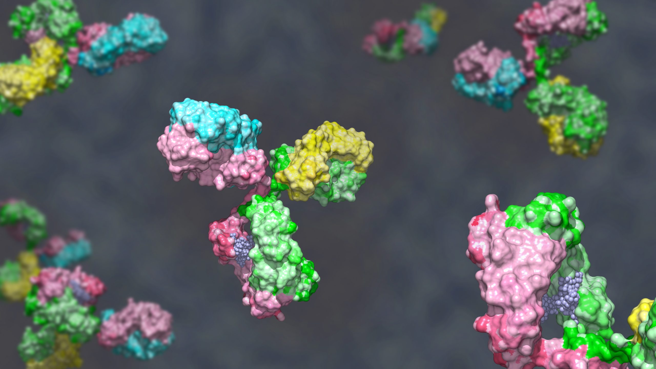
However, as for many antibody-based experiments, labeling challenges related to brightness, signal stability, target detection, and background noise are common roadblocks in flow cytometry. Scientists can overcome many of these challenges with high-quality fluorescently conjugated antibodies and appropriate controls. Tracking Targets with Flow Cytometry Fluorescently labeled antibodies enable scientists to precisely detect cell surface or intracellular proteins. Small organic molecules such as allophycocyanin (APC), fluorescein isothiocyanate (FITC), and phycoerythrin (PE) are commonly used for antibody conjugation because they have consistent emission spectra and the difference between excitation and emission wavelengths is small, making it easy to detect multiple labels with a single instrument. <sup>1</sup> These fluorophores are also stable and reasonably easy to conjugate to antibodies. Certain conjugates were designed to be more resistant to photobleaching, making them ideal choices for imaging or other fluorescence-based analyses on the same samples. Antibody labeling tools have been fundamental to research characterizing many different cell populations and biomolecule targets, including disease-relevant antibodies, nucleic acids, and cell surface markers.<sup>2</sup> Still, some targets remain out of reach for translational applications due to low abundance and lack of reliable flow cytometry-specific antibodies.
Flow Cytometry Challenges
When a target of interest is in low abundance, such as a rare cell population, it can be challenging to investigate through flow cytometry because it may fall below the lower threshold limit. <sup>3</sup> This limit depends on the ability to distinguish a target from normal background cells and the total cell count in the sample. Factors such as the immunophenotype of other cell populations, target autofluorescence, antigen expression level, fluorophore brightness, and other technical aspects can influence the flow cytometry detection limit. The use of the FMO or Fluorescence Minus One control has been demonstrated time and again as a critical control for setting specificity. FMO control is especially critical for determining the effects of the spillovers from other fluorochromes into the channel of interest and helps to establish positivity. 4 Background staining depends on several different factors, including the conjugated antibody's specificity, concentration, and fluorophore-to-antibody ratio.
To address these hurdles in flow cytometry, scientists turn to monoclonal antibodies that are conjugated to high-quality fluorescent dyes validated for use in both flow cytometry and other antibody-based assays. FlowMAb™ is conjugated to a high-quality fluorescent dye, such as APC, FITC, or PE, ensuring stable signals and the precise detection of target antigens. These highly specific, bright fluorophores enable FlowMAb™ antibodies to minimize background noise caused by autofluorescence, ensuring accurate detection even in complex tissue environments.
FlowMAb™ targets include expert-validated immune cell lineage markers and immune checkpoint markers, among other critical targets for precise cellular identification in immunology, oncology, stem cell biology, neuroscience, and infectious disease research. Whether researchers seek to identify known cell types or explore novel therapeutic targets, these conjugated antibodies offer high performance and reliability for flow cytometry, fluorescence- activated cell sorting (FACS), immunofluorescence, immunohistochemistry, or immunocytochemistry experiments.
FlowMAb™ antibodies are high-quality fluorescently conjugated antibodies designed for use in flow cytometry and other immunological applications. They offer several key features:
- High Photostability: Ensures stable signals during experiments.
- Range of Fluorophore Options: Provides flexibility in experimental design.
- Precise Detection: Helps in accurately identifying target cells or biomarkers.
- Versatility: Suitable for various applications, including flow cytometry, FACS, immunofluorescence, immunohistochemistry, and immunocytochemistry.
These antibodies are particularly useful in fields like immunology, oncology, stem cell biology, neuroscience, and infectious disease research.
References
- McKinnon KM. Flow cytometry: An overview. Curr Protoc Immunol. 2019;120(1):5.1.1- 5.1.11.
- Robinson JP, et al. Flow Cytometry: the next revolution. Cells. 2023;12(14):1875.
- Brestoff JR, Frater JL. Contemporary challenges in clinical flow cytometry: small samples, big data, little time. J Appl Lab Med. 2021;7(4):931-944.
- Maecker HT, et al. Flow cytometry controls, instrument setup, and the determination of positivity. Cytometry A. 2006;69(9):1037-42. 5. Song Y, Lee Y. Brief guide to flow cytometry. Mol Cells. 2024;47(11):100129