InVivoPlus anti-mouse IL-4
Product Description
Specifications
| Isotype | Rat IgG1, κ |
|---|---|
| Recommended Isotype Control(s) | InVivoPlus rat IgG1 isotype control, anti-horseradish peroxidase |
| Recommended Dilution Buffer | InVivoPure pH 7.0 Dilution Buffer |
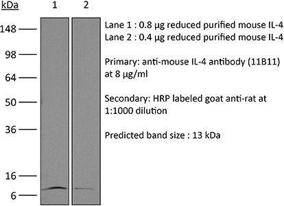
| Conjugation | This product is unconjugated. Conjugation is available via our Antibody Conjugation Services. |
| Immunogen | Partially purified native mouse IL-4 |
| Reported Applications |
in vivo IL-4 neutralization in vitro IL-4 neutralization in vivo IL-4 receptor stimulation (as a complex with IL-4) Flow cytometry Western blot |
| Formulation |
PBS, pH 7.0 Contains no stabilizers or preservatives |
| Endotoxin* |
≤0.5EU/mg (≤0.0005EU/μg) Determined by LAL assay |
| Aggregation* |
<5% Determined by SEC |
| Purity |
≥95% Determined by SDS-PAGE |
| Sterility | 0.2 µm filtration |
| Production | Purified from cell culture supernatant in an animal-free facility |
| Purification | Protein G |
| RRID | AB_1107707 |
| Molecular Weight | 150 kDa |
| Murine Pathogen Tests* |
Ectromelia/Mousepox Virus: Negative Hantavirus: Negative K Virus: Negative Lactate Dehydrogenase-Elevating Virus: Negative Lymphocytic Choriomeningitis virus: Negative Mouse Adenovirus: Negative Mouse Cytomegalovirus: Negative Mouse Hepatitis Virus: Negative Mouse Minute Virus: Negative Mouse Norovirus: Negative Mouse Parvovirus: Negative Mouse Rotavirus: Negative Mycoplasma Pulmonis: Negative Pneumonia Virus of Mice: Negative Polyoma Virus: Negative Reovirus Screen: Negative Sendai Virus: Negative Theiler’s Murine Encephalomyelitis: Negative |
| Storage | The antibody solution should be stored at the stock concentration at 4°C. Do not freeze. |
| Need a Custom Formulation? | See All Antibody Customization Options |
Application References
-
Burton, B. R., et al (2014). "Sequential transcriptional changes dictate safe and effective antigen-specific immunotherapy" Nat Commun 5: 4741.
PubMed
Antigen-specific immunotherapy combats autoimmunity or allergy by reinstating immunological tolerance to target antigens without compromising immune function. Optimization of dosing strategy is critical for effective modulation of pathogenic CD4(+) T-cell activity. Here we report that dose escalation is imperative for safe, subcutaneous delivery of the high self-antigen doses required for effective tolerance induction and elicits anergic, interleukin (IL)-10-secreting regulatory CD4(+) T cells. Analysis of the CD4(+) T-cell transcriptome, at consecutive stages of escalating dose immunotherapy, reveals progressive suppression of transcripts positively regulating inflammatory effector function and repression of cell cycle pathways. We identify transcription factors, c-Maf and NFIL3, and negative co-stimulatory molecules, LAG-3, TIGIT, PD-1 and TIM-3, which characterize this regulatory CD4(+) T-cell population and whose expression correlates with the immunoregulatory cytokine IL-10. These results provide a rationale for dose escalation in T-cell-directed immunotherapy and reveal novel immunological and transcriptional signatures as surrogate markers of successful immunotherapy.
-
Tang, W., et al (2014). "The oncoprotein and transcriptional regulator Bcl-3 governs plasticity and pathogenicity of autoimmune T cells" Immunity 41(4): 555-566.
PubMed
Bcl-3 is an atypical member of the IkappaB family that modulates transcription in the nucleus via association with p50 (NF-kappaB1) or p52 (NF-kappaB2) homodimers. Despite evidence attesting to the overall physiologic importance of Bcl-3, little is known about its cell-specific functions or mechanisms. Here we demonstrate a T-cell-intrinsic function of Bcl-3 in autoimmunity. Bcl-3-deficient T cells failed to induce disease in T cell transfer-induced colitis and experimental autoimmune encephalomyelitis. The protection against disease correlated with a decrease in Th1 cells that produced the cytokines IFN-gamma and GM-CSF and an increase in Th17 cells. Although differentiation into Th1 cells was not impaired in the absence of Bcl-3, differentiated Th1 cells converted to less-pathogenic Th17-like cells, in part via mechanisms involving expression of the RORgammat transcription factor. Thus, Bcl-3 constrained Th1 cell plasticity and promoted pathogenicity by blocking conversion to Th17-like cells, revealing a unique type of regulation that shapes adaptive immunity.
-
Gu, A. D., et al (2015). "A critical role for transcription factor Smad4 in T cell function that is independent of transforming growth factor beta receptor signaling" Immunity 42(1): 68-79.
PubMed
Transforming growth factor-beta (TGF-beta) suppresses T cell function to maintain self-tolerance and to promote tumor immune evasion. Yet how Smad4, a transcription factor component of TGF-beta signaling, regulates T cell function remains unclear. Here we have demonstrated an essential role for Smad4 in promoting T cell function during autoimmunity and anti-tumor immunity. Smad4 deletion rescued the lethal autoimmunity resulting from transforming growth factor-beta receptor (TGF-betaR) deletion and compromised T-cell-mediated tumor rejection. Although Smad4 was dispensable for T cell generation, homeostasis, and effector function, it was essential for T cell proliferation after activation in vitro and in vivo. The transcription factor Myc was identified to mediate Smad4-controlled T cell proliferation. This study thus reveals a requirement of Smad4 for T-cell-mediated autoimmunity and tumor rejection, which is beyond the current paradigm. It highlights a TGF-betaR-independent role for Smad4 in promoting T cell function, autoimmunity, and anti-tumor immunity.
-
Clever, D., et al (2016). "Oxygen Sensing by T Cells Establishes an Immunologically Tolerant Metastatic Niche" Cell 166(5): 1117-1131 e1114.
PubMed
Cancer cells must evade immune responses at distant sites to establish metastases. The lung is a frequent site for metastasis. We hypothesized that lung-specific immunoregulatory mechanisms create an immunologically permissive environment for tumor colonization. We found that T-cell-intrinsic expression of the oxygen-sensing prolyl-hydroxylase (PHD) proteins is required to maintain local tolerance against innocuous antigens in the lung but powerfully licenses colonization by circulating tumor cells. PHD proteins limit pulmonary type helper (Th)-1 responses, promote CD4(+)-regulatory T (Treg) cell induction, and restrain CD8(+) T cell effector function. Tumor colonization is accompanied by PHD-protein-dependent induction of pulmonary Treg cells and suppression of IFN-gamma-dependent tumor clearance. T-cell-intrinsic deletion or pharmacological inhibition of PHD proteins limits tumor colonization of the lung and improves the efficacy of adoptive cell transfer immunotherapy. Collectively, PHD proteins function in T cells to coordinate distinct immunoregulatory programs within the lung that are permissive to cancer metastasis.
Product Citations
-
Cardiolipin preserves Treg metabolic fitness and immune homeostasis in the gut.
In Nat Metab on 18 May 2026 by Regina, A., Solagna, F., et al.
PubMed
Loss of host-microbiota balance promotes gut inflammation, colitis and inflammatory bowel disease. Yet, whether host or microbial factors are the critical driver of the pathology remains unclear. Here, we investigate how cardiolipin maintains metabolic fitness of regulatory T (Treg) cells to preserve gut-immune homeostasis. We discover that deleting the cardiolipin-synthesizing enzyme protein tyrosine phosphatase mitochondrial 1 (PTPMT1) in T cells predisposes mice to colitis due to impaired Treg cell function in the absence of dysbiosis. Subsequent pathobiont infections accelerate the progression and severity of gut inflammation. Mechanistically, the absence of cardiolipin impairs Treg cell metabolic fitness and triggers a maladaptive integrated stress response, which can be reversed pharmacologically or genetically, restoring gut homeostasis and extending lifespan in PTPMT1 ΔT mice. Barth syndrome, a genetic disorder marked by severe cardiolipin deficiency, also exhibits gastrointestinal symptoms and inflammation associated with helper T cell imbalance and an active integrated stress response signature. Overall, these results suggest that a cardiolipin-mediated mitonuclear axis in T cells preserves gut-immune homeostasis and dictates outcome in pathobiont infections.
-
IL-4 treatment induces apoptosis of blood monocytes and proliferation of recruited injury-associated macrophages to resolve liver injury.
In Cell Rep on 20 March 2026 by Lynch, R. W., Louwe, P. A., et al.
PubMed
IL-4 can have significant therapeutic benefit in many injury settings, but its mechanism of action is unclear. Using a model of carbon tetrachloride (CCl4)-mediated acute liver injury, we find that exogenous IL-4 causes a dramatic shift from recruited Ly6Chi monocytes to an abundance of monocyte-derived macrophages (MoMFs) within the injured tissue that is accompanied by reduced indices of hepatic damage and enhanced hepatic regeneration. Rather than altering the recruitment or differentiation of monocytes, treatment with IL-4 triggers monocyte apoptosis alongside proliferation of MoMFs. Single-cell RNA sequencing reveals injury and cell-type-specific responses to IL-4 treatment across hepatic myeloid lineages and a largely pro-reparative gene signature in the expanded pool of MoMFs. IL-4 treatment fails to enhance hepatic repair when the accrual of MoMFs is limited using Ccr2-deficient monocytopenic mice. Together, these data reveal a pathway through which therapeutic IL-4 alters the composition, number, and function of injury-associated myeloid cells to resolve liver injury.
-
IL-4 and TGF-β regulate inflammatory cytokines and cellular infiltration in the lung and systemic IL-6 in mouse-adapted SARS-CoV-2 infection.
In Immunohorizons on 25 August 2025 by Taye Sima, S., Puebla-Clark, L., et al.
PubMed
The pathology of severe COVID-19 is due to a hyperinflammatory immune response persisting after viral clearance. To understand how the immune response to SARS-CoV-2 is regulated to avoid severe COVID-19, we tested relevant immunoregulatory cytokines. Transforming growth factor β (TGF-β), interleukin (IL)-10, and IL-4 were neutralized upon infection with mouse-adapted SARS-CoV-2 (CMA3p20), a model of mild disease; lung inflammation was quantified by histology and flow cytometry at early and late time points. Mild weight loss and lung inflammation including consolidation and alveolar thickening were evident 3 d postinfection (dpi), and inflammation persisted to 7 dpi. Coinciding with early monocytic infiltrates, CCL2 and granulocyte colony-stimulating factor were transiently produced 3 dpi, while IL-12 and CCL5 persisted to 7 dpi, modeling viral and inflammatory phases of disease. Neutralization of TGF-β, but not IL-10 or IL-4, significantly increased lung inflammatory monocytes and elevated serum but not lung IL-6. Neutralization of IL-4 prolonged weight loss and increased early perivascular infiltration without changing viral titer. Anti-IL-4 reduced expression of Arg1, a gene associated with alternative activation of macrophages. Neutralizing TGF-β and IL-4 had differential effects on pathology after virus control. Lung perivascular infiltration was reduced 7 dpi by neutralization of IL-4 or TGF-β, and periairway inflammation was affected by anti-TGF-β, while alveolar infiltrates were not affected by either. Anti-IL-4 prolonged IL-12 to 7 dpi along with reduced IL-10 in lungs. Overall, the immunoregulatory cytokines TGF-β and IL-4 dampen initial inflammation in this mouse-adapted SARS-CoV-2 infection, suggesting that promotion of immunoregulation could help patients in early stages of disease.
-
Mitochondrial fatty acid synthesis and MECR regulate CD4+ T cell function and oxidative metabolism.
In J Immunol on 1 May 2025 by Steiner, K. K., Young, A. C., et al.
PubMed
Imbalanced effector and regulatory CD4+ T cell subsets drive many inflammatory diseases. These T cell subsets rely on distinct metabolic programs, modulation of which differentially affects T cell fate and function. Lipid metabolism is fundamental yet remains poorly understood across CD4+ T cell subsets. Therefore, we performed targeted in vivo CRISPR/Cas9 screens to identify lipid metabolism genes and pathways essential for T cell functions. These screens established mitochondrial fatty acid synthesis genes Mecr, Mcat, and Oxsm as key metabolic regulators. Of these, the inborn error of metabolism gene Mecr was most dynamically regulated. Mecrfl/fl; Cd4cre mice had normal naïve CD4+ and CD8+ T cell numbers, demonstrating that MECR is not essential in homeostatic conditions. However, effector and memory T cells were reduced in Mecr knockout and MECR-deficient CD4+ T cells and proliferated, differentiated, and survived less well than control T cells. Interestingly, T cells ultimately showed signs of mitochondrial stress and dysfunction in the absence of MECR. Mecr-deficient T cells also had decreased mitochondrial respiration, reduced tricarboxylic acid intermediates, and accumulated intracellular iron, which appeared to contribute to increased cell death and sensitivity to ferroptosis. Importantly, MECR-deficient T cells exhibited fitness disadvantages and were less effective at driving disease in an in vivo model of inflammatory bowel disease. Thus, MECR-mediated metabolism broadly supports CD4+ T cell proliferation and survival in vivo. These findings may also provide insight to the immunological state of MECR- and other mitochondrial fatty acid synthesis-deficient patients.