InVivoMAb anti-mouse IL-4

Catalog #BE0045
Product Citations:
155
Clone:
11B11
Reactivities:
Mouse

$164.00 - $4,280.00

Choose an Option...
  • 100 mg - $4,280.00
  • 50 mg - $3,024.00
  • 25 mg - $2,009.00
  • 5 mg - $600.00
  • 1 mg - $164.00
  • Custom Amount (Quotes Only)
In stock
Only %1 left

Product Details

The 11B11 monoclonal antibody reacts with mouse IL-4 (interleukin-4) a multifunctional 14 kDa cytokine. IL-4 is expressed primarily by activated Th2 cells and NK cells, and at lower levels by mast cells, and basophils. IL-4 signals through the IL-4RĪ±. Upon receptor binding IL-4 stimulates activated B and T lymphocyte proliferation, and the differentiation of B cells into plasma cells. It also induces B cell class switching to IgE, and up-regulates MHC class II production while decreasing the production of Th1 cells, macrophages, IFNĪ³, and dendritic cell IL-12. Like other Th2 associated cytokines, IL-4 is involved in the airway inflammation observed in the lungs of patients with allergic asthma. The 11B11 monoclonal antibody has been shown to neutralize the bioactivity of natural or recombinant IL-4.

Specifications

Isotype Rat IgG1,Ā Īŗ
Recommended Isotype Control(s) InVivoMAb rat IgG1 isotype control, anti-horseradish peroxidase
Recommended Dilution Buffer InVivoPure pH 7.0 Dilution Buffer
Conjugation This product is unconjugated. Conjugation is available via our Antibody Conjugation Services.
Immunogen Partially purified native mouse IL-4
Reported Applications 
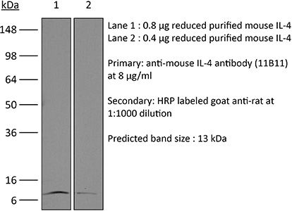
in vivo IL-4 neutralization
in vitro IL-4 neutralization
in vivoĀ IL-4 receptor stimulation (as a complex with IL-4)
Flow cytometry
Western blot
Formulation PBS, pH 7.0
Contains no stabilizers or preservatives
Endotoxin <2EU/mg (<0.002EU/Ī¼g)
Determined by LAL gel clotting assay
Purity >95%
Determined by SDS-PAGE
Sterility 0.2 Āµm filtration
Production Purified from cell culture supernatant in an animal-free facility
Purification Protein G
RRID AB_1107707
Molecular Weight 150 kDa
Storage The antibody solution should be stored at the stock concentration at 4Ā°C. Do not freeze.

Additional Formats

in vivo IL-4 neutralization, in vivo IL-4 receptor stimulation (as a complex with IL-4)
Gaya, M., et al. (2018). "Initiation of Antiviral B Cell Immunity Relies on Innate Signals from Spatially Positioned NKT Cells" Cell 172(3): 517-533 e520. PubMed

B cells constitute an essential line of defense from pathogenic infections through the generation of class-switched antibody-secreting cells (ASCs) in germinal centers. Although this process is known to be regulated by follicular helper T (TfH) cells, the mechanism by which B cells initially seed germinal center reactions remains elusive. We found that NKT cells, a population of innate-like T lymphocytes, are critical for the induction of B cell immunity upon viral infection. The positioning of NKT cells at the interfollicular areas of lymph nodes facilitates both their direct priming by resident macrophages and the localized delivery of innate signals to antigen-experienced B cells. Indeed, NKT cells secrete an early wave of IL-4 and constitute up to 70% of the total IL-4-producing cells during the initial stages of infection. Importantly, the requirement of this innate immunity arm appears to be evolutionarily conserved because early NKT and IL-4 gene signatures also positively correlate with the levels of neutralizing antibodies in Zika-virus-infected macaques. In conclusion, our data support a model wherein a pre-TfH wave of IL-4 secreted by interfollicular NKT cells triggers the seeding of germinal center cells and serves as an innate link between viral infection and B cell immunity.

in vitro IL-4 neutralization
Clever, D., et al. (2016). "Oxygen Sensing by T Cells Establishes an Immunologically Tolerant Metastatic Niche" Cell 166(5): 1117-1131 e1114. PubMed

Cancer cells must evade immune responses at distant sites to establish metastases. The lung is a frequent site for metastasis. We hypothesized that lung-specific immunoregulatory mechanisms create an immunologically permissive environment for tumor colonization. We found that T-cell-intrinsic expression of the oxygen-sensing prolyl-hydroxylase (PHD) proteins is required to maintain local tolerance against innocuous antigens in the lung but powerfully licenses colonization by circulating tumor cells. PHD proteins limit pulmonary type helper (Th)-1 responses, promote CD4(+)-regulatory T (Treg) cell induction, and restrain CD8(+) T cell effector function. Tumor colonization is accompanied by PHD-protein-dependent induction of pulmonary Treg cells and suppression of IFN-gamma-dependent tumor clearance. T-cell-intrinsic deletion or pharmacological inhibition of PHD proteins limits tumor colonization of the lung and improves the efficacy of adoptive cell transfer immunotherapy. Collectively, PHD proteins function in T cells to coordinate distinct immunoregulatory programs within the lung that are permissive to cancer metastasis.

in vitro IL-4 neutralization, Flow Cytometry
Gu, A. D., et al. (2015). "A critical role for transcription factor Smad4 in T cell function that is independent of transforming growth factor beta receptor signaling" Immunity 42(1): 68-79. PubMed

Transforming growth factor-beta (TGF-beta) suppresses T cell function to maintain self-tolerance and to promote tumor immune evasion. Yet how Smad4, a transcription factor component of TGF-beta signaling, regulates T cell function remains unclear. Here we have demonstrated an essential role for Smad4 in promoting T cell function during autoimmunity and anti-tumor immunity. Smad4 deletion rescued the lethal autoimmunity resulting from transforming growth factor-beta receptor (TGF-betaR) deletion and compromised T-cell-mediated tumor rejection. Although Smad4 was dispensable for T cell generation, homeostasis, and effector function, it was essential for T cell proliferation after activation in vitro and in vivo. The transcription factor Myc was identified to mediate Smad4-controlled T cell proliferation. This study thus reveals a requirement of Smad4 for T-cell-mediated autoimmunity and tumor rejection, which is beyond the current paradigm. It highlights a TGF-betaR-independent role for Smad4 in promoting T cell function, autoimmunity, and anti-tumor immunity.

in vitro IL-4 neutralization
Hill, E. V., et al. (2015). "Glycogen synthase kinase-3 controls IL-10 expression in CD4(+) effector T-cell subsets through epigenetic modification of the IL-10 promoter" Eur J Immunol 45(4): 1103-1115. PubMed

The serine/threonine kinase glycogen synthase kinase-3 (GSK3) plays an important role in balancing pro- and anti-inflammatory cytokines. We have examined the role of GSK3 in production of IL-10 by subsets of CD4(+) T helper cells. Treatment of naive murine CD4(+) T cells with GSK3 inhibitors did not affect their production of IL-10. However, treatment of Th1 and Th2 cells with GSK3 inhibitors dramatically increased production of IL-10. GSK3 inhibition also led to upregulation of IL-10 among Th1, Th2, and Th17 subsets isolated from human blood. The encephalitogenic potential of GSK3 inhibitor treated murine Th1 cells was significantly reduced in adoptive transfer experiments by an IL-10-dependent mechanism. Analysis of the murine IL-10 promoter in response to inhibition of GSK3 in Th1 cells showed modification to a transcriptionally active state indicated by changes in histone H3 acetylation and methylation. Additionally, GSK3 inhibition increased expression of the transcription factors c-Maf, Nfil3, and GATA3, correlating with the increase in IL-10. These findings are important in the context of autoimmune disease since they show that it is possible to reprogram disease-causing cells through GSK3 inhibition.

in vitro IL-4 neutralization
Choi, Y. S., et al. (2015). "LEF-1 and TCF-1 orchestrate TFH differentiation by regulating differentiation circuits upstream of the transcriptional repressor Bcl6" Nat Immunol 16(9): 980-990. PubMed

Follicular helper T cells (TFH cells) are specialized effector CD4(+) T cells that help B cells develop germinal centers (GCs) and memory. However, the transcription factors that regulate the differentiation of TFH cells remain incompletely understood. Here we report that selective loss of Lef1 or Tcf7 (which encode the transcription factor LEF-1 or TCF-1, respectively) resulted in TFH cell defects, while deletion of both Lef1 and Tcf7 severely impaired the differentiation of TFH cells and the formation of GCs. Forced expression of LEF-1 enhanced TFH differentiation. LEF-1 and TCF-1 coordinated such differentiation by two general mechanisms. First, they established the responsiveness of naive CD4(+) T cells to TFH cell signals. Second, they promoted early TFH differentiation via the multipronged approach of sustaining expression of the cytokine receptors IL-6Ralpha and gp130, enhancing expression of the costimulatory receptor ICOS and promoting expression of the transcriptional repressor Bcl6.

in vitro IL-4 neutralization
Kim, Y. U., et al. (2015). "Regulation of autoimmune germinal center reactions in lupus-prone BXD2 mice by follicular helper T cells" PLoS One 10(3): e0120294. PubMed

BXD2 mice spontaneously develop autoantibodies and subsequent glomerulonephritis, offering a useful animal model to study autoimmune lupus. Although initial studies showed a critical contribution of IL-17 and Th17 cells in mediating autoimmune B cell responses in BXD2 mice, the role of follicular helper T (Tfh) cells remains incompletely understood. We found that both the frequency of Th17 cells and the levels of IL-17 in circulation in BXD2 mice were comparable to those of wild-type. By contrast, the frequency of PD-1+ CXCR5+ Tfh cells was significantly increased in BXD2 mice compared with wild-type mice, while the frequency of PD-1+ CXCR5+ Foxp3+ follicular regulatory T (Tfr) cells was reduced in the former group. The frequency of Tfh cells rather than that of Th17 cells was positively correlated with the frequency of germinal center B cells as well as the levels of autoantibodies to dsDNA. More importantly, CXCR5+ CD4+ T cells isolated from BXD2 mice induced the production of IgG from naive B cells in an IL-21-dependent manner, while CCR6+ CD4+ T cells failed to do so. These results together demonstrate that Tfh cells rather than Th17 cells contribute to the autoimmune germinal center reactions in BXD2 mice.

in vitro IL-4 neutralization
Hou, L., et al. (2015). "The protease cathepsin L regulates Th17 cell differentiation" J Autoimmun. S 0896-8411(15): 30024-X. PubMed

Previously we reported that IL-17+ T cells, primarily IL-17+ gammadelta cells, are increased in mice lacking the protease inhibitor serpinB1 (serpinb1-/- mice). Here we show that serpinB1-deficient CD4 cells exhibit a cell-autonomous and selective deficiency in suppressing T helper 17 (Th17) cell differentiation. This suggested an opposing role for one or more protease in promoting Th17 differentiation. We found that several SerpinB1-inhibitable cysteine cathepsins are induced in Th17 cells, most prominently cathepsin L (catL); this was verified by peptidase assays, active site labeling and Western blots. Moreover, Th17 differentiation was suppressed by both broad cathepsin inhibitors and catL selective inhibitors. CatL is present in Th17 cells as single chain (SC)- and two-chain (TC)-forms. Inhibiting asparagine endopeptidase (AEP) blocked conversion of SC-catL to TC-catL and increased generation of serpinb1-/- Th17 cells, but not wild-type Th17 cells. These findings suggest that SC-catL is biologically active in promoting Th17 generation and is counter-regulated by serpinB1 and secondarily by AEP. Thus, in addition to regulation by cytokines and transcription factors, differentiation of CD4 cells to Th17 cells is actively regulated by a catL-serpinB1-AEP module. Targeting this protease regulatory module could be an approach to treating Th17 cell-driven autoimmune disorders.

in vitro IL-4 neutralization
McKinstry, K. K., et al. (2014). "Effector CD4 T-cell transition to memory requires late cognate interactions that induce autocrine IL-2" Nat Commun 5: 5377. PubMed

It is unclear how CD4 T-cell memory formation is regulated following pathogen challenge, and when critical mechanisms act to determine effector T-cell fate. Here, we report that following influenza infection most effectors require signals from major histocompatibility complex class II molecules and CD70 during a late window well after initial priming to become memory. During this timeframe, effector cells must produce IL-2 or be exposed to high levels of paracrine or exogenously added IL-2 to survive an otherwise rapid default contraction phase. Late IL-2 promotes survival through acute downregulation of apoptotic pathways in effector T cells and by permanently upregulating their IL-7 receptor expression, enabling IL-7 to sustain them as memory T cells. This new paradigm defines a late checkpoint during the effector phase at which cognate interactions direct CD4 T-cell memory generation.

in vitro IL-4 neutralization
Heinemann, C., et al. (2014). "IL-27 and IL-12 oppose pro-inflammatory IL-23 in CD4+ T cells by inducing Blimp1" Nat Commun 5: 3770. PubMed

Central nervous system (CNS) autoimmunity is regulated by the balance of pro-inflammatory cytokines and IL-10. Here we identify the transcriptional regulator Blimp1 as crucial to induce IL-10 in inflammatory T helper cells. Pre-committed Th17 cells respond to IL-27 and IL-12 by upregulating Blimp1 and adopt a Tr-1-like phenotype characterized by IL-10 and IFN-gamma production. Accordingly, Blimp1-deficient effector T cells fail to produce IL-10, and deficiency in Tr-1 cell function leads to uncontrolled Th17 cell-driven CNS pathology without the need to stabilize the Th17 phenotype with IL-23. IL-23 counteracts IL-27 and IL-12-mediated effects on Tr-1-development reinforcing the pro-inflammatory phenotype of Th17 cells. Thus, the balance of IL-23 vs IL-12/IL-27 signals into CD4(+) effector T cells determines whether tissue inflammation is perpetuated or resolves.

in vitro IL-4 neutralization
Burton, B. R., et al. (2014). "Sequential transcriptional changes dictate safe and effective antigen-specific immunotherapy" Nat Commun 5: 4741. PubMed

Antigen-specific immunotherapy combats autoimmunity or allergy by reinstating immunological tolerance to target antigens without compromising immune function. Optimization of dosing strategy is critical for effective modulation of pathogenic CD4(+) T-cell activity. Here we report that dose escalation is imperative for safe, subcutaneous delivery of the high self-antigen doses required for effective tolerance induction and elicits anergic, interleukin (IL)-10-secreting regulatory CD4(+) T cells. Analysis of the CD4(+) T-cell transcriptome, at consecutive stages of escalating dose immunotherapy, reveals progressive suppression of transcripts positively regulating inflammatory effector function and repression of cell cycle pathways. We identify transcription factors, c-Maf and NFIL3, and negative co-stimulatory molecules, LAG-3, TIGIT, PD-1 and TIM-3, which characterize this regulatory CD4(+) T-cell population and whose expression correlates with the immunoregulatory cytokine IL-10. These results provide a rationale for dose escalation in T-cell-directed immunotherapy and reveal novel immunological and transcriptional signatures as surrogate markers of successful immunotherapy.

in vitro IL-4 neutralization
Tang, W., et al. (2014). "The oncoprotein and transcriptional regulator Bcl-3 governs plasticity and pathogenicity of autoimmune T cells" Immunity 41(4): 555-566. PubMed

Bcl-3 is an atypical member of the IkappaB family that modulates transcription in the nucleus via association with p50 (NF-kappaB1) or p52 (NF-kappaB2) homodimers. Despite evidence attesting to the overall physiologic importance of Bcl-3, little is known about its cell-specific functions or mechanisms. Here we demonstrate a T-cell-intrinsic function of Bcl-3 in autoimmunity. Bcl-3-deficient T cells failed to induce disease in T cell transfer-induced colitis and experimental autoimmune encephalomyelitis. The protection against disease correlated with a decrease in Th1 cells that produced the cytokines IFN-gamma and GM-CSF and an increase in Th17 cells. Although differentiation into Th1 cells was not impaired in the absence of Bcl-3, differentiated Th1 cells converted to less-pathogenic Th17-like cells, in part via mechanisms involving expression of the RORgammat transcription factor. Thus, Bcl-3 constrained Th1 cell plasticity and promoted pathogenicity by blocking conversion to Th17-like cells, revealing a unique type of regulation that shapes adaptive immunity.

in vivo IL-4 neutralization
Mishra, P. K., et al. (2013). "Prevention of type 1 diabetes through infection with an intestinal nematode parasite requires IL-10 in the absence of a Th2-type response" Mucosal Immunol 6(2): 297-308. PubMed

Helminth infection can prevent type 1 diabetes (T1D); however, the regulatory mechanisms inhibiting disease remain largely undefined. In these studies, nonobese diabetic (NOD) IL-4(-/-) mice were infected with the strictly enteric nematode parasite, Heligmosomoides polygyrus. Short-term infection, 5-7 weeks of age, inhibited T1D onset, as late as 40 weeks of age. CD4(+) T-cell STAT6 phosphorylation was inhibited, while suppressed signal transducer and activator of transcription 1 phosphorylation was sustained, as were increases in FOXP3(-), CD4(+) T-cell interleukin (IL)-10 production. Blockade of IL-10 signaling in NOD-IL-4(-/-), but not in NOD, mice during this short interval abrogated protective effects resulting in pancreatic beta-cell destruction and ultimately T1D. Transfer of CD4(+) T cells from H. polygyrus (Hp)-inoculated NOD IL-4(-/-) mice to NOD mice blocked the onset of T1D. These studies indicate that Hp infection induces non-T-regulatory cells to produce IL-10 independently of STAT6 signaling and that in this Th2-deficient environment IL-10 is essential for T1D inhibition.

in vitro IL-4 neutralization
Li, X., et al. (2012). "Divergent requirement for Galphas and cAMP in the differentiation and inflammatory profile of distinct mouse Th subsets" J Clin Invest 122(3): 963-973. PubMed

cAMP, the intracellular signaling molecule produced in response to GPCR signaling, has long been recognized as an immunosuppressive agent that inhibits T cell receptor activation and T cell function. However, recent studies show that cAMP also promotes T cell-mediated immunity. Central to cAMP production downstream of GPCR activation is the trimeric G protein Gs. In order to reconcile the reports of divergent effects of cAMP in T cells and to define the direct effect of cAMP in T cells, we engineered mice in which the stimulatory Galpha subunit of Gs (Galphas) could be deleted in T cells using CD4-Cre (Gnas(DeltaCD4)). Gnas(DeltaCD4) CD4(+) T cells had reduced cAMP accumulation and Ca2(+) influx. In vitro and in vivo, Gnas(DeltaCD4) CD4(+) T cells displayed impaired differentiation to specific Th subsets: Th17 and Th1 cells were reduced or absent, but Th2 and regulatory T cells were unaffected. Furthermore, Gnas(DeltaCD4) CD4(+) T cells failed to provoke colitis in an adoptive transfer model, indicating reduced inflammatory function. Restoration of cAMP levels rescued the impaired phenotype of Gnas(DeltaCD4) CD4(+) T cells, reinstated the PKA-dependent influx of Ca2(+), and enhanced the ability of these cells to induce colitis. Our findings thus define an important role for cAMP in the differentiation of Th subsets and their subsequent inflammatory responses, and provide evidence that altering cAMP levels in CD4(+) T cells could provide an immunomodulatory approach targeting specific Th subsets.

in vitro IL-4 neutralization
Ueda, A., et al. (2012). "Fyn promotes Th17 differentiation by regulating the kinetics of RORgammat and Foxp3 expression" J Immunol 188(11): 5247-5256. PubMed

Th17 cells constitute a proinflammatory CD4(+) T cell subset that is important for microbial clearance, but also are implicated as propagators of various autoimmune pathologies. Evidence suggests that Th17 cells share common progenitors with immunosuppressive CD4(+) inducible regulatory T cells (T(REG)) and that the developmental pathways of these two subsets are reciprocally regulated. In this study, we show evidence that the Src family tyrosine kinase Fyn helps regulate this Th17/T(REG) balance. When placed under Th17-skewing conditions, CD4(+) T cells from fyn(-/-) mice had decreased levels of IL-17, but increased expression of the T(REG) transcription factor Foxp3. The defect in IL-17 expression occurred independently of the ectopic Foxp3 expression and correlated with a delay in retinoic acid-related orphan receptor gammat upregulation and an inability to maintain normal STAT3 activation. Fyn-deficient Th17 cells also exhibited delayed upregulation of Il23r, Il21, Rora, and Irf4, as well as aberrant expression of Socs3, suggesting that Fyn may function upstream of a variety of molecular pathways that contribute to Th17 polarization. The fyn(-/-) mice had fewer IL-17(+)CD4(+) T cells in the large intestinal lamina propria compared with littermate controls. Furthermore, after transfer of either wild-type or fyn(-/-) naive CD4(+) T cells into Rag1(-/-) hosts, recipients receiving fyn(-/-) cells had fewer IL-17-producing T cells, indicating that Fyn may also regulate Th17 differentiation in vivo. These results identify Fyn as a possible novel regulator of the developmental balance between the Th17 cell and T(REG) subsets.

    • Genetics
    • ,
    • Immunology and Microbiology
    Mice with FVB-derived sequence on chromosome 17 succumb to disseminated virus infection due to aberrant NK cell and TĀ cell responses.

    In IScience on 17 November 2023 by Tibbs, T. N., Donoghue, L. J., et al.

    PubMed

    Zoonotic arenavirus infections can result in viral hemorrhagic disease, characterized by platelet loss, petechia, and multi-organ injury. The mechanisms governing these outcomes are likely impacted by virus strain and infection dose, as well as an individual's genetic background and immune constitution. To better understand the processes leading to severe pathogenesis, we compared two strains of inbred mice, C57BL/6J (B6) and FVB/NJ (FVB), that have diametrically opposed outcomes during disseminated lymphocytic choriomeningitis virus (LCMV) infection. Infection caused minimal pathogenesis in B6 mice, whereas FVB mice developed acute hepatitis and perished due, in part, to aberrant NK cell and TĀ cell responses. Susceptible mice showed an outgrowth of cytolytic CD4+ TĀ cells and loss of Treg cells. B6 congenic mice with the FVB allele at a 25Mb locus on chromosome 17 recapitulated FVB pathogenesis upon infection. A locus containing a limited number of variants in immune-related genes greatly impacts survival during infection. Ā© 2023 The Author(s).

    • In Vitro
    • ,
    • Mus musculus (House mouse)
    • ,
    • Immunology and Microbiology
    • ,
    • Pathology
    Loss of CD4+ TĀ cell-intrinsic arginase 1 accelerates Th1 response kinetics and reduces lung pathology during influenza infection.

    In Immunity on 12 September 2023 by West, E. E., Merle, N. S., et al.

    PubMed

    Arginase 1 (Arg1), the enzyme catalyzing the conversion of arginine to ornithine, is a hallmark of IL-10-producing immunoregulatory M2 macrophages. However, its expression in TĀ cells is disputed. Here, we demonstrate that induction of Arg1 expression is a key feature of lung CD4+ TĀ cells during mouse inĀ vivo influenza infection. Conditional ablation of Arg1 in CD4+ TĀ cells accelerated both virus-specific T helper 1 (Th1) effector responses and its resolution, resulting in efficient viral clearance and reduced lung pathology. Using unbiased transcriptomics and metabolomics, we found that Arg1-deficiency was distinct from Arg2-deficiency and caused altered glutamine metabolism. Rebalancing this perturbed glutamine flux normalized the cellular Th1 response. CD4+ TĀ cells from rare ARG1-deficient patients or CRISPR-Cas9-mediated ARG1-deletion in healthy donor cells phenocopied the murine cellular phenotype. Collectively, CD4+ TĀ cell-intrinsic Arg1 functions as an unexpected rheostat regulating the kinetics of the mammalian Th1 lifecycle with implications for Th1-associated tissue pathologies. Published by Elsevier Inc.

    • Mus musculus (House mouse)
    • ,
    • Immunology and Microbiology
    Quantitative control of Ets1 dosage by a multi-enhancer hub promotes Th1 cell differentiation and protects from allergic inflammation.

    In Immunity on 11 July 2023 by Chandra, A., Yoon, S., et al.

    PubMed

    Multi-enhancer hubs are spatial clusters of enhancers present across numerous developmental programs. Here, we studied the functional relevance of these three-dimensional structures in TĀ cell biology. Mathematical modeling identified a highly connected multi-enhancer hub at the Ets1 locus, comprising a noncoding regulatory element that was a hotspot for sequence variation associated with allergic disease in humans. Deletion of this regulatory element in mice revealed that the multi-enhancer connectivity was dispensable for TĀ cell development but required for CD4+ T helper 1 (Th1) differentiation. These mice were protected from Th1-mediated colitis but exhibited overt allergic responses. Mechanistically, the multi-enhancer hub controlled the dosage of Ets1 that was required for CTCF recruitment and assembly of Th1-specific genome topology. Our findings establish a paradigm wherein multi-enhancer hubs control cellular competence to respond to an inductive cue through quantitative control of gene dosage and provide insight into how sequence variation within noncoding elements at the Ets1 locus predisposes individuals to allergic responses. Copyright Ā© 2023 Elsevier Inc. All rights reserved.

    • Immunology and Microbiology
    Lysophosphatidylcholine facilitates the pathogenesis of psoriasis through activating keratinocytes and T cells differentiation via glycolysis.

    In Journal of the European Academy of Dermatology and Venereology : JEADV on 1 July 2023 by Liu, P., Zhou, Y., et al.

    PubMed

    Although abnormal metabolism plays a critical role in the pathogenesis of psoriasis, the details are unclear. Here, we identified to explore the role and mechanism of lysophosphatidylcholine (LPC) on the pathogenesis of psoriasis. The level of LPC in plasma and skin lesions and the expression of G2A on skin lesions of psoriasis patients were detected by enzyme-linked immunosorbent assay, liquid chromatography-tandem mass spectrometry, or immunohistochemistry, respectively. The glycolysis in the skin lesions of imiquimod (IMQ)-induced psoriasis-like mouse model was detected by extracellular acidification rate. LPC was subcutaneously injected into IMQ-treated mouse ears, and the phenotype as well as the glycolysis were evaluated. Exploring the effects and mechanism of LPC on keratinocytes and CD4+ T cells by culturing primary keratinocytes and CD4+ T in vitro. We found that LPC was significantly increased both in the plasma and skin lesions of psoriatic patients, while G2A, exerting an essential role in LPC-inducing biological functions, was increased in psoriatic lesions. The abundance of LPC was positively correlated with glycolytic activity in the psoriasis-like mouse model. LPC treatment facilitated psoriasis-like inflammation and glycolytic activity in skin lesions. Mechanistically, the LPC/G2A axis significantly triggered glycolytic activity and produced inflammatory factors in keratinocytes, and blockade of glycolysis abrogated LPC-induced expression of inflammatory mediators in keratinocytes. LPC activated STAT1, resulting in recognition and binding to the promoters of GCK and PKLR, which are glycolytic rate-limiting enzymes. Furthermore, the LPC/G2A axis directly benefited Th1 differentiation, which was dependent on LPC-induced glycolytic activity. Notably, LPC indirectly facilitated Th17 differentiation by inducing the secretion of IL-1Ī² in keratinocytes-T cells coculture system. Taken together, our findings revealed the role of the LPC/G2A axis in the pathogenesis of psoriasis; targeting LPC/G2A is a potential strategy for psoriasis therapy. Ā© 2023 European Academy of Dermatology and Venereology.

    • FC/FACS
    • ,
    • Mus musculus (House mouse)
    • ,
    • Cancer Research
    • ,
    • Immunology and Microbiology
    Enhancement of T cell infiltration via tumor-targeted Th9 cell delivery improves the efficacy of antitumor immunotherapy of solid tumors.

    In Bioactive Materials on 1 May 2023 by Chen, T., Xue, Y., et al.

    PubMed

    Insufficient infiltration of T cells severely compromises the antitumor efficacy of adoptive cell therapy (ACT) against solid tumors. Here, we present a facile immune cell surface engineering strategy aiming to substantially enhance the anti-tumor efficacy of Th9-mediated ACT by rapidly identifying tumor-specific binding ligands and improving the infiltration of infused cells into solid tumors. Non-genetic decoration of Th9 cells with tumor-targeting peptide screened from phage display not only allowed precise targeted ACT against highly heterogeneous solid tumors but also substantially enhanced infiltration of CD8+ T cells, which led to improved antitumor outcomes. Mechanistically, infusion of Th9 cells modified with tumor-specific binding ligands facilitated the enhanced distribution of tumor-killing cells and remodeled the immunosuppressive microenvironment of solid tumors via IL-9 mediated immunomodulation. Overall, we presented a simple, cost-effective, and cell-friendly strategy to enhance the efficacy of ACT against solid tumors with the potential to complement the current ACT. Ā© 2022 The Authors.

    • Immunology and Microbiology
    Multidimensional Analysis of Lung Lymph Nodes in a Mouse Model of Allergic Lung Inflammation following PM2.5 and Indeno[1,2,3-cd]pyrene Exposure.

    In Environmental Health Perspectives on 1 March 2023 by Liu, K. Y., Gao, Y., et al.

    PubMed

    Ambient particulate matter with an aerodynamic diameter of ā‰¤2.5ā€‚Ī¼m (PM2.5) is suggested to act as an adjuvant for allergen-mediated sensitization and recent evidence suggests the importance of T follicular helper (Tfh) cells in allergic diseases. However, the impact of PM2.5 exposure and its absorbed polycyclic aromatic hydrocarbon (PAHs) on Tfh cells and humoral immunity remains unknown. We aimed to explore the impact of environmental PM2.5 and indeno[1,2,3-cd]pyrene (IP), a prominent PAH, as a model, on Tfh cells and the subsequent pulmonary allergic responses. PM2.5- or IP-mediated remodeling of cellular composition in lung lymph nodes (LNs) was determined by mass cytometry in a house dust mite (HDM)-induced mouse allergic lung inflammation model. The differentiation and function of Tfh cells in vitro were analyzed by flow cytometry, quantitative reverse transcription polymerase chain reaction, enzyme-linked immunosorbent assay, chromatin immunoprecipitation, immunoprecipitation, and western blot analyses. Mice exposed to PM2.5 during the HDM sensitization period demonstrated immune cell population shifts in lung LNs as compared with those sensitized with HDM alone, with a greater number of differentiated Tfh2 cells, enhanced allergen-induced immunoglobulin E (IgE) response and pulmonary inflammation. Similarly enhanced phenotypes were also found in mice exposed to IP and sensitized with HDM. Further, IP administration was found to induce interleukin-21 (Il21) and Il4 expression and enhance Tfh2 cell differentiation in vitro, a finding which was abrogated in aryl hydrocarbon receptor (AhR)-deficient CD4+ T cells. Moreover, we showed that IP exposure increased the interaction of AhR and cellular musculoaponeurotic fibrosarcoma (c-Maf) and its occupancy on the Il21 and Il4 promoters in differentiated Tfh2 cells. These findings suggest that the PM2.5 (IP)-AhR-c-Maf axis in Tfh2 cells was important in allergen sensitization and lung inflammation, thus adding a new dimension in the understanding of Tfh2 cell differentiation and function and providing a basis for establishing the environment-disease causal relationship. https://doi.org/10.1289/EHP11580.

    • Immunology and Microbiology
    IL-6/ERK signaling pathway participates in type I IFN-programmed, unconventional M2-like macrophage polarization.

    In Scientific Reports on 1 February 2023 by Yang, L., Guo, P., et al.

    PubMed

    Type I interferons (IFN-Is) have been harnessed for cancer therapies due to their immunostimulatory functions. However, certain tumor-tolerating activities by IFN-Is also exist, and may potentially thwart their therapeutic effects. In this respect, our previous studies have demonstrated a monocyte-orchestrated, IFN-I-to-IL-4 cytokine axis, which can subsequently drive M2-skewed pro-tumoral polarization of macrophages. Whether other IFN-dependent signals may also contribute to such an unconventional circumstance of M2-like macrophage skewing remain unexplored. Herein, we first unveil IL-6 as another ligand that participates in IFN-dependent induction of a typical M2 marker (ARG1) in transitional monocytes. Indeed, IL-6 significantly promotes IL-4-dependent induction of a major group of prominent M2 markers in mouse bone marrow-derived macrophages (BMDMs) and human peripheral blood-derived macrophages, while it alone does not engage marked increases of these markers. Such a pattern of regulation is confirmed globally by RNAseq analyses in BMDMs, which in turn suggests an association of IL-6-amplified subset of M2 genes with the ERK1/2 signaling pathway. Interestingly, pharmacological experiments establish the role of SHP2-ERK cascade in mediating IL-6's enhancement effect on these M2 targets. Similar approaches also validate the involvement of IL-6/ERK signaling in promoting the IFN-dependent, unconventional M2-skewing phenotype in transitional monocytes. Furthermore, an inhibitor of ERK signaling cooperates with an IFN-I inducer to enable a greater antitumor effect, which correlates with suppression of treatment-elicited ARG1. The present work establishes a role of IL-6/ERK signaling in promoting M2-like macrophage polarization, and suggests this axis as a potential therapeutic target for combination with IFN-I-based cancer treatments. Ā© 2023. The Author(s).

    • Biochemistry and Molecular biology
    • ,
    • Cell Biology
    • ,
    • Immunology and Microbiology
    Lipidomic profiling reveals metabolic signatures in psoriatic skin lesions.

    In Clinical Immunology (Orlando, Fla.) on 1 January 2023 by Liu, P., Hou, G., et al.

    PubMed

    Psoriasis is a chronic immune-mediated inflammatory disease. Lipids play an important role in regulating the inflammatory response. However, the alteration of lipids involved in psoriasis particular in skin lesions remain unclear. Here, we performed the lipidomics to investigate lipid profiling in the skin lesions of the imiquimod-induced psoriasis-like dermatitis and psoriasis patients. The findings showed that ceramides phosphate (CerP) and ceramides were enriched in psoriatic lesions compared with controls from both psoriasis patients and psoriasis-like mouse model. Psoriasis patients were classified into two subtypes, the CC1 and CC2, by consensus clustering of these lipid signatures. The CC1 was characterized by the higher levels of CerP, uric acid, and more severe psoriasis, compared with CC2 subtype. Interestingly, ceramide-1-phosphate (C1P), dramatically enriched in CC1 subtype, facilitated imiquimod-induced psoriasis-like inflammatory responses. Mechanistically, C1P induced the expression of inflammatory factors and activated DNA replication and cell cycle signaling pathways in the primary keratinocytes. Inhibiting the production of C1P with ceramide kinase inhibitor effectively alleviated the imiquimod-induced psoriasis-like inflammation. Taken together, we described the landscape of lipids alteration and established lipids classification based on pattern of abundance of lipids in psoriatic skin lesions. Suppression of C1P pathway is a novel potential strategy for psoriasis treatment. Copyright Ā© 2022. Published by Elsevier Inc.

    • FC/FACS
    • ,
    • Mus musculus (House mouse)
    • ,
    • Immunology and Microbiology
    • ,
    • Neuroscience
    T cells modulate the microglial response to brain ischemia.

    In eLife on 13 December 2022 by Benakis, C., Simats, A., et al.

    PubMed

    Neuroinflammation after stroke is characterized by the activation of resident microglia and the invasion of circulating leukocytes into the brain. Although lymphocytes infiltrate the brain in small number, they have been consistently demonstrated to be the most potent leukocyte subpopulation contributing to secondary inflammatory brain injury. However, the exact mechanism of how this minimal number of lymphocytes can profoundly affect stroke outcome is still largely elusive. Here, using a mouse model for ischemic stroke, we demonstrated that early activation of microglia in response to stroke is differentially regulated by distinct T cell subpopulations - with TH1 cells inducing a type I INF signaling in microglia and regulatory T cells (TREG) cells promoting microglial genes associated with chemotaxis. Acute treatment with engineered T cells overexpressing IL-10 administered into the cisterna magna after stroke induces a switch of microglial gene expression to a profile associated with pro-regenerative functions. Whereas microglia polarization by T cell subsets did not affect the acute development of the infarct volume, these findings substantiate the role of T cells in stroke by polarizing the microglial phenotype. Targeting T cell-microglia interactions can have direct translational relevance for further development of immune-targeted therapies for stroke and other neuroinflammatory conditions. Ā© 2022, Benakis et al.

    • Cancer Research
    • ,
    • Neuroscience
    • ,
    • Mus musculus (House mouse)
    Nociceptor neurons affect cancer immunosurveillance.

    In Nature on 1 November 2022 by Balood, M., Ahmadi, M., et al.

    PubMed

    Solid tumours are innervated by nerve fibres that arise from the autonomic and sensory peripheral nervous systems1-5. Whether the neo-innervation of tumours by pain-initiating sensory neurons affects cancer immunosurveillance remains unclear. Here we show that melanoma cells interact with nociceptor neurons, leading to increases in their neurite outgrowth, responsiveness to noxious ligands and neuropeptide release. Calcitonin gene-related peptide (CGRP)-one such nociceptor-produced neuropeptide-directly increases the exhaustion of cytotoxic CD8+ T cells, which limits their capacity to eliminate melanoma. Genetic ablation of the TRPV1 lineage, local pharmacological silencing of nociceptors and antagonism of the CGRP receptor RAMP1 all reduced the exhaustion of tumour-infiltrating leukocytes and decreased the growth of tumours, nearly tripling the survival rate of mice that were inoculated with B16F10 melanoma cells. Conversely, CD8+ T cell exhaustion was rescued in sensory-neuron-depleted mice that were treated with local recombinant CGRP. As compared with wild-type CD8+ T cells, Ramp1-/- CD8+ T cells were protected against exhaustion when co-transplanted into tumour-bearing Rag1-deficient mice. Single-cell RNA sequencing of biopsies from patients with melanoma revealed that intratumoral RAMP1-expressing CD8+ T cells were more exhausted than their RAMP1-negative counterparts, whereas overexpression of RAMP1 correlated with a poorer clinical prognosis. Overall, our results suggest that reducing the release of CGRP from tumour-innervating nociceptors could be a strategy to improve anti-tumour immunity by eliminating the immunomodulatory effects of CGRP on cytotoxic CD8+ T cells. Ā© 2022. The Author(s).

    • Immunology and Microbiology
    A multi-enhancer hub at theEts1locus controls T cell differentiation and allergic inflammation through 3D genome topology

    Preprint on BioRxiv : the Preprint Server for Biology on 28 October 2022 by Chandra, A., Yoon, S., et al.

    PubMed

    Multi-enhancer hubs are spatial clusters of enhancers which have been recently characterized across numerous developmental programs. Yet, the functional relevance of these three-dimensional (3D) structures is poorly understood. Here we show that the multiplicity of enhancers interacting with the transcription factor Ets1 is essential to control the precise expression level of this gene in response to cellular cues, and the failure to do so can lead to allergic diseases. Focusing on T cells as a model, we identified a highly connected multi-enhancer hub at the Ets1 locus, comprising a noncoding regulatory element that is a hotspot for sequence variation associated with allergic diseases. We deleted this hotspot and found that the multi-enhancer connectivity is dispensable for T cell development but required for CD4 + T helper (Th1) differentiation in response to changes in the cytokine milieu. Mice lacking this hotspot are thus protected from Th1-mediated colitis but demonstrate an overt allergic response to house dust mites, a T cell-mediated response which is dampened by Th1 cells. Mechanistically, the multi-enhancer hub controls the expression level of Ets1 that is dispensable for the active enhancer landscape but required for the Th1-specific genome topology through recruitment of CTCF. Together, we establish a paradigm for the functional and mechanistic relevance of multi-enhancer hubs controlling cellular competence to respond specifically to an inductive cue.

    • In Vitro
    • ,
    • Mus musculus (House mouse)
    • ,
    • Immunology and Microbiology
    Innate type 2 immunity controls hair follicle commensalism by Demodex mites.

    In Immunity on 11 October 2022 by Ricardo-Gonzalez, R. R., Kotas, M. E., et al.

    PubMed

    Demodex mites are commensal parasites of hair follicles (HFs). Normally asymptomatic, inflammatory outgrowth of mites can accompany malnutrition, immune dysfunction, and aging, but mechanisms restricting Demodex outgrowth are not defined. Here, we show that control of mite HF colonization in mice required group 2 innate lymphoid cells (ILC2s), interleukin-13 (IL-13), and its receptor, IL-4Ra-IL-13Ra1. HF-associated ILC2s elaborated IL-13 that attenuated HFs and epithelial proliferation at anagen onset; in their absence, Demodex colonization led to increased epithelial proliferation and replacement of gene programs for repair by aberrant inflammation, leading to the loss of barrier function and HF exhaustion. Humans with rhinophymatous acne rosacea, an inflammatory condition associated with Demodex, had increased HF inflammation with decreased type 2 cytokines, consistent with the inverse relationship seen in mice. Our studies uncover a key role for skin ILC2s and IL-13, which comprise an immune checkpoint that sustains cutaneous integrity and restricts pathologic infestation by colonizing HF mites.Copyright Ā© 2022 The Author(s). Published by Elsevier Inc. All rights reserved.

    • Cancer Research
    • ,
    • Immunology and Microbiology
    Targeting interleukin 4 receptor alpha on tumor-associated macrophages reduces the pro-tumor macrophage phenotype.

    In Neoplasia (New York, N.Y.) on 1 October 2022 by de Groot, A. E., Myers, K. V., et al.

    PubMed

    Tumor-associated macrophages (TAMs) are an abundant tumor-promoting cell type in the tumor microenvironment (TME). Most TAMs exhibit a pro-tumor M2-like phenotype supportive of tumor growth, immune evasion, and metastasis. IL-4 and IL-13 are major cytokines that polarize macrophages to an M2 subset and share a common receptor, IL-4 receptor alpha (IL-4R alpha). Treatment of human ex vivo polarized M2 macrophages and M2 macrophage precursors with IL-4R alpha antagonist antibody Dupilumab (Dupixentā“‡) reduces M2 macrophage features, including a shift in cell surface marker protein expression and gene expression. In animal models of prostate cancer, both pharmacologic inhibition of IL-4R alpha and genetic deletion of IL-4R alpha utilizing an Il4ra -/- mouse model result in decreased CD206 on TAMs. These data support IL-4R alpha as a target to reduce the pro-tumor, M2-like macrophage phenotype as a novel adjunct cancer therapy. Copyright Ā© 2022. Published by Elsevier Inc.

    • Immunology and Microbiology
    • ,
    • Mus musculus (House mouse)
    Glucose promotes regulatory TĀ cell differentiation to maintain intestinal homeostasis.

    In IScience on 16 September 2022 by Yu, Y., Yang, W., et al.

    PubMed

    Glucose, the critical energy source in the human body, is considered a potential risk factor in various autoimmune diseases when consumed in high amounts. However, the roles of glucose at moderate doses in the regulation of autoimmune inflammatory diseases and CD4+ TĀ cell responses are controversial. Here, we show that while glucose at a high concentration (20% w/v) promotes intestinal inflammation, it suppresses colitis at a moderate dose (6% w/v), which increases the proportion of intestinal regulatory T (Treg) cells but does not affect effector CD4+ TĀ cells. Glucose treatment promotes Treg cell differentiation but it does not affect Treg stability. Feeding glucose alters gut microbiota compositions, which are not involved in the glucose induction of Treg cells. Glucose promotes aryl hydrocarbon receptor (AhR) activation to induce Treg polarization. These findings reveal the different effects of glucose at different doses on the intestinal immune response. Ā© 2022 The Author(s).

    • Immunology and Microbiology
    Evaluation of Glutaminolysis in T Cells.

    In Current Protocols on 1 September 2022 by Tajima, M. & Strober, W.

    PubMed

    The activity of living cells is necessarily dependent on the amount of available bioenergy. In T cells, the latter is mainly derived from ATP, a molecular energy "coin" generated by one of several metabolic processes that differ in their ability to satisfy energy demand. Thus, whereas naĆÆve or quiescent T cells efficiently utilize oxidative phosphorylation to generate ATP, T cells subjected to antigenic stimulation followed by clonal expansion and cytokine production meet their increased need for energy by supplementing ATP generation by oxidative phosphorylation with ATP generation by glycolysis. Yet additional need for ATP can be met by other basic biologic sources of energy such as glutamine, an amino acid that is metabolized through a process called glutaminolysis to result in end products that flows into the TCA cycle and augment ATP generation by oxidative phosphorylation. It is now possible to track the dominant energy supplying processes (i.e., the ATP generation process) in differentiating or activated T cells in a real-time manner. Here, we provide one element of such tracking by describing protocols for the assessment of the contribution of glutaminolysis to overall ATP production within different T cell subsets. Ā© 2022 Wiley Periodicals LLC. This article has been contributed to by US Government employees and their work is in the public domain in the USA. Basic Protocol 1: Evaluation of the role of glutaminolysis during T cell activation/differentiation Basic Protocol 2: Evaluation of the role of glutaminolysis in T cell responses utilizing glutaminolysis inhibitors Basic Protocol 3: Evaluation of the effect of glutaminolysis on cellular oxidative phosphorylation/glycolysis. Ā© 2022 Wiley Periodicals LLC. This article has been contributed to by US Government employees and their work is in the public domain in the USA.

    • Mus musculus (House mouse)
    IL-24 intrinsically regulates Th17 cell pathogenicity in mice.

    In The Journal of Experimental Medicine on 1 August 2022 by Sie, C., Kant, R., et al.

    PubMed

    In certain instances, Th17 responses are associated with severe immunopathology. T cell-intrinsic mechanisms that restrict pathogenic effector functions have been described for type 1 and 2 responses but are less well studied for Th17 cells. Here, we report a cell-intrinsic feedback mechanism that controls the pathogenicity of Th17 cells. Th17 cells produce IL-24, which prompts them to secrete IL-10. The IL-10-inducing function of IL-24 is independent of the cell surface receptor of IL-24 on Th17 cells. Rather, IL-24 is recruited to the inner mitochondrial membrane, where it interacts with the NADH dehydrogenase (ubiquinone) 1 Ī± subcomplex subunit 13 (also known as Grim19), a constituent of complex I of the respiratory chain. Together, Grim19 and IL-24 promote the accumulation of STAT3 in the mitochondrial compartment. We propose that IL-24-guided mitochondrial STAT3 constitutes a rheostat to blunt extensive STAT3 deflections in the nucleus, which might then contribute to a robust IL-10 response in Th17 cells and a restriction of immunopathology in experimental autoimmune encephalomyelitis. Ā© 2022 Sie et al.

    • Biochemistry and Molecular biology
    • ,
    • Cell Biology
    Lipidomic profiling reveals metabolic signatures in psoriatic skin lesions

    Preprint on Research Square on 27 July 2022 by Peng, C., Liu, P., et al.

    PubMed

    Psoriasis is a chronic immune-mediated inflammatory disease. Lipids play an important role in regulating the inflammatory response. However, the alteration of lipids involved in psoriasis particular in skin lesions remain unclear. Here, we performed the lipidomics to investigate lipid profiling in the skin lesions of the imiquimod-induced psoriasis-like dermatitis and psoriasis patients. The findings showed that ceramides phosphate (CerP) and ceramides were enriched in psoriatic lesions compared with controls from both psoriasis patients and psoriasis-like mouse model. Psoriasis patients were classified into two subtypes, the CC1 and CC2, by consensus clustering of these lipid signatures. The CC1 was characterized by the higher levels of CerP, uric acid, and more severe psoriasis, compared with CC2 subtype. Interestingly, ceramide-1-phosphate (C1P), dramatically enriched in CC1 subtype, facilitated imiquimod-induced psoriasis-like inflammatory responses. Mechanistically, C1P induced the expression of inflammatory factors and activated DNA replication and cell cycle signaling pathways in the primary keratinocytes. Inhibiting the production of C1P with ceramide kinase inhibitor effectively alleviated the imiquimod-induced psoriasis-like inflammation. Taken together, we described the landscape of lipids alteration and established lipids classification based on pattern of abundance of lipids in psoriatic skin lesions. Suppression of C1P pathway is a novel potential strategy for psoriasis treatment.

    • Immunology and Microbiology
    Gut Microflora Modulates Th17/Treg Cell Differentiation in Experimental Autoimmune Prostatitis via the Short-Chain Fatty Acid Propionate.

    In Frontiers in Immunology on 22 July 2022 by Du, H. X., Yue, S. Y., et al.

    PubMed

    Chronic prostatitis/chronic pelvic pain syndrome (CP/CPPS) is a very common urological disorder and has been gradually regarded as an immune-mediated disease. Multiple studies have indicated that the gut microflora plays a pivotal part in immune homeostasis and autoimmune disorder development. However, whether the gut microflora affects the CP/CPPS, and the underlying mechanism behind them remain unclear. Here, we built an experimental autoimmune prostatitis (EAP) mouse model by subcutaneous immunity and identified that its Th17/Treg frequency was imbalanced. Using fecal 16s rRNA sequencing and untargeted/targeted metabolomics, we discovered that the diversity and relative abundance of gut microflora and their metabolites were obviously different between the control and the EAP group. Propionic acid, a kind of short-chain fatty acid (SCFA), was decreased in EAP mice compared to that in controls, and supplementation with propionic acid reduced susceptibility to EAP and corrected the imbalance of Th17/Treg cell differentiation in vivo and in vitro. Furthermore, SCFA receptor G-protein-coupled receptor 43 and intracellular histone deacetylase 6 regulated by propionic acid in Th17 and Treg cells were also evaluated. Lastly, we observed that fecal transplantation from EAP mice induced the decrease of Treg cell frequency in recipient mice. Our data showed that gut dysbiosis contributed to a Th17/Treg differentiation imbalance in EAP via the decrease of metabolite propionic acid and provided valuable immunological groundwork for further intervention in immunologic derangement of CP/CPPS by targeting propionic acid. Copyright Ā© 2022 Du, Yue, Niu, Liu, Zhang, Chen, Chen, Guan, Hua, Li, Chen, Zhang and Liang.

    • Immunology and Microbiology
    • ,
    • Neuroscience
    • ,
    • In Vitro
    • ,
    • Mus musculus (House mouse)
    Viral infection engenders bona fide and bystander subsets of lung-resident memory B cells through a permissive mechanism.

    In Immunity on 12 July 2022 by GrƩgoire, C., Spinelli, L., et al.

    PubMed

    Lung-resident memory B cells (MBCs) provide localized protection against reinfection in respiratory airways. Currently, the biology of these cells remains largely unexplored. Here, we combined influenza and SARS-CoV-2 infection with fluorescent-reporter mice to identify MBCs regardless of antigen specificity. We found that two main transcriptionally distinct subsets of MBCs colonized the lung peribronchial niche after infection. These subsets arose from different progenitors and were both class switched, somatically mutated, and intrinsically biased in their differentiation fate toward plasma cells. Combined analysis of antigen specificity and B cell receptor repertoire segregated these subsets into "bona fide" virus-specific MBCs and "bystander" MBCs with no apparent specificity for eliciting viruses generated through an alternative permissive process. Thus, diverse transcriptional programs in MBCs are not linked to specific effector fates but rather to divergent strategies of the immune system to simultaneously provide rapid protection from reinfection while diversifying the initial B cell repertoire. Copyright Ā© 2022 Elsevier Inc. All rights reserved.

    • Immunology and Microbiology
    • ,
    • Stem Cells and Developmental Biology
    The ubiquitin ligase Cul5 regulates CD4+ T cell fate choice and allergic inflammation.

    In Nature Communications on 19 May 2022 by Kumar, B., Field, N. S., et al.

    PubMed

    Antigen encounter directs CD4+ T cells to differentiate into T helper or regulatory cells. This process focuses the immune response on the invading pathogen and limits tissue damage. Mechanisms that govern T helper cell versus T regulatory cell fate remain poorly understood. Here, we show that the E3 ubiquitin ligase Cul5 determines fate selection in CD4+ T cells by regulating IL-4 receptor signaling. Mice lacking Cul5 in T cells develop Th2 and Th9 inflammation and show pathophysiological features of atopic asthma. Following T cell activation, Cul5 forms a complex with CIS and pJak1. Cul5 deletion reduces ubiquitination and subsequent degradation of pJak1, leading to an increase in pJak1 and pSTAT6 levels and reducing the threshold of IL-4 receptor signaling. As a consequence, Cul5 deficient CD4+ T cells deviate from Treg to Th9 differentiation in low IL-4 conditions. These data support the notion that Cul5 promotes a tolerogenic T cell fate choice and reduces susceptibility to allergic asthma. Ā© 2022. The Author(s).

1 2 3 4 5 6 7 8 9