InVivoMAb anti-mouse TNFα
Product Description
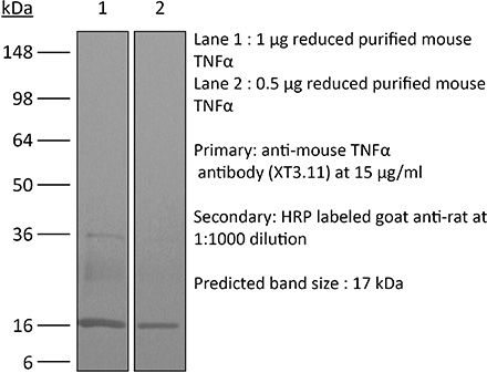
Specifications
| Isotype | Rat IgG1 |
|---|---|
| Recommended Isotype Control(s) | InVivoMAb rat IgG1 isotype control, anti-horseradish peroxidase |
| Recommended Dilution Buffer | InVivoPure pH 8.0 Dilution Buffer |
| Conjugation | This product is unconjugated. Conjugation is available via our Antibody Conjugation Services. |
| Immunogen | Recombinant mouse TNFα |
| Reported Applications |
in vivo TNFα neutralization in vitro TNFα neutralization Western blot |
| Formulation |
PBS, pH 8.0 Contains no stabilizers or preservatives |
| Endotoxin |
≤1EU/mg (≤0.001EU/μg) Determined by LAL assay |
| Purity |
≥95% Determined by SDS-PAGE |
| Sterility | 0.2 µm filtration |
| Production | Purified from cell culture supernatant in an animal-free facility |
| Purification | Protein G |
| RRID | AB_1107764 |
| Molecular Weight | 150 kDa |
| Storage | The antibody solution should be stored at the stock concentration at 4°C. Do not freeze. |
| Need a Custom Formulation? | See All Antibody Customization Options |
Application References
-
Deng, L., et al (2014). "Irradiation and anti-PD-L1 treatment synergistically promote antitumor immunity in mice" J Clin Invest 124(2): 687-695.
PubMed
High-dose ionizing irradiation (IR) results in direct tumor cell death and augments tumor-specific immunity, which enhances tumor control both locally and distantly. Unfortunately, local relapses often occur following IR treatment, indicating that IR-induced responses are inadequate to maintain antitumor immunity. Therapeutic blockade of the T cell negative regulator programmed death-ligand 1 (PD-L1, also called B7-H1) can enhance T cell effector function when PD-L1 is expressed in chronically inflamed tissues and tumors. Here, we demonstrate that PD-L1 was upregulated in the tumor microenvironment after IR. Administration of anti-PD-L1 enhanced the efficacy of IR through a cytotoxic T cell-dependent mechanism. Concomitant with IR-mediated tumor regression, we observed that IR and anti-PD-L1 synergistically reduced the local accumulation of tumor-infiltrating myeloid-derived suppressor cells (MDSCs), which suppress T cells and alter the tumor immune microenvironment. Furthermore, activation of cytotoxic T cells with combination therapy mediated the reduction of MDSCs in tumors through the cytotoxic actions of TNF. Our data provide evidence for a close interaction between IR, T cells, and the PD-L1/PD-1 axis and establish a basis for the rational design of combination therapy with immune modulators and radiotherapy.
-
Shaabani, N., et al (2018). "The probacterial effect of type I interferon signaling requires its own negative regulator USP18" Sci Immunol 3(27).
PubMed
Type I interferon (IFN-I) signaling paradoxically impairs host immune responses during many primary and secondary bacterial infections. Lack of IFN-I receptor reduces bacterial replication and/or bacterial persistence during infection with several bacteria. However, the mechanisms that mediate the adverse IFN-I effect are incompletely understood. Here, we show that Usp18, an interferon-stimulated gene that negatively regulates IFN-I signaling, is primarily responsible for the deleterious effect of IFN-I signaling during infection of mice with Listeria monocytogenes or Staphylococcus aureus Mechanistically, USP18 promoted bacterial replication by inhibiting antibacterial tumor necrosis factor-alpha (TNF-alpha) signaling. Deleting IFNAR1 or USP18 in CD11c-Cre(+) cells similarly reduced bacterial titers in multiple organs and enhanced survival. Our results demonstrate that inhibiting USP18 function can promote control of primary and secondary bacterial infection by enhancing the antibacterial effect of TNF-alpha, which correlates with induction of reactive oxygen species (ROS). These findings suggest that USP18 could be targeted therapeutically in patients to ameliorate disease caused by serious bacterial infections.
-
Baeyens, A., et al (2015). "Effector T cells boost regulatory T cell expansion by IL-2, TNF, OX40, and plasmacytoid dendritic cells depending on the immune context" J Immunol 194(3): 999-1010.
PubMed
CD4(+)CD25(+)Foxp3(+) regulatory T (Treg) cells play a major role in peripheral tolerance. Multiple environmental factors and cell types affect their biology. Among them, activated effector CD4(+) T cells can boost Treg cell expansion through TNF or IL-2. In this study, we further characterized this effector T (Teff) cell-dependent Treg cell boost in vivo in mice. This phenomenon was observed when both Treg and Teff cells were activated by their cognate Ag, with the latter being the same or different. Also, when Treg cells highly proliferated on their own, there was no additional Treg cell boost by Teff cells. In a condition of low inflammation, the Teff cell-mediated Treg cell boost involved TNF, OX40L, and plasmacytoid dendritic cells, whereas in a condition of high inflammation, it involved TNF and IL-2. Thus, this feedback mechanism in which Treg cells are highly activated by their Teff cell counterparts depends on the immune context for its effectiveness and mechanism. This Teff cell-dependent Treg cell boost may be crucial to limit inflammatory and autoimmune responses.
-
Christensen, A. D., et al (2015). "Depletion of regulatory T cells in a hapten-induced inflammation model results in prolonged and increased inflammation driven by T cells" Clin Exp Immunol 179(3): 485-499.
PubMed
Regulatory T cells (Tregs ) are known to play an immunosuppressive role in the response of contact hypersensitivity (CHS), but neither the dynamics of Tregs during the CHS response nor the exaggerated inflammatory response after depletion of Tregs has been characterized in detail. In this study we show that the number of Tregs in the challenged tissue peak at the same time as the ear-swelling reaches its maximum on day 1 after challenge, whereas the number of Tregs in the draining lymph nodes peaks at day 2. As expected, depletion of Tregs by injection of a monoclonal antibody to CD25 prior to sensitization led to a prolonged and sustained inflammatory response which was dependent upon CD8 T cells, and co-stimulatory blockade with cytotoxic T lymphocyte antigen-4-immunoglobulin (CTLA-4-Ig) suppressed the exaggerated inflammation. In contrast, blockade of the interleukin (IL)-10-receptor (IL-10R) did not further increase the exaggerated inflammatory response in the Treg -depleted mice. In the absence of Tregs , the response changed from a mainly acute reaction with heavy infiltration of neutrophils to a sustained response with more chronic characteristics (fewer neutrophils and dominated by macrophages). Furthermore, depletion of Tregs enhanced the release of cytokines and chemokines locally in the inflamed ear and augmented serum levels of the systemic inflammatory mediators serum amyloid (SAP) and haptoglobin early in the response.
Product Citations
-
IL-9 and Blimp-1 protect the transcriptional identity of group 2 innate lymphocytes in allergic asthma.
In Nat Immunol on 1 June 2026 by Zheng, Y., Giri, S., et al.
PubMed
Allergic asthma is driven by type 2 immune responses, including type 2 innate lymphoid cells (ILC2s). Although ILC2s are activated by the tissue alarmins interleukin (IL)-33 and IL-25, these signals do not intrinsically enforce type 2 identity and the mechanisms that maintain type 2 cytokine expression remain unclear. Here we show that allergen-induced IL-33 and IL-25 rapidly induce IL-9, which in turn upregulates the transcriptional repressor Blimp-1 in ILC2s. Blimp-1 sustains type 2 immunity by directly repressing type 1 inflammatory programs, including expression of interferon-γ and tumor necrosis factor. Deletion of Blimp-1 in ILC2s increased type 1 cytokine production and reduced IL-5 and IL-13 expression, eosinophil recruitment and mucus production in the lung. In contrast, IL-9 expression was enhanced in the absence of Blimp-1, leading to increased mast cell recruitment. Together, these findings identify Blimp-1 as a key regulator of ILC2 transcriptional fidelity that stabilizes type 2 inflammation while constraining divergent inflammatory programs during allergic responses.
-
Granzyme B PET Imaging Uncovers Dynamic Patterns of Disease Activity and Therapeutic Response in a Murine Colitis Model.
In Int J Mol Sci on 8 May 2026 by Haj-Mirzaian, A., Ma, M., et al.
PubMed
The evaluation of therapeutic response is essential in disease monitoring both for disease status and treatment efficacy in inflammatory bowel disease. Here, we focused on the use of positron emission tomography directed towards granzyme B, a serine protease released by activated cytotoxic T cells and natural killer cells, to evaluate the dynamics of therapeutic response in a colitis model. The goal was to explore the use of granzyme B positron emission tomography as a non-invasive biomarker to monitor disease activity and therapeutic response across several treatments in a dextran sulfate sodium-induced colitis model. C57BL/6 interleukin-10 knockout mice were divided into five groups, including a negative control, positive control and three treatment arms (antitumor necrosis factor, prednisolone, and anti-interleukin-23). The negative control group received regular water, while all other groups were induced with colitis via 3% DSS water for 1 week followed by normal water. Treatments were initiated after colitis was induced (anti-TNF antibody, prednisolone, or anti-IL-23 antibody). Positron emission tomography/computed tomography imaging with 68Ga-NOTA-GZP was performed at baseline (after colitis induction, before therapy), and at 1 and 2 weeks after treatment initiation. Histological analyses were also performed at 1 and 2 weeks after treatment initiation. Gzmb expression and histological changes were also assessed with immunofluorescence staining and bulk ribonucleic acid sequencing. Gzmb-targeted PET imaging revealed distinct longitudinal patterns of colonic tracer uptake related to treatment response. In positive control mice with DSS colitis (no treatment), bowel uptake of 68Ga-NOTA-GZP increased significantly from baseline to week 2. Anti-TNF treatment reduced granzyme B positron emission tomography uptake significantly at week 2, approaching levels seen in negative controls. In prednisolone-treated mice, 68Ga-NOTA-GZP uptake decreased at week 1 but rose significantly by week 2 but still was in normal range. Anti-IL-23 therapy produced a significantly elevated Gzmb PET signal at week 1, followed by a significant decline by week 2 of treatment. The imaging trends were corroborated by tissue analyses and IF staining for Gzmb, which revealed no colonic expression in negative controls and strong Gzmb elevation in positive controls and the prednisolone group but a decreased Gzmb signal in the anti-TNF and late anti-IL-23 groups. Bulk RNA sequencing also supported these findings, with Gzmb gene expression tracking with inflammation severity and NK/T cell abundance and decreasing after effective therapy. Gzmb-targeted PET/CT allows for dynamic and non-invasive assessment of intestinal immune compartment activity and an assessment of therapy in colitis. Gzmb PET was able to detect initial treatment responses of anti-TNF, steroid and anti-IL-23 based on changes in the Gzmb PET signal. This suggests that clinical Gzmb PET imaging may serve as precision imaging for monitoring disease activity with treatment in IBD and help improve patient care by identifying responders and non-responders in real time.