InVivoMAb anti-mouse LAG-3
Product Description
Specifications
| Isotype | Rat IgG1, κ |
|---|---|
| Recommended Isotype Control(s) | InVivoMAb rat IgG1 isotype control, anti-horseradish peroxidase |
| Recommended Dilution Buffer | InVivoPure pH 7.0 Dilution Buffer |
| Conjugation | This product is unconjugated. Conjugation is available via our Antibody Conjugation Services. |
| Immunogen | Mouse CD223-Ig fusion protein |
| Reported Applications |
in vivo LAG-3 neutralization in vitro LAG-3 neutralization Flow cytometry Western blot |
| Formulation |
PBS, pH 7.0 Contains no stabilizers or preservatives |
| Endotoxin |
≤1EU/mg (≤0.001EU/μg) Determined by LAL assay |
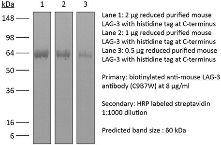
| Purity |
≥95% Determined by SDS-PAGE |
| Sterility | 0.2 µm filtration |
| Production | Purified from cell culture supernatant in an animal-free facility |
| Purification | Protein G |
| RRID | AB_10949602 |
| Molecular Weight | 150 kDa |
| Storage | The antibody solution should be stored at the stock concentration at 4°C. Do not freeze. |
| Need a Custom Formulation? | See All Antibody Customization Options |
Application References
-
Bauche, D., et al (2018). "LAG3(+) Regulatory T Cells Restrain Interleukin-23-Producing CX3CR1(+) Gut-Resident Macrophages during Group 3 Innate Lymphoid Cell-Driven Colitis" Immunity 49(2): 342-352 e345.
PubMed
Interleukin-22 (IL-22)-producing group 3 innate lymphoid cells (ILC3) maintains gut homeostasis but can also promote inflammatory bowel disease (IBD). The regulation of ILC3-dependent colitis remains to be elucidated. Here we show that Foxp3(+) regulatory T cells (Treg cells) prevented ILC3-mediated colitis in an IL-10-independent manner. Treg cells inhibited IL-23 and IL-1beta production from intestinal-resident CX3CR1(+) macrophages but not CD103(+) dendritic cells. Moreover, Treg cells restrained ILC3 production of IL-22 through suppression of CX3CR1(+) macrophage production of IL-23 and IL-1beta. This suppression was contact dependent and was mediated by latent activation gene-3 (LAG-3)-an immune checkpoint receptor-expressed on Treg cells. Engagement of LAG-3 on MHC class II drove profound immunosuppression of CX3CR1(+) tissue-resident macrophages. Our study reveals that the health of the intestinal mucosa is maintained by an axis driven by Treg cells communication with resident macrophages that withhold inflammatory stimuli required for ILC3 function.
-
Erickson, J. J., et al (2014). "Programmed death-1 impairs secondary effector lung CD8(+) T cells during respiratory virus reinfection" J Immunol 193(10): 5108-5117.
PubMed
Reinfections with respiratory viruses are common and cause significant clinical illness, yet precise mechanisms governing this susceptibility are ill defined. Lung Ag-specific CD8(+) T cells (T(CD8)) are impaired during acute viral lower respiratory infection by the inhibitory receptor programmed death-1 (PD-1). To determine whether PD-1 contributes to recurrent infection, we first established a model of reinfection by challenging B cell-deficient mice with human metapneumovirus (HMPV) several weeks after primary infection, and found that HMPV replicated to high titers in the lungs. A robust secondary effector lung TCD8 response was generated during reinfection, but these cells were more impaired and more highly expressed the inhibitory receptors PD-1, LAG-3, and 2B4 than primary T(CD8). In vitro blockade demonstrated that PD-1 was the dominant inhibitory receptor early after reinfection. In vivo therapeutic PD-1 blockade during HMPV reinfection restored lung T(CD8) effector functions (i.e., degranulation and cytokine production) and enhanced viral clearance. PD-1 also limited the protective efficacy of HMPV epitope-specific peptide vaccination and impaired lung T(CD8) during heterotypic influenza virus challenge infection. Our results indicate that PD-1 signaling may contribute to respiratory virus reinfection and evasion of vaccine-elicited immune responses. These results have important implications for the design of effective vaccines against respiratory viruses.
-
McGray, A. J., et al (2014). "Immunotherapy-induced CD8+ T cells instigate immune suppression in the tumor" Mol Ther 22(1): 206-218.
PubMed
Despite clear evidence of immunogenicity, cancer vaccines only provide a modest clinical benefit. To evaluate the mechanisms that limit tumor regression following vaccination, we have investigated the weak efficacy of a highly immunogenic experimental vaccine using a murine melanoma model. We discovered that the tumor adapts rapidly to the immune attack instigated by tumor-specific CD8+ T cells in the first few days following vaccination, resulting in the upregulation of a complex set of biological networks, including multiple immunosuppressive processes. This rapid adaptation acts to prevent sustained local immune attack, despite continued infiltration by increasing numbers of tumor-specific T cells. Combining vaccination with adoptive transfer of tumor-specific T cells produced complete regression of the treated tumors but did not prevent the adaptive immunosuppression. In fact, the adaptive immunosuppressive pathways were more highly induced in regressing tumors, commensurate with the enhanced level of immune attack. Examination of tumor infiltrating T-cell functionality revealed that the adaptive immunosuppression leads to a progressive loss in T-cell function, even in tumors that are regressing. These novel observations that T cells produced by therapeutic intervention can instigate a rapid adaptive immunosuppressive response within the tumor have important implications for clinical implementation of immunotherapies.
-
Verhagen, J. and D. C. Wraith (2014). "Blockade of LFA-1 augments in vitro differentiation of antigen-induced Foxp3(+) Treg cells" J Immunol Methods 414: 58-64.
PubMed
Adoptive transfer of antigen-specific, in vitro-induced Foxp3(+) Treg (iTreg) cells protects against autoimmune disease. To generate antigen-specific iTreg cells at high purity, however, remains a challenge. Whereas polyclonal T cell stimulation with anti-CD3 and anti-CD28 antibody yields Foxp3(+) iTreg cells at a purity of 90-95%, antigen-induced iTreg cells typically do not exceed a purity of 65-75%, even in a TCR-transgenic model. In a similar vein to thymic Treg cell selection, iTreg cell differentiation is influenced not only by antigen recognition and the availability of TGF-beta but also by co-factors including costimulation and adhesion molecules. In this study, we demonstrate that blockade of the T cell integrin Leukocyte Function-associated Antigen-1 (LFA-1) during antigen-mediated iTreg cell differentiation augments Foxp3 induction, leading to approximately 90% purity of Foxp3(+) iTreg cells. This increased efficacy not only boosts the yield of Foxp3(+) iTreg cells, it also reduces contamination with activated effector T cells, thus improving the safety of adoptive transfer immunotherapy.
Product Citations
-
Galectin-3 exacerbates autoimmune diabetes by limiting regulatory T cell differentiation and function.
In Sci Adv on 2 January 2026 by Xie, L., Zhang, R., et al.
PubMed
Galectin-3, a β-galactoside-binding lectin, has been implicated in several inflammatory and autoimmune diseases. However, the significance of circulating Galectin-3 in type 1 diabetes (T1D) remains unclear. Here, we report that compared to healthy controls, patients with T1D and their first-degree relatives (FDRs) exhibited significantly increased serum Galectin-3 levels primarily produced and secreted by monocytes/macrophages. Pharmacological inhibition (TD139) as well as knockout of Galectin-3 gene both attenuated Galectin-3-mediated suppression of regulatory T cells (Treg cells) and protected from insulitis and diabetes onset in NOD mice. Mechanistically, Galectin-3 bound to and activated lymphocyte activation gene 3 (LAG3), a receptor expressed on activated T cells, subsequently suppressing the MEK/ERK signaling pathway and thereby hindering Treg cell differentiation and function. In summary, our study identifies Galectin-3 as a potential biomarker for T1D and suggests that TD139 holds promise as a therapeutic candidate for patients with T1D and high serum Galectin-3 levels.
-
Dysfunctional CD4 T cells in an oncovirus-specific TCR-transgenic in vivo model.
In Nat Commun on 18 December 2025 by Spitzer, F. S., Camps, M. G. M., et al.
PubMed
T cell exhaustion has been implicated in cancer and infectious diseases. In this study, we report a novel mouse model, "MolT-II", with T cells expressing a transgenic T cell receptor (TCR) specific for a Moloney virus envelope-derived, MHC class II-presented peptide epitope. Characterization of MolT-II CD4 T cells revealed that they are dysfunctional, showing severely impaired effector functions, reduced proliferation and increased baseline expression of co-inhibitory receptors such as PD-1, LAG-3 and CTLA-4, likely due to chronic exposure to a self-antigen. We further show that epitope-specific peptide vaccination combined with immune checkpoint blockade is able to restore the function of MolT-II CD4 T cells in vivo, associated with enhanced tumor control in mice. The MolT-II mouse strain thus represents an in vivo model for reversible CD4 T cell dysfunction, allowing the study of the role of CD4 T cell regulation in cancer, mechanisms underlying CD4 T cell dysfunction and exhaustion, and novel immunomodulatory therapies aiming to rescue dysfunctional T cells.
-
Coordinated macrophage and T cell interactions mediate response to checkpoint blockade in colorectal cancer
In Research Square on 8 December 2025 by Mestrallet, G., Brown, M., et al.
-
Efficacy of anti-LAG3 and anti-PD-1 combination checkpoint inhibitor therapy against head and neck squamous cell carcinoma in a genetically engineered mouse model.
In Oncoimmunology on 1 December 2025 by Lamenza, F. F., Roth, P., et al.
PubMed
Head and neck squamous cell carcinoma (HNSCC) continues to be among the most common malignancies worldwide with limited treatment options for patients. Targeting the PD-1/PDL-1 axis is currently the only FDA approved immune checkpoint inhibitor treatment for HNSCC. Novel therapies targeting other pathways are needed along with testing a combinational approach to find new and more efficient ways to treat this disease. We utilized a tamoxifen inducible TgfβR1/Pten deletion mouse model to explore the efficacy of combined anti-LAG-3 and anti-PD-1 therapy against tongue HNSCC and determine underlying immunological mechanisms. Combined anti-LAG-3/anti-PD-1 therapy was effective at decreasing the tumor burden and lymphatic metastasis compared to anti-LAG-3 treatment but not when compared to the anti-PD-1 treatment alone. Anti-tumoral effects of anti-PD1 and anti-LAG-3/anti-PD-1 combined therapy were associated with increased CD4+ and CD8+ T-cell proliferative responses in secondary lymphoid organs along with increased CD8+ T-cell tumor infiltration. Anti-LAG-3 treatment potentiated the anti-tumoral properties of CD4+ T-cells treated with anti-PD-1, including enhanced systemic IFN-γ production and TNF-α production in the tumor microenvironment. Further, anti-tumoral cytotoxic CD8+ T-cell effector function and granzyme B production were enhanced by anti-PD-1 and combinatorial anti-LAG-3/anti-PD-1 immunotherapy, resulting in greater tumor cell death. Our results demonstrate that anti-LAG-3 has the potential to enhance the efficacy of anti-PD-1 therapy; however, humanized mouse models that better recapitulate the human disease with FDA approved antibodies are needed to further characterize the efficacy of this treatment as a viable treatment option for HNSCC patients.