
InVivoMAb anti-mouse CD40L (CD154)
in vitro blocking of CD40/CD40L signaling
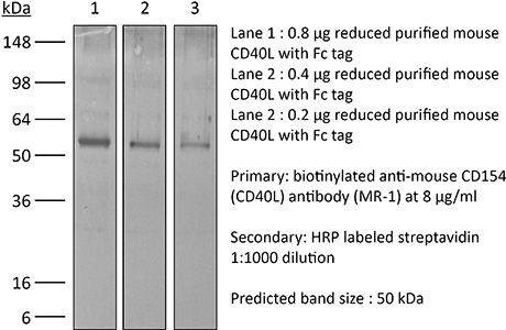
Western blotin vitro Organoids/Organ-on-Chip
Product Description
Specifications
| Isotype | Armenian hamster IgG |
|---|---|
| Recommended Isotype Control(s) | InVivoMAb polyclonal Armenian hamster IgG |
| Recommended Dilution Buffer | InVivoPure pH 7.0 Dilution Buffer |
| Conjugation | This product is unconjugated. Conjugation is available via our Antibody Conjugation Services. |
| Immunogen | Activated mouse Th1 clone D1.6 |
| Reported Applications |
in vivo blocking of CD40/CD40L signaling in vitro blocking of CD40/CD40L signaling Western blotin vitro Organoids/Organ-on-Chip |
| Formulation |
PBS, pH 7.0 Contains no stabilizers or preservatives |
| Endotoxin |
≤1EU/mg (≤0.001EU/μg) Determined by LAL assay |
| Purity |
≥95% Determined by SDS-PAGE |
| Sterility | 0.2 µm filtration |
| Production | Purified from cell culture supernatant in an animal-free facility |
| Purification | Protein A |
| RRID | AB_1107601 |
| Molecular Weight | 150 kDa |
| Storage | The antibody solution should be stored at the stock concentration at 4°C. Do not freeze. |
| Need a Custom Formulation? | See All Antibody Customization Options |
Application References
-
Pasqual, G., et al (2018). "Monitoring T cell-dendritic cell interactions in vivo by intercellular enzymatic labelling" Nature 553(7689): 496-500.
PubMed
Interactions between different cell types are essential for multiple biological processes, including immunity, embryonic development and neuronal signalling. Although the dynamics of cell-cell interactions can be monitored in vivo by intravital microscopy, this approach does not provide any information on the receptors and ligands involved or enable the isolation of interacting cells for downstream analysis. Here we describe a complementary approach that uses bacterial sortase A-mediated cell labelling across synapses of immune cells to identify receptor-ligand interactions between cells in living mice, by generating a signal that can subsequently be detected ex vivo by flow cytometry. We call this approach for the labelling of ‘kiss-and-run’ interactions between immune cells ‘Labelling Immune Partnerships by SorTagging Intercellular Contacts’ (LIPSTIC). Using LIPSTIC, we show that interactions between dendritic cells and CD4(+) T cells during T-cell priming in vivo occur in two distinct modalities: an early, cognate stage, during which CD40-CD40L interactions occur specifically between T cells and antigen-loaded dendritic cells; and a later, non-cognate stage during which these interactions no longer require prior engagement of the T-cell receptor. Therefore, LIPSTIC enables the direct measurement of dynamic cell-cell interactions both in vitro and in vivo. Given its flexibility for use with different receptor-ligand pairs and a range of detectable labels, we expect that this approach will be of use to any field of biology requiring quantification of intercellular communication.
-
Awe, O., et al (2015). "PU.1 Expression in T Follicular Helper Cells Limits CD40L-Dependent Germinal Center B Cell Development" J Immunol .
PubMed
PU.1 is an ETS family transcription factor that is important for the development of multiple hematopoietic cell lineages. Previous work demonstrated a critical role for PU.1 in promoting Th9 development and in limiting Th2 cytokine production. Whether PU.1 has functions in other Th lineages is not clear. In this study, we examined the effects of ectopic expression of PU.1 in CD4+ T cells and observed decreased expression of genes involved with the function of T follicular helper (Tfh) cells, including Il21 and Tnfsf5 (encoding CD40L). T cells from conditional mutant mice that lack expression of PU.1 in T cells (Sfpi1lck-/-) demonstrated increased production of CD40L and IL-21 in vitro. Following adjuvant-dependent or adjuvant-independent immunization, we observed that Sfpi1lck-/- mice had increased numbers of Tfh cells, increased germinal center B cells (GCB cells), and increased Ab production in vivo. This correlated with increased expression of IL-21 and CD40L in Tfh cells from Sfpi1lck-/- mice compared with control mice. Finally, although blockade of IL-21 did not affect GCB cells in Sfpi1lck-/- mice, anti-CD40L treatment of immunized Sfpi1lck-/- mice decreased GCB cell numbers and Ag-specific Ig concentrations. Together, these data indicate an inhibitory role for PU.1 in the function of Tfh cells, germinal centers, and Tfh-dependent humoral immunity.
-
Brasseit, J., et al (2015). "CD4 T cells are required for both development and maintenance of disease in a new mouse model of reversible colitis" Mucosal Immunol. doi : 10.1038/mi.2015.93.
PubMed
Current therapies to treat inflammatory bowel diseases have limited efficacy, significant side effects, and often wane over time. Little is known about the cellular and molecular mechanisms operative in the process of mucosal healing from colitis. To study such events, we developed a new model of reversible colitis in which adoptive transfer of CD4+CD45RBhi T cells into Helicobacter typhlonius-colonized lymphopenic mice resulted in a rapid onset of colonic inflammation that was reversible through depletion of colitogenic T cells. Remission was associated with an improved clinical and histopathological score, reduced immune cell infiltration to the intestinal mucosa, altered intestinal gene expression profiles, regeneration of the colonic mucus layer, and the restoration of epithelial barrier integrity. Notably, colitogenic T cells were not only critical for induction of colitis but also for maintenance of disease. Depletion of colitogenic T cells resulted in a rapid drop in tumor necrosis factor alpha (TNFalpha) levels associated with reduced infiltration of inflammatory immune cells to sites of inflammation. Although neutralization of TNFalpha prevented the onset of colitis, anti-TNFalpha treatment of mice with established disease failed to resolve colonic inflammation. Collectively, this new model of reversible colitis provides an important research tool to study the dynamics of mucosal healing in chronic intestinal remitting-relapsing disorders.
-
Conde, P., et al (2015). "DC-SIGN(+) Macrophages Control the Induction of Transplantation Tolerance" Immunity 42(6): 1143-1158.
PubMed
Tissue effector cells of the monocyte lineage can differentiate into different cell types with specific cell function depending on their environment. The phenotype, developmental requirements, and functional mechanisms of immune protective macrophages that mediate the induction of transplantation tolerance remain elusive. Here, we demonstrate that costimulatory blockade favored accumulation of DC-SIGN-expressing macrophages that inhibited CD8(+) T cell immunity and promoted CD4(+)Foxp3(+) Treg cell expansion in numbers. Mechanistically, that simultaneous DC-SIGN engagement by fucosylated ligands and TLR4 signaling was required for production of immunoregulatory IL-10 associated with prolonged allograft survival. Deletion of DC-SIGN-expressing macrophages in vivo, interfering with their CSF1-dependent development, or preventing the DC-SIGN signaling pathway abrogated tolerance. Together, the results provide new insights into the tolerogenic effects of costimulatory blockade and identify DC-SIGN(+) suppressive macrophages as crucial mediators of immunological tolerance with the concomitant therapeutic implications in the clinic.
Product Citations
-
Antimetabolites synergize with non-genotoxic antibody drug conjugate conditioning in hematopoietic stem cell lentiviral gene therapy.
In Mol Ther Adv on 10 September 2026 by Okalova, J., Alexander, J. S., et al.
PubMed
Hematopoietic stem cell transplantation (HSCT) of genetically engineered cells is a promising treatment modality for monogenic diseases. However, hematopoietic stem cell (HSC)-directed lentiviral vector (LV) gene therapies remain limited by genotoxicities associated with standard non-targeted conditioning using irradiation or alkylating chemotherapy. We and others developed an antibody drug conjugate (ADC) with anti-CD117 and saporin (sap), which selectively depletes CD117-expressing HSC and progenitor cells (HSPCs). Since anti-CD117-sap does not generally provide engraftment comparable to conventional conditioning, we investigated the hypothesis anti-CD117-sap is not effective as a single conditioning agent due to insufficient HSPC depletion. We show anti-CD117-sap induces nearly complete residual HSPC entry into a non-G0 proliferative state and postulated agents targeting cycling cells would enhance engraftment. When we administered the antimetabolite 5-fluorouracil with anti-CD117-sap, anti-thymocyte globulin, and an immunoglobulin-degrading enzyme, we achieved >90% peripheral blood chimerism of unmodified cultured donor cells. Additional immunosuppression combined with an increase in transplanted cells further permitted ∼70% engraftment of HSPCs transduced with an LV encoding a high-expression, immunogenic factor VIII (FVIII) transgene. Copy numbers of ∼1.0 and normal FVIII plasma activity levels (>50%) by 6 months post-HSCT were achieved. Collectively, we show ADC and antimetabolite administration can augment LV-engineered HSPC engraftment by targeting cycling cells.
-
Antimetabolites synergize with non-genotoxic antibody drug conjugate conditioning in hematopoietic stem cell lentiviral gene therapy.
In Mol Ther Adv on 10 September 2026 by Okalova, J., Alexander, J. S., et al.
PubMed
Hematopoietic stem cell transplantation (HSCT) of genetically engineered cells is a promising treatment modality for monogenic diseases. However, hematopoietic stem cell (HSC)-directed lentiviral vector (LV) gene therapies remain limited by genotoxicities associated with standard non-targeted conditioning using irradiation or alkylating chemotherapy. We and others developed an antibody drug conjugate (ADC) with anti-CD117 and saporin (sap), which selectively depletes CD117-expressing HSC and progenitor cells (HSPCs). Since anti-CD117-sap does not generally provide engraftment comparable to conventional conditioning, we investigated the hypothesis anti-CD117-sap is not effective as a single conditioning agent due to insufficient HSPC depletion. We show anti-CD117-sap induces nearly complete residual HSPC entry into a non-G0 proliferative state and postulated agents targeting cycling cells would enhance engraftment. When we administered the antimetabolite 5-fluorouracil with anti-CD117-sap, anti-thymocyte globulin, and an immunoglobulin-degrading enzyme, we achieved >90% peripheral blood chimerism of unmodified cultured donor cells. Additional immunosuppression combined with an increase in transplanted cells further permitted ∼70% engraftment of HSPCs transduced with an LV encoding a high-expression, immunogenic factor VIII (FVIII) transgene. Copy numbers of ∼1.0 and normal FVIII plasma activity levels (>50%) by 6 months post-HSCT were achieved. Collectively, we show ADC and antimetabolite administration can augment LV-engineered HSPC engraftment by targeting cycling cells.
-
Functional role of skull lymphoid structures in CNS immunosurveillance.
In Nature on 19 August 2026 by Park, J. H., Abramishvili, D., et al.
PubMed
Accumulating evidence demonstrates that the central nervous system (CNS) is not disconnected from the peripheral immune system; however, precisely how the adaptive immune system surveils the CNS remains a critical question. Recent findings reveal that channels between the skull and the dura mater facilitate the exchange of cerebrospinal fluid and immune cells between the CNS and skull bone marrow of mice under both homeostatic and disease conditions1-6. Skull bone marrow functions as a source of immune cells for the CNS5, yet its role in CNS antigen-specific adaptive immune responses remains unclear. Here we identify lymphoid structures within the skull bone marrow, featuring germinal-centre-like formations and containing a distinct population of follicular-helper-like T cells that promote B cell activation and humoral immunity through CD40L, IL-21 and IFNγ signalling. Adaptive immune cells within these skull bone marrow lymphoid structures surveil and respond to CNS-derived antigens and contribute to anti-tumour immune responses in mouse brain cancer models. Together, our findings show that the skull bone marrow is a site of CNS immunosurveillance that may influence immune responses across diverse neurological diseases.
-
Requirements for development of T helper 1 and T follicular helper cells from a common precursor.
In Cell Rep on 26 May 2026 by Bosma, D. M. T., Busselaar, J., et al.
PubMed
T helper (Th) and T follicular helper (Tfh) cells support cellular and humoral immunity, respectively. How activated CD4+ T cells commit to these fates remains unclear. Using a mouse vaccination model, we traced endogenous, polyclonal CD4+ T cells during bifurcation into Th1 and Tfh lineages. We found that Th1 and Tfh cells originate from shared, highly proliferative TCF1+SLAMF6+PD-1+ precursor clones that co-express Th1- and Tfh-related transcription factors and chemokine receptors, including T-bet, BCL6, CXCR3, and CXCR5. The generation of common Th1/Tfh precursors from naive CD4+ T cells requires CD28 costimulation but occurs independently of CD40, ICOS, and interaction with type 1 conventional dendritic cells (cDC1s) or B cells. Lineage commitment subsequently diverges: differentiation into Th1 cells relies on CD40 costimulation and cDC1s, whereas differentiation into Tfh cells requires ICOS costimulation and B cells. Activated CD4+ T cells thus give rise to a common Th1/Tfh precursor whose fate depends on interactions with distinct antigen-presenting cells.