InVivoMAb anti-mouse CD3ε
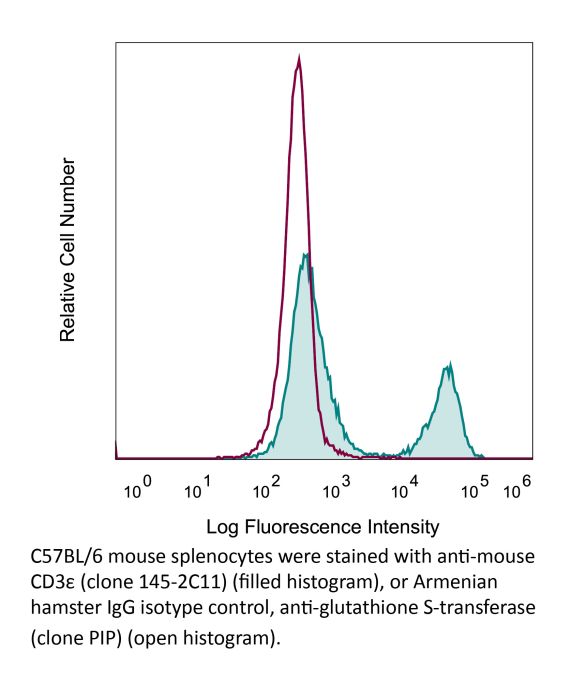
Product Description
Specifications
| Isotype | Armenian Hamster IgG1 |
|---|---|
| Recommended Isotype Control(s) | InVivoMAb polyclonal Armenian hamster IgG |
| Recommended Dilution Buffer | InVivoPure pH 7.0 Dilution Buffer |
| Conjugation | This product is unconjugated. Conjugation is available via our Antibody Conjugation Services. |
| Immunogen | Mouse BM10-37 cytotoxic T cells |
| Reported Applications |
in vivo T cell depletion in vitro T cell stimulation/activation Immunofluorescence Flow cytometry Western blot |
| Formulation |
PBS, pH 7.0 Contains no stabilizers or preservatives |
| Endotoxin |
≤1EU/mg (≤0.001EU/μg) Determined by LAL assay |
| Purity |
≥95% Determined by SDS-PAGE |
| Sterility | 0.2 µm filtration |
| Production | Purified from cell culture supernatant in an animal-free facility |
| Purification | Protein A |
| RRID | AB_1107634 |
| Molecular Weight | 150 kDa |
| Storage | The antibody solution should be stored at the stock concentration at 4°C. Do not freeze. |
| Need a Custom Formulation? | See All Antibody Customization Options |
Application References
in vivo T cell depletion
Glasner, A., et al (2018). "NKp46 Receptor-Mediated Interferon-gamma Production by Natural Killer Cells Increases Fibronectin 1 to Alter Tumor Architecture and Control Metastasis" Immunity 48(1): 107-119 e104.
PubMed
Natural killer (NK) cells are innate lymphoid cells, and their presence within human tumors correlates with better prognosis. However, the mechanisms by which NK cells control tumors in vivo are unclear. Here, we used reflectance confocal microscopy (RCM) imaging in humans and in mice to visualize tumor architecture in vivo. We demonstrated that signaling via the NK cell receptor NKp46 (human) and Ncr1 (mouse) induced interferon-gamma (IFN-gamma) secretion from intratumoral NK cells. NKp46- and Ncr1-mediated IFN-gamma production led to the increased expression of the extracellular matrix protein fibronectin 1 (FN1) in the tumors, which altered primary tumor architecture and resulted in decreased metastases formation. Injection of IFN-gamma into tumor-bearing mice or transgenic overexpression of Ncr1 in NK cells in mice resulted in decreased metastasis formation. Thus, we have defined a mechanism of NK cell-mediated control of metastases in vivo that may help develop NK cell-dependent cancer therapies.
in vitro T cell stimulation/activation
Lacher, S. M., et al (2018). "NF-kappaB inducing kinase (NIK) is an essential post-transcriptional regulator of T-cell activation affecting F-actin dynamics and TCR signaling" J Autoimmun 94: 110-121.
PubMed
NF-kappaB inducing kinase (NIK) is the key protein of the non-canonical NF-kappaB pathway and is important for the development of lymph nodes and other secondary immune organs. We elucidated the specific role of NIK in T cells using T-cell specific NIK-deficient (NIK(DeltaT)) mice. Despite showing normal development of lymphoid organs, NIK(DeltaT) mice were resistant to induction of CNS autoimmunity. T cells from NIK(DeltaT) mice were deficient in late priming, failed to up-regulate T-bet and to transmigrate into the CNS. Proteomic analysis of activated NIK(-/-) T cells showed de-regulated expression of proteins involved in the formation of the immunological synapse: in particular, proteins involved in cytoskeleton dynamics. In line with this we found that NIK-deficient T cells were hampered in phosphorylation of Zap70, LAT, AKT, ERK1/2 and PLCgamma upon TCR engagement. Hence, our data disclose a hitherto unknown function of NIK in T-cell priming and differentiation.
in vitro T cell stimulation/activation
Wendland, K., et al (2018). "Retinoic Acid Signaling in Thymic Epithelial Cells Regulates Thymopoiesis" J Immunol 201(2): 524-532.
PubMed
Despite the essential role of thymic epithelial cells (TEC) in T cell development, the signals regulating TEC differentiation and homeostasis remain incompletely understood. In this study, we show a key in vivo role for the vitamin A metabolite, retinoic acid (RA), in TEC homeostasis. In the absence of RA signaling in TEC, cortical TEC (cTEC) and CD80(lo)MHC class II(lo) medullary TEC displayed subset-specific alterations in gene expression, which in cTEC included genes involved in epithelial proliferation, development, and differentiation. Mice whose TEC were unable to respond to RA showed increased cTEC proliferation, an accumulation of stem cell Ag-1(hi) cTEC, and, in early life, a decrease in medullary TEC numbers. These alterations resulted in reduced thymic cellularity in early life, a reduction in CD4 single-positive and CD8 single-positive numbers in both young and adult mice, and enhanced peripheral CD8(+) T cell survival upon TCR stimulation. Collectively, our results identify RA as a regulator of TEC homeostasis that is essential for TEC function and normal thymopoiesis.
in vitro T cell stimulation/activation
Ron-Harel, N., et al (2016). "Mitochondrial Biogenesis and Proteome Remodeling Promote One-Carbon Metabolism for T Cell Activation" Cell Metab 24(1): 104-117.
PubMed
Naive T cell stimulation activates anabolic metabolism to fuel the transition from quiescence to growth and proliferation. Here we show that naive CD4(+) T cell activation induces a unique program of mitochondrial biogenesis and remodeling. Using mass spectrometry, we quantified protein dynamics during T cell activation. We identified substantial remodeling of the mitochondrial proteome over the first 24 hr of T cell activation to generate mitochondria with a distinct metabolic signature, with one-carbon metabolism as the most induced pathway. Salvage pathways and mitochondrial one-carbon metabolism, fed by serine, contribute to purine and thymidine synthesis to enable T cell proliferation and survival. Genetic inhibition of the mitochondrial serine catabolic enzyme SHMT2 impaired T cell survival in culture and antigen-specific T cell abundance in vivo. Thus, during T cell activation, mitochondrial proteome remodeling generates specialized mitochondria with enhanced one-carbon metabolism that is critical for T cell activation and survival.
in vitro T cell stimulation/activation
Gu, A. D., et al (2015). "A critical role for transcription factor Smad4 in T cell function that is independent of transforming growth factor beta receptor signaling" Immunity 42(1): 68-79.
PubMed
Transforming growth factor-beta (TGF-beta) suppresses T cell function to maintain self-tolerance and to promote tumor immune evasion. Yet how Smad4, a transcription factor component of TGF-beta signaling, regulates T cell function remains unclear. Here we have demonstrated an essential role for Smad4 in promoting T cell function during autoimmunity and anti-tumor immunity. Smad4 deletion rescued the lethal autoimmunity resulting from transforming growth factor-beta receptor (TGF-betaR) deletion and compromised T-cell-mediated tumor rejection. Although Smad4 was dispensable for T cell generation, homeostasis, and effector function, it was essential for T cell proliferation after activation in vitro and in vivo. The transcription factor Myc was identified to mediate Smad4-controlled T cell proliferation. This study thus reveals a requirement of Smad4 for T-cell-mediated autoimmunity and tumor rejection, which is beyond the current paradigm. It highlights a TGF-betaR-independent role for Smad4 in promoting T cell function, autoimmunity, and anti-tumor immunity.
in vitro T cell stimulation/activation
Awe, O., et al (2015). "PU.1 Expression in T Follicular Helper Cells Limits CD40L-Dependent Germinal Center B Cell Development" J Immunol .
PubMed
PU.1 is an ETS family transcription factor that is important for the development of multiple hematopoietic cell lineages. Previous work demonstrated a critical role for PU.1 in promoting Th9 development and in limiting Th2 cytokine production. Whether PU.1 has functions in other Th lineages is not clear. In this study, we examined the effects of ectopic expression of PU.1 in CD4+ T cells and observed decreased expression of genes involved with the function of T follicular helper (Tfh) cells, including Il21 and Tnfsf5 (encoding CD40L). T cells from conditional mutant mice that lack expression of PU.1 in T cells (Sfpi1lck-/-) demonstrated increased production of CD40L and IL-21 in vitro. Following adjuvant-dependent or adjuvant-independent immunization, we observed that Sfpi1lck-/- mice had increased numbers of Tfh cells, increased germinal center B cells (GCB cells), and increased Ab production in vivo. This correlated with increased expression of IL-21 and CD40L in Tfh cells from Sfpi1lck-/- mice compared with control mice. Finally, although blockade of IL-21 did not affect GCB cells in Sfpi1lck-/- mice, anti-CD40L treatment of immunized Sfpi1lck-/- mice decreased GCB cell numbers and Ag-specific Ig concentrations. Together, these data indicate an inhibitory role for PU.1 in the function of Tfh cells, germinal centers, and Tfh-dependent humoral immunity.
in vitro T cell stimulation/activation
Xu, H., et al (2015). "Regulation of bifurcating B cell trajectories by mutual antagonism between transcription factors IRF4 and IRF8" Nat Immunol .
PubMed
Upon recognition of antigen, B cells undertake a bifurcated response in which some cells rapidly differentiate into plasmablasts while others undergo affinity maturation in germinal centers (GCs). Here we identified a double-negative feedback loop between the transcription factors IRF4 and IRF8 that regulated the initial developmental bifurcation of activated B cells as well as the GC response. IRF8 dampened signaling via the B cell antigen receptor (BCR), facilitated antigen-specific interaction with helper T cells, and promoted antibody affinity maturation while antagonizing IRF4-driven differentiation of plasmablasts. Genomic analysis revealed concentration-dependent actions of IRF4 and IRF8 in regulating distinct gene-expression programs. Stochastic modeling suggested that the double-negative feedback was sufficient to initiate bifurcation of the B cell developmental trajectories.
in vitro T cell stimulation/activation
Huang, Y., et al (2015). "CRK proteins selectively regulate T cell migration into inflamed tissues" J Clin Invest 125(3): 1019-1032.
PubMed
Effector T cell migration into inflamed sites greatly exacerbates tissue destruction and disease severity in inflammatory diseases, including graft-versus-host disease (GVHD). T cell migration into such sites depends heavily on regulated adhesion and migration, but the signaling pathways that coordinate these functions downstream of chemokine receptors are largely unknown. Using conditional knockout mice, we found that T cells lacking the adaptor proteins CRK and CRK-like (CRKL) exhibit reduced integrin-dependent adhesion, chemotaxis, and diapedesis. Moreover, these two closely related proteins exhibited substantial functional redundancy, as ectopic expression of either protein rescued defects in T cells lacking both CRK and CRKL. We determined that CRK proteins coordinate with the RAP guanine nucleotide exchange factor C3G and the adhesion docking molecule CASL to activate the integrin regulatory GTPase RAP1. CRK proteins were required for effector T cell trafficking into sites of inflammation, but not for migration to lymphoid organs. In a murine bone marrow transplantation model, the differential migration of CRK/CRKL-deficient T cells resulted in efficient graft-versus-leukemia responses with minimal GVHD. Together, the results from our studies show that CRK family proteins selectively regulate T cell adhesion and migration at effector sites and suggest that these proteins have potential as therapeutic targets for preventing GVHD.
in vitro T cell stimulation/activation
Immunofluorescence
Kim, Y. U., et al (2015). "Regulation of autoimmune germinal center reactions in lupus-prone BXD2 mice by follicular helper T cells" PLoS One 10(3): e0120294.
PubMed
BXD2 mice spontaneously develop autoantibodies and subsequent glomerulonephritis, offering a useful animal model to study autoimmune lupus. Although initial studies showed a critical contribution of IL-17 and Th17 cells in mediating autoimmune B cell responses in BXD2 mice, the role of follicular helper T (Tfh) cells remains incompletely understood. We found that both the frequency of Th17 cells and the levels of IL-17 in circulation in BXD2 mice were comparable to those of wild-type. By contrast, the frequency of PD-1+ CXCR5+ Tfh cells was significantly increased in BXD2 mice compared with wild-type mice, while the frequency of PD-1+ CXCR5+ Foxp3+ follicular regulatory T (Tfr) cells was reduced in the former group. The frequency of Tfh cells rather than that of Th17 cells was positively correlated with the frequency of germinal center B cells as well as the levels of autoantibodies to dsDNA. More importantly, CXCR5+ CD4+ T cells isolated from BXD2 mice induced the production of IgG from naive B cells in an IL-21-dependent manner, while CCR6+ CD4+ T cells failed to do so. These results together demonstrate that Tfh cells rather than Th17 cells contribute to the autoimmune germinal center reactions in BXD2 mice.
in vitro T cell stimulation/activation
Liu, H., et al (2015). "The Immune Adaptor SLP-76 Binds to SUMO-RANGAP1 at Nuclear Pore Complex Filaments to Regulate Nuclear Import of Transcription Factors in T Cells" Mol Cell 59(5): 840-849.
PubMed
While immune cell adaptors regulate proximal T cell signaling, direct regulation of the nuclear pore complex (NPC) has not been reported. NPC has cytoplasmic filaments composed of RanGAP1 and RanBP2 with the potential to interact with cytoplasmic mediators. Here, we show that the immune cell adaptor SLP-76 binds directly to SUMO-RanGAP1 of cytoplasmic fibrils of the NPC, and that this interaction is needed for optimal NFATc1 and NF-kappaB p65 nuclear entry in T cells. Transmission electron microscopy showed anti-SLP-76 cytoplasmic labeling of the majority of NPCs in anti-CD3 activated T cells. Further, SUMO-RanGAP1 bound to the N-terminal lysine 56 of SLP-76 where the interaction was needed for optimal RanGAP1-NPC localization and GAP exchange activity. While the SLP-76-RanGAP1 (K56E) mutant had no effect on proximal signaling, it impaired NF-ATc1 and p65/RelA nuclear entry and in vivo responses to OVA peptide. Overall, we have identified SLP-76 as a direct regulator of nuclear pore function in T cells.
in vitro T cell stimulation/activation
Flow Cytometry
Tang, W., et al (2014). "The oncoprotein and transcriptional regulator Bcl-3 governs plasticity and pathogenicity of autoimmune T cells" Immunity 41(4): 555-566.
PubMed
Bcl-3 is an atypical member of the IkappaB family that modulates transcription in the nucleus via association with p50 (NF-kappaB1) or p52 (NF-kappaB2) homodimers. Despite evidence attesting to the overall physiologic importance of Bcl-3, little is known about its cell-specific functions or mechanisms. Here we demonstrate a T-cell-intrinsic function of Bcl-3 in autoimmunity. Bcl-3-deficient T cells failed to induce disease in T cell transfer-induced colitis and experimental autoimmune encephalomyelitis. The protection against disease correlated with a decrease in Th1 cells that produced the cytokines IFN-gamma and GM-CSF and an increase in Th17 cells. Although differentiation into Th1 cells was not impaired in the absence of Bcl-3, differentiated Th1 cells converted to less-pathogenic Th17-like cells, in part via mechanisms involving expression of the RORgammat transcription factor. Thus, Bcl-3 constrained Th1 cell plasticity and promoted pathogenicity by blocking conversion to Th17-like cells, revealing a unique type of regulation that shapes adaptive immunity.
in vitro T cell stimulation/activation
Rabenstein, H., et al (2014). "Differential kinetics of antigen dependency of CD4+ and CD8+ T cells" J Immunol 192(8): 3507-3517.
PubMed
Ag recognition via the TCR is necessary for the expansion of specific T cells that then contribute to adaptive immunity as effector and memory cells. Because CD4+ and CD8+ T cells differ in terms of their priming APCs and MHC ligands we compared their requirements of Ag persistence during their expansion phase side by side. Proliferation and effector differentiation of TCR transgenic and polyclonal mouse T cells were thus analyzed after transient and continuous TCR signals. Following equally strong stimulation, CD4+ T cell proliferation depended on prolonged Ag presence, whereas CD8+ T cells were able to divide and differentiate into effector cells despite discontinued Ag presentation. CD4+ T cell proliferation was neither affected by Th lineage or memory differentiation nor blocked by coinhibitory signals or missing inflammatory stimuli. Continued CD8+ T cell proliferation was truly independent of self-peptide/MHC-derived signals. The subset divergence was also illustrated by surprisingly broad transcriptional differences supporting a stronger propensity of CD8+ T cells to programmed expansion. These T cell data indicate an intrinsic difference between CD4+ and CD8+ T cells regarding the processing of TCR signals for proliferation. We also found that the presentation of a MHC class II-restricted peptide is more efficiently prolonged by dendritic cell activation in vivo than a class I bound one. In summary, our data demonstrate that CD4+ T cells require continuous stimulation for clonal expansion, whereas CD8+ T cells can divide following a much shorter TCR signal.
in vitro T cell stimulation/activation
Vegran, F., et al (2014). "The transcription factor IRF1 dictates the IL-21-dependent anticancer functions of TH9 cells" Nat Immunol 15(8): 758-766.
PubMed
The TH9 subset of helper T cells was initially shown to contribute to the induction of autoimmune and allergic diseases, but subsequent evidence has suggested that these cells also exert antitumor activities. However, the molecular events that account for their effector properties are elusive. Here we found that the transcription factor IRF1 enhanced the effector function of TH9 cells and dictated their anticancer properties. Under TH9-skewing conditions, interleukin 1beta (IL-1beta) induced phosphorylation of the transcription factor STAT1 and subsequent expression of IRF1, which bound to the promoters of Il9 and Il21 and enhanced secretion of the cytokines IL-9 and IL-21 from TH9 cells. Furthermore, IL-1beta-induced TH9 cells exerted potent anticancer functions in an IRF1- and IL-21-dependent manner. Our findings thus identify IRF1 as a target for controlling the function of TH9 cells.
in vitro T cell stimulation/activation
Bertin, S., et al (2014). "The ion channel TRPV1 regulates the activation and proinflammatory properties of CD4(+) T cells" Nat Immunol 15(11): 1055-1063.
PubMed
TRPV1 is a Ca(2+)-permeable channel studied mostly as a pain receptor in sensory neurons. However, its role in other cell types is poorly understood. Here we found that TRPV1 was functionally expressed in CD4(+) T cells, where it acted as a non-store-operated Ca(2+) channel and contributed to T cell antigen receptor (TCR)-induced Ca(2+) influx, TCR signaling and T cell activation. In models of T cell-mediated colitis, TRPV1 promoted colitogenic T cell responses and intestinal inflammation. Furthermore, genetic and pharmacological inhibition of TRPV1 in human CD4(+) T cells recapitulated the phenotype of mouse Trpv1(-/-) CD4(+) T cells. Our findings suggest that inhibition of TRPV1 could represent a new therapeutic strategy for restraining proinflammatory T cell responses.
in vitro T cell stimulation/activation
Berger, H., et al (2013). "SOCS3 transactivation by PPARgamma prevents IL-17-driven cancer growth" Cancer Res 73(12): 3578-3590.
PubMed
Activation of the transcription factor PPARgamma by the n-3 fatty acid docosahexaenoic acid (DHA) is implicated in controlling proinflammatory cytokine secretion, but the intracellular signaling pathways engaged by PPARgamma are incompletely characterized. Here, we identify the adapter-encoding gene SOCS3 as a critical transcriptional target of PPARgamma. SOCS3 promoter binding and gene transactivation by PPARgamma was associated with a repression in differentiation of proinflammatory T-helper (TH)17 cells. Accordingly, TH17 cells induced in vitro displayed increased SOCS3 expression and diminished capacity to produce interleukin (IL)-17 following activation of PPARgamma by DHA. Furthermore, naive CD4 T cells derived from mice fed a DHA-enriched diet displayed less capability to differentiate into TH17 cells. In two different mouse models of cancer, DHA prevented tumor outgrowth and angiogenesis in an IL-17-dependent manner. Altogether, our results uncover a novel molecular pathway by which PPARgamma-induced SOCS3 expression prevents IL-17-mediated cancer growth.
in vitro T cell stimulation/activation
Sledzinska, A., et al (2013). "TGF-beta signalling is required for CD4(+) T cell homeostasis but dispensable for regulatory T cell function" PLoS Biol 11(10): e1001674.
PubMed
TGF-beta is widely held to be critical for the maintenance and function of regulatory T (T(reg)) cells and thus peripheral tolerance. This is highlighted by constitutive ablation of TGF-beta receptor (TR) during thymic development in mice, which leads to a lethal autoimmune syndrome. Here we describe that TGF-beta-driven peripheral tolerance is not regulated by TGF-beta signalling on mature CD4(+) T cells. Inducible TR2 ablation specifically on CD4(+) T cells did not result in a lethal autoinflammation. Transfer of these TR2-deficient CD4(+) T cells to lymphopenic recipients resulted in colitis, but not overt autoimmunity. In contrast, thymic ablation of TR2 in combination with lymphopenia led to lethal multi-organ inflammation. Interestingly, deletion of TR2 on mature CD4(+) T cells does not result in the collapse of the T(reg) cell population as observed in constitutive models. Instead, a pronounced enlargement of both regulatory and effector memory T cell pools was observed. This expansion is cell-intrinsic and seems to be caused by increased T cell receptor sensitivity independently of common gamma chain-dependent cytokine signals. The expression of Foxp3 and other regulatory T cells markers was not dependent on TGF-beta signalling and the TR2-deficient T(reg) cells retained their suppressive function both in vitro and in vivo. In summary, absence of TGF-beta signalling on mature CD4(+) T cells is not responsible for breakdown of peripheral tolerance, but rather controls homeostasis of mature T cells in adult mice.
in vitro T cell stimulation/activation
Goswami, R., et al (2012). "STAT6-dependent regulation of Th9 development" J Immunol 188(3): 968-975.
PubMed
Th cell effector subsets develop in response to specific cytokine environments. The development of a particular cytokine-secreting pattern requires an integration of signals that may promote the development of opposing pathways. A recent example of this paradigm is the IL-9-secreting Th9 cell that develops in response to TGF-beta and IL-4, cytokines that, in isolation, promote the development of inducible regulatory T cells and Th2 cells, respectively. To determine how the balance of these factors results in priming for IL-9 secretion, we examined the effects of each pathway on transcription factors that regulate Th cell differentiation. We demonstrated that TGF-beta induces the PU.1-encoding Sfpi1 locus and that this is independent of IL-4-induced STAT6 activation. IL-4-activated STAT6 is required for repressing the expression of T-bet and Foxp3 in Th9 cells, transcription factors that inhibit IL-9 production, and STAT6 is required for the induction of IRF4, which promotes Th9 development. These data established a transcription factor network that regulates IL-9 and demonstrated how combinations of cytokine signals generate cytokine-secreting potential by altering the expression of a panel of transcription factors.
in vivo T cell depletion
Peng, B., et al (2009). "Anti-CD3 antibodies modulate anti-factor VIII immune responses in hemophilia A mice after factor VIII plasmid-mediated gene therapy" Blood 114(20): 4373-4382.
PubMed
One major obstacle in gene therapy is the generation of immune responses directed against transgene product. Five consecutive anti-CD3 treatments concomitant with factor VIII (FVIII) plasmid injection prevented the formation of inhibitory antibodies against FVIII and achieved persistent, therapeutic levels of FVIII gene expression in treated hemophilia A mice. Repeated plasmid gene transfer is applicable in tolerized mice without eliciting immune responses. Anti-CD3 treatment significantly depleted both CD4+ and CD8+ T cells, whereas increased transforming growth factor-beta levels in plasma and the frequency of both CD4+CD25+FoxP3+ and CD4+CD25-Foxp3+ regulatory T cells in the initial few weeks after treatment. Although prior depletion of CD4+CD25+ cells did not abrogate tolerance induction, adoptive transfer of CD4+ cells from tolerized mice at 6 weeks after treatment protected recipient mice from anti-FVIII immune responses. Anti-CD3-treated mice mounted immune responses against both T-dependent and T-independent neo-antigens, indicating that anti-CD3 did not hamper the immune systems in the long term. Concomitant FVIII plasmid + anti-CD3 treatment induced long-term tolerance specific to FVIII via a mechanism involving the increase in transforming growth factor-beta levels and the generation of adaptive FVIII-specific CD4+Foxp3+ regulatory T cells at the periphery. Furthermore, anti-CD3 can reduce the titers of preexisting anti-FVIII inhibitory antibodies in hemophilia A mice.
in vitro T cell stimulation/activation
Dardalhon, V., et al (2008). "IL-4 inhibits TGF-beta-induced Foxp3+ T cells and, together with TGF-beta, generates IL-9+ IL-10+ Foxp3(-) effector T cells" Nat Immunol 9(12): 1347-1355.
PubMed
Transcription factor Foxp3 is critical for generating regulatory T cells (T(reg) cells). Transforming growth factor-beta (TGF-beta) induces Foxp3 and suppressive T(reg) cells from naive T cells, whereas interleukin 6 (IL-6) inhibits the generation of inducible T(reg) cells. Here we show that IL-4 blocked the generation of TGF-beta-induced Foxp3(+) T(reg) cells and instead induced a population of T helper cells that produced IL-9 and IL-10. The IL-9(+)IL-10(+) T cells demonstrated no regulatory properties despite producing abundant IL-10. Adoptive transfer of IL-9(+)IL-10(+) T cells into recombination-activating gene 1-deficient mice induced colitis and peripheral neuritis, the severity of which was aggravated if the IL-9(+)IL-10(+) T cells were transferred with CD45RB(hi) CD4(+) effector T cells. Thus IL-9(+)IL-10(+) T cells lack suppressive function and constitute a distinct population of helper-effector T cells that promote tissue inflammation.
Product Citations
-
-
Immunology and Microbiology
-
Biochemistry and Molecular biology
-
Genetics
Uridine depletion impairs CD8⁺ T cell antitumor activity through N-glycosylation.
In Cell Metab on 3 March 2026 by Xiao, J., Li, Z., et al.
PubMed
Immune checkpoint blockade (ICB) faces limitations owing to high cost and restricted efficacy. This study identifies SNX17 as a key mediator of ICB resistance. Elevated SNX17 correlates with poor anti-PD-1 response in humans and mice. SNX17 deletion in tumor cells inhibits tumor growth via CD8+ T cell-dependent mechanisms. SNX17 reduces uridine in the tumor microenvironment (TME), suppressing IFN-γ and upregulating PD1 in CD8+ T cells. Exogenous uridine shows antitumor efficacy comparable to anti-PD-1/PD-L1 in low-SNX17 tumors and overcomes resistance in high-SNX17 models. Uridine enhances CD8+ T cell function by promoting CD45 N-glycosylation and LCK phosphorylation. Mechanistically, SNX17 stabilizes RUNX2, promoting UPP1 transcription and uridine degradation in the TME. These findings position SNX17 as an ICB response biomarker and nominate uridine as a cost-effective immunotherapeutic strategy.
-
-
-
Flow cytometry/Cell sorting
IFITM1 is required for epithelial mesenchymal transition in airway remodeling of allergic asthma.
In World Allergy Organ J on 1 March 2026 by Zhu, M., Weng, X., et al.
PubMed
Interferon-induced transmembrane protein 1 (IFITM1) restricts virus infection. IFITM proteins are involved in Th2 cell differentiation in allergic asthma. The epithelial‒mesenchymal transition (EMT) regulates allergic airway remodeling. We sought to explore the functional contributions and underlying mechanisms of IFITM1 in the EMT associated with allergic asthma.
-
-
-
Immunology and Microbiology
-
Cancer Research
CEBPB Expression in Tumor Cells Drives Immune Evasion in Colorectal Cancer via CTLA4 Up-regulation in T Cells.
In Cancer Commun (Lond) on 26 February 2026 by Yun, H. J., Park, C. H., et al.
PubMed
Background: Immune checkpoint inhibitors are ineffective in the majority of colorectal cancers (CRCs) that are microsatellite stable. However, the underlying reasons for their unresponsiveness and mechanisms of immune evasion are poorly understood. In the present study, we aimed to elucidate the mechanisms underlying the immune evasion driven by CRC cells. Methods: We performed single-cell RNA sequencing of tumor tissues from 30 CRC patients and syngeneic mice implanted with transformation-related protein 53 (Trp53) knockout CT26 cells. Gene expression and correlations of individual tumor microenvironment (TME) components were analyzed, and their functional significance was investigated using syngeneic mouse models and cell line co-culture experiments. Results: CCAAT enhancer-binding protein beta (CEBPB) expression was increased in tumor protein 53 (TP53)-mutated CRCs. We confirmed that wild-type TP53 negatively regulated CEBPB expression in CRC cell lines. CEBPB expression was associated with decreased intratumoral T cell infiltration and negatively impacted survival in CRC patients. In the intercellular correlation analysis of gene expression, tumor epithelial cell CEBPB expression was significantly correlated with cytotoxic T-lymphocyte associated protein 4 (CTLA4) expression in T cells, especially in regulatory and exhausted T cells. Cebpb overexpression promoted tumor growth in the immunocompetent syngeneic mouse models, which was accompanied by increased CTLA-4 expression in tumor-infiltrating CD4+ T cells. In vitro co-culture experiments also showed that tumor cell CEBPB overexpression increased CTLA4 in T cells. Conclusions: Tumor cell CEBPB expression, up-regulated by TP53 mutation, can increase CTLA4 expression in T cells and negatively affect patient outcomes. These findings suggested a central role of tumor cell CEBPB in shaping an immunosuppressive TME.
-
-
-
Immunology and Microbiology
-
Neuroscience
-
Veterinary Research
Single-nucleus transcriptomics of an engineered pig model reveals microglia-T cell interactions driving Huntington's disease neurodegeneration.
In Nat Biomed Eng on 24 February 2026 by Li, J., Lin, Y., et al.
PubMed
Immune cell infiltration into the central nervous system is increasingly recognized as a driver of neurodegeneration, yet its role in Huntington's disease remains unresolved. Addressing this question requires models that replicate the selective vulnerability of striatal neurons observed in patients, a challenge unmet by rodent systems. Here we use a previously established and genetically engineered pig model carrying the human huntingtin mutation with an expanded cytosine-adenine-guanine repeat, enabling investigation of immune-neural interactions in a physiologically relevant context. Using single-nucleus and spatial transcriptomics, integrated with immunohistochemistry and T cell receptor sequencing, we constructed a cellular map of the striatum. We identified an interferon-responsive microglial state that secretes chemokine ligand eight, recruiting cytotoxic CD8-positive T cells that release perforin and granzyme, thereby accelerating neuronal loss. Functional experiments confirmed the pathogenic role of chemokine ligand eight and demonstrated that its neutralization mitigates neurodegeneration. These findings uncover a species-dependent immune mechanism in Huntington's disease and nominate chemokine ligand eight-mediated T cell infiltration as a therapeutic target.
-
-
Sustained monocyte activation by persistent challenges with either oxLDL or free cholesterol and underlying mechanisms.
In Immunohorizons on 12 February 2026 by Wu, Y., Geng, S., et al.
PubMed
Chronic low-grade inflammation is a hallmark of atherosclerosis and cardiovascular diseases, with monocytes playing a central role in sustaining this pathological state. In this study, we demonstrate that prolonged exposure to oxidized low-density lipoprotein (oxLDL) or cholesterol reprograms murine bone marrow-derived monocytes into a persistent pro-inflammatory phenotype. This is characterized by elevated surface markers (CD49d, CD74, CD38, CD86), enhanced endothelial and T cell interactions, and sustained activation of the Src-SYK-mTORC1-STAT3/5 signaling axis. Notably, the inflammatory state persisted even after stimulus withdrawal, suggesting the establishment of an immune memory-like phenotype. Mechanistically, we defined the membrane clustering of Src is responsible for the generation of intra-cellular stress signaling and sustained monocyte activation, which can be alleviated by the administration of fumagillin, a selective inhibitor of protein myristoylation and Src membrane clustering. Our findings uncover mechanistic insights into the generation of sustained monocyte low-grade inflammatory memory and pinpoint potential therapeutic strategies in erasing low-grade inflammation related to chronic diseases.
-
CXCR3+ LEF1low NK cells cause immunopathological hepatic damage in MASH
In Research Square on 11 February 2026 by Bo, J., Yang, J., et al.
-
-
Immunology and Microbiology
-
Cancer Research
Escherichia coli promotes colorectal cancer metastasis by maintaining enhancer-promoter loops through releasing neutrophil extracellular traps.
In Nat Commun on 3 February 2026 by Pan, B., Yao, Y., et al.
PubMed
The involvement of intestinal microbiota in the process of neutrophil-mediated colorectal cancer liver metastasis (CRCLM) is not yet fully understood. Here, we show that Escherichia coli (E. coli) is prevalent in CRC tissues with LM using 2bRAD-M-Seq and is linked to the release of neutrophil extracellular traps (NETs). Utilizing multi-omics and molecular techniques, we establish that E. coli recruits RIPK2, which promotes the binding of HNRNPK to the Atf3/Relb promoters in neutrophils, thereby enhancing their transcription. This process results in the upregulation of Ncf4, which triggers p-MLKL-mediated NET formation. NETs, in turn, increase the expression of TRPC1 and NFATC3 in CRC cells, promoting the calcium-dependent assembly of the STAT3/S100A8/9 heterotrimer. This trimer stabilizes STAT3-enhancer-promoter loops (EPLs), thereby reinforcing the Tns1 transcription and facilitating CRCLM. Our findings elucidate the mechanism by which E. coli-induced NETs promote CRCLM through epigenetic modifications, offering an insight into the role of EPLs in immune regulation and tumor progression.
-
-
-
Cancer Research
-
Immunology and Microbiology
Reprogramming the melanoma tumor immune microenvironment via combinatorial signal 2/3 gene delivery.
In J Immunother Cancer on 27 January 2026 by Luly, K. M., Zhou, X. M. M., et al.
PubMed
An adaptive immune response to cancer requires three main signals: antigen presentation and recognition ("signal 1"), costimulation ("signal 2"), and secreted immunostimulatory cytokines ("signal 3"). Expression of these signals in tumors via non-viral gene delivery represents a promising strategy to reprogram the tumor microenvironment (TME) and prime antitumor immunity.
-
-
-
Immunology and Microbiology
The E3 ligases Itch and WWP2 regulate autoimmune neuroinflammation by controlling TH2 to TH17 cell conversion via interleukin-4-STAT5 axis in mice.
In Nat Commun on 23 January 2026 by Zhao, M., Zhang, C., et al.
PubMed
Multiple sclerosis (MS) is a neurodegenerative autoimmune disease primarily mediated by T helper 17 (TH17) cells. We previously showed that Itch/WWP2 double knockout (DKO) T cells produce high levels of type 2 cytokines, driving spontaneous autoinflammation. Here, we report that DKO TH2-high carrying autoantigen-specific TCR (2D2) develop atypical spontaneous experimental autoimmune encephalomyelitis (EAE), with CD4+ T cells simultaneously producing IL-4 and GM-CSF, directly causing neuroinflammation. Unexpectedly, IL-4 deletion in DKO TH2-high 2D2 mice exacerbates TH17-driven classical EAE, indicating a TH2 to TH17 conversion. Furthermore, we show that the JAK3/STAT5 signaling pathway is critical for maintaining TH2 lineage stability by modulating Blimp1 and c-Maf thereby suppressing TH17 differentiation. Importantly, we find that this phenomenon can also be observed in dupilumab-treated patients with atopic dermatitis who develop psoriasis. Thus, our findings uncover the molecular antagonism and plasticity in the TH2 and TH17 cell programs and identify potential therapeutic targets for modulating TH2 and TH17 cell responses in autoimmune diseases.
-
-
-
Cancer Research
-
Immunology and Microbiology
DF6215, an α-optimized IL-2-Fc fusion, expands immune effectors and drives robust preclinical anti-tumor activity.
In Cell Rep Med on 20 January 2026 by Stockmann, A. P., Vincent, S., et al.
PubMed
DF6215 is a rationally engineered interleukin-2 (IL-2) Fc-fusion protein developed to overcome efficacy and safety limitations of traditional IL-2 cancer immunotherapy. Unlike non-alpha (non-α) IL-2 variants that eliminate CD25 binding and underperform clinically, DF6215 retains moderate IL-2 receptor α (IL-2Rα) affinity while enhancing IL-2Rβγ signaling and extending the half-life via an engineered immunoglobulin (Ig)G1 Fc domain. This design preferentially expands cytotoxic CD8+ T cells and natural killer cells over regulatory T cells, resulting in favorable effector-to-regulatory cell ratios, enhanced immune activation, and robust tumor regression in mouse models. In poorly immunogenic tumors, DF6215 synergized with PD-1 blockade to achieve durable responses without added toxicity. Cynomolgus monkey studies confirm DF6215's pharmacodynamics and favorable safety profile, with no signs of vascular leak syndrome or cytokine release syndrome. These findings position DF6215 as a differentiated IL-2 capable of modulating the tumor microenvironment and achieving potent anti-tumor immunity with improved tolerability, supporting its advancement into clinical trials for solid tumors.
-
-
-
Biochemistry and Molecular biology
-
Cell Biology
CCL20-CCR6 signaling alters the metabolic reprogramming to promote the pathogenic Th17 cell differentiation.
In iScience on 16 January 2026 by Meitei, H. T., Kulkarni, N., et al.
PubMed
CCR6 is a G-protein-coupled receptor that binds to its ligand CCL20. Th17 and Foxp3+CD4+ T cells express CCR6, enabling their migration to inflamed tissues with unique metabolic environments. The factors that regulate their metabolic adaptation and functional roles in these tissues remain unclear. Using inflammatory bowel disease patient samples and experimental colitis models in mice, we demonstrated that the intrinsic signaling of CCL20-CCR6 in CD4+ T cells promotes the differentiation of inflammatory Th1-like Th17 cells (T-bet+RORγt+) during colitis. This signaling induces the rapamycin-sensitive phosphorylation of PI3K, Akt, mTORC1, and STAT3 in a CCR6-dependent manner. RNA-seq and proteomics analysis revealed that the addition of CCL20 during Th17 differentiation affects several metabolic pathways, including energy metabolism. CCL20 significantly increased glycolysis and inhibited oxidative phosphorylation, thereby driving the differentiation of pathogenic Th17 cells. Our findings suggest that alterations in CCR6-induced changes in Th17 metabolism offer an interesting therapeutic target for gut inflammation and autoimmunity.
-
-
-
Immunology and Microbiology
The ionotropic AMPA receptor contributes to autoimmunity via altered regulatory T cell differentiation.
In iScience on 16 January 2026 by Mitchell-Flack, M., Higgins, M., et al.
PubMed
The AMPA receptor (AMPAR) is an ionotropic glutamate receptor that is essential for neuronal communication, yet its role in the immune system remains poorly understood. Here, using a CD4Cre selective deletion mouse model, we provide the first functional characterization of AMPAR deficient T cells. We demonstrate that AMPAR deletion in T cells significantly protects against severe paralysis in an experimental autoimmune encephalomyelitis (EAE) model, and this protection is associated with increased regulatory T cell (Treg) presence within the spinal cord. In vitro studies reveal that the deletion of the AMPAR intrinsically promotes Treg generation. Mechanistically, AMPAR deletion increases IL2 signaling and activates the mTORC1 pathway, supporting Treg development and function. These novel findings suggest that a function of the AMPAR in CD4 T cells is to limit immune suppression by restricting Treg differentiation. Targeting AMPARs on T cells could offer a novel therapeutic approach for the treatment of autoimmune disease.
-
-
-
Biochemistry and Molecular biology
-
Immunology and Microbiology
FOXP3 expression depends on cell-type-specific cis-regulatory elements and transcription factor circuitry.
In Immunity on 13 January 2026 by Umhoefer, J. M., Arce, M. M., et al.
PubMed
FOXP3 is a lineage-defining transcription factor (TF) for immune-suppressive regulatory T cells (Treg cells). Although mice exclusively express FOXP3 in Treg cells, stimulated conventional CD4+ T cells (Tconv cells) also transiently express FOXP3 in humans. Mechanisms governing these distinct expression patterns need elucidation. Here, we performed CRISPR screens tiling the FOXP3 locus and targeting TFs in human Treg and Tconv cells to identify cis-regulatory elements (CREs) and trans-regulators of FOXP3. Tconv cell FOXP3 expression depended on a subset of Treg cell CREs, as well as Tconv-cell-selective positive (NS+) and negative (NS-) CREs. Combinatorial silencing of Tconv cell CREs revealed their epistatic logic. These CREs are occupied and regulated by TFs that we identified as FOXP3 regulators. Finally, mutagenesis of murine NS- CRE revealed its essentiality for restricting FOXP3 expression to Treg cells. We map CRE and TF circuitry to reveal distinct cell- and species-specific regulation of FOXP3 expression.
-
-
-
Cell Biology
-
Biochemistry and Molecular biology
Antibiotic cocktail-induced changes in gut microbiota drive alteration of bile acid metabolism to restrain Th17 differentiation through the FXR-NLRP3 axis.
In Gut Microbes on 31 December 2025 by Shao-Yu, Y., Niu, D., et al.
PubMed
Antibiotics influence both gut microbial composition and immune regulation, but the detailed mechanisms are still undefined. Shifts in the microbiome caused by antibiotic exposure can modulate immune activity through various pathways. Therefore, we aimed to explore how antibiotics affect immune-inflammation by regulating Th17 cells through the gut microbiota of mice with experimental autoimmune prostatitis (EAP). Antibiotic-driven shifts in gut microbial communities and metabolite profiling in EAP mice were performed by integrating 16S rRNA sequencing with mass spectrometry-driven metabolomic analysis. Antibiotic cocktail (ABX) therapy mitigated EAP, modified the gut microbiome composition, and influenced bile acid metabolism. Fecal microbiota transplantation (FMT) using microbiota from ABX-treated feces into EAP mice effectively altered gut microbiome composition and alleviated disease symptoms, indicating that microbiome intervention reduces autoimmune inflammation and decreases deoxycholic acid (DCA) in mice. Subsequent experiments demonstrated that DCA suppresses farnesol X receptor (FXR) expression which can inhibit the NLRP3‒ IL17A axis, thus promoting Th17 cell development and exacerbating inflammatory cell infiltration of the prostate. Our initial clinical examination of patients with prostatitis and antibiotic treatment indicated that bile acid metabolism and Th17 cell development are affected by antibiotic therapy. This work revealed that antibiotic-induced gut microbiota dysbiosis decreases the bile acid metabolite DCA, further restraining Th17 cell differentiation via the FXR‒NLRP3 axis to alleviate autoimmune prostatitis. Our results reveal new perspectives regarding the interconnected dynamics of antibiotics, gut microbiota, bile acid metabolism, and immune regulation, with potential relevance for therapies targeting immune-mediated diseases.
-
-
-
Endocrinology and Physiology
Propagation of monocyte exhaustion memory and underlying mechanisms.
In Cell Commun Signal on 5 December 2025 by Wang, J., Caldwell, B. A., et al.
PubMed
Monocyte exhaustion is a dysfunctional state characterized by prolonged pathogenic inflammation and immune suppression, commonly observed in chronic infections and sepsis. However, the mechanisms underlying the generation and propagation of exhausted monocytes remain poorly understood. In this study, we investigate the impacts of exhausted monocytes on neighboring naïve monocytes, endothelial cells, and T cell function. Using an in vitro co-culture system, we demonstrate that exhausted monocytes induced by prolonged LPS stimulation propagate the exhaustion phenotype to neighboring naïve monocytes. Meanwhile these exhausted monocytes can promote endothelial apoptosis, upregulate adhesion molecules ICAM-1 and VCAM-1, and enhance monocyte transmigration, contributing to endothelial dysfunction. Pharmacological inhibition of CD38, a key marker of monocyte exhaustion, significantly mitigates these effects, highlighting its critical role in monocyte-driven endothelial alterations. Furthermore, we show that exhausted monocytes suppress T cell proliferation and activation, a process reversed by CD38 inhibition. We also identify mTOR signaling as a key regulator of monocyte exhaustion and its propagation, with mTOR inhibition partially restoring monocyte functionality by downregulating exhaustion markers and STAT1/STAT3/S6K signaling. Collectively, our findings highlight the CD38-mTOR axis as a central driver of monocyte exhaustion and its pathological consequences, offering potential therapeutic targets for reversing immune dysfunction in inflammatory diseases.
-
-
A glucose kinase-independent HK2 activity prevents TNF-induced cell death by phosphorylating RIPK1.
In Nat Commun on 13 November 2025 by Zou, T., Liu, R., et al.
PubMed
Tumor necrosis factor (TNF)-induced RIPK1-mediated cell death is implicated in various human diseases. However, the mechanisms RIPK1-mediated cell death is regulated by metabolic processes remain unclear. Here, we identify hexokinase 2 (HK2), a critical regulator of glycolysis, as a suppressor of TNF-induced RIPK1 kinase-dependent cell death through its non-metabolic function. HK2 inhibits RIPK1 kinase activity through constitutively phosphorylation at serine 32 of RIPK1. Inhibition of RIPK1 S32-phosphorylation results in RIPK1 kinase activation and subsequent cell death in response to TNFα stimulation. We further show that HK2 is elevated under pathological conditions including liver ischemia-reperfusion (IR) injury and hepatocellular carcinoma (HCC) via the transcriptional factor HMGA1. Moreover, the upregulation of HK2 in the liver confers protection against liver IR injury mediated by RIPK1 kinase, while depleting HK2 in HCC cells enhances TNFα-induced cell death and synergistically improves the efficacy of anti-PD1 therapy in an HCC model. Thus, the findings reveal a potential therapeutic avenue for RIPK1-related diseases through manipulating HK2 non-metabolic function.
-
-
Cell Biology
IRF7 drives macrophages to kill bacteria and improves septic outcomes via autophagy.
In JCI Insight on 10 November 2025 by Chen, G., Li, K., et al.
PubMed
Sepsis contributes substantially to mortality rates worldwide, yet clinical trials that have focused on its underlying pathogenesis have failed to demonstrate benefits. Recently, enhancing self-defense has been regarded as an emerging therapeutic approach. Autophagy is a self-defense mechanism that protects septic mice, but its regulatory factor is still unknown. Moreover, the role of interferon regulatory factor 7 (IRF7) in sepsis has been debated. Here, we showed that Irf7 deficiency increased mortality during polymicrobial sepsis. Furthermore, IRF7 drove macrophages to protect against sepsis. Mechanistically, IRF7 is a transcription factor that upregulates the expression of autophagy-related genes responsible for autophagosome formation and autolysosome maturation, induces autophagic killing of bacteria, and ultimately reduces septic organ injury. Recombinant adeno-associated virus 9-Irf7-mediated IRF7 overexpression promoted the autophagic clearance of pathogens and improved sepsis outcomes, which may be the mechanism underlying the observed improvement in bacterial clearance. These findings provide evidence that IRF7 is the underlying regulatory factor that drives autophagy to eliminate pathogens in macrophages during sepsis. Collectively, IRF7 overexpression represents a potential host-directed therapeutic strategy for preclinical sepsis models, operating independently of antibiotic mechanisms.
-
-
-
Immunology and Microbiology
-
Genetics
Myeloperoxidase-anchored ENO1 mediates neutrophil extracellular trap DNA to enhance Treg differentiation via IFITM2 during sepsis.
In J Clin Invest on 3 November 2025 by Jiang, Y., Gao, S., et al.
PubMed
Sepsis is a life-threatening disease caused by a dysfunctional host response to infection. During sepsis, inflammation-related immunosuppression is the critical factor causing secondary infection and multiple organ dysfunction syndrome. The regulatory mechanisms underlying Treg differentiation and function, which significantly contribute to septic immunosuppression, require further clarification. In this study, we found that neutrophil extracellular traps (NETs) participated in the development of sepsis-induced immunosuppression by enhancing Treg differentiation and function via direct interaction with CD4+ T cells. Briefly, NETs anchored enolase 1 (ENO1) on the membrane of CD4+ T cells through its key protein myeloperoxidase (MPO) and subsequently recruited interferon-induced transmembrane protein 2 (IFITM2). IFITM2 acted as a DNA receptor that sensed NET-DNA and activated intracellular RAS-associated protein 1B (RAP1B) and its downstream ERK signaling pathway to promote Treg differentiation and function. ENO1 inhibition significantly attenuated NET-induced Treg differentiation and alleviated sepsis in mice. Overall, we demonstrated the role of NETs in sepsis-induced immunosuppression by enhancing Treg differentiation, identified ENO1 as an anchor of NET-MPO, and elucidated the downstream molecular mechanism by which IFITM2-RAP1B-ERK regulates Treg differentiation. These findings improve our understanding of the immunopathogenesis of sepsis and provide potential therapeutic targets for sepsis-induced immunosuppression.
-
-
-
Immunology and Microbiology
-
Biochemistry and Molecular biology
Temporal and context-dependent requirements for the transcription factor Foxp3 expression in regulatory T cells.
In Nat Immunol on 1 November 2025 by Hu, W., Dolsten, G. A., et al.
PubMed
Regulatory T (Treg) cells, expressing the transcription factor Foxp3, are obligatory gatekeepers of immune responsiveness, yet the mechanisms by which Foxp3 governs the Treg transcriptional network remain incompletely understood. Using a novel chemogenetic system of inducible Foxp3 protein degradation in vivo, we found that while Foxp3 was indispensable for the establishment of transcriptional and functional programs of newly generated Treg cells, Foxp3 loss in mature Treg cells resulted in minimal functional and transcriptional changes under steady state. This resilience of the Foxp3-dependent program in mature Treg cells was acquired over an unexpectedly long timescale; however, in settings of severe inflammation, Foxp3 loss led to a pronounced perturbation of Treg cell transcriptome and fitness. Furthermore, tumoral Treg cells were uniquely sensitive to Foxp3 degradation, which led to impairment in their suppressive function and tumor shrinkage in the absence of pronounced adverse effects. These studies demonstrate a context-dependent differential requirement for Foxp3 for Treg transcriptional and functional programs.
-
-
-
Immunology and Microbiology
Heme and iron toxicity in the aged spleen impairs T cell immunity through iron deprivation.
In Nat Aging on 1 November 2025 by Ezuz, D., Ombashe, H., et al.
PubMed
Mechanisms of T cell aging involve cell-intrinsic alterations and interactions with immune and stromal cells. Here we found that splenic T cells exhibit greater functional decline than lymph node T cells within the same aged mouse, prompting investigation into how the aged spleen contributes to T cell aging. Proteomic analysis revealed increased expression of heme detoxification in aged spleen-derived lymphocytes. Exposure to the heme- and iron-rich aged splenic microenvironment induced aging phenotypes in young T cells, including reduced proliferation and CD39 upregulation. T cells survived this hostile niche by maintaining a low labile iron pool, at least in part, via IRP2 downregulation to resist ferroptosis but failed to induce sufficient iron uptake for activation. Iron supplementation enhanced antigen-specific T cell responses in aged mice. This study identifies the aged spleen as a source of hemolytic signals that systemically impair T cell function, underscoring a trade-off between T cell survival and function and implicating iron metabolism in immune aging.
-