InVivoMAb anti-mouse CD28
Product Description
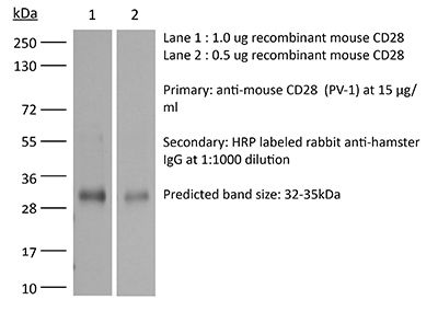
Specifications
| Isotype | Armenian Hamster IgG, κ |
|---|---|
| Recommended Isotype Control(s) | InVivoMAb polyclonal Armenian hamster IgG |
| Recommended Dilution Buffer | InVivoPure pH 7.0 Dilution Buffer |
| Conjugation | This product is unconjugated. Conjugation is available via our Antibody Conjugation Services. |
| Immunogen | C57BL/6 mouse T cell lymphoma EL-4 cells |
| Reported Applications | in vitro T cell stimulation/activation |
| Formulation |
PBS, pH 7.0 Contains no stabilizers or preservatives |
| Endotoxin |
≤1EU/mg (≤0.001EU/μg) Determined by LAL assay |
| Purity |
≥95% Determined by SDS-PAGE |
| Sterility | 0.2 µm filtration |
| Production | Purified from cell culture supernatant in an animal-free facility |
| Purification | Protein G |
| RRID | AB_1107628 |
| Molecular Weight | 150 kDa |
| Storage | The antibody solution should be stored at the stock concentration at 4°C. Do not freeze. |
| Need a Custom Formulation? | See All Antibody Customization Options |
Application References
-
Berger, H., et al (2013). "SOCS3 transactivation by PPARgamma prevents IL-17-driven cancer growth" Cancer Res 73(12): 3578-3590.
PubMed
Activation of the transcription factor PPARgamma by the n-3 fatty acid docosahexaenoic acid (DHA) is implicated in controlling proinflammatory cytokine secretion, but the intracellular signaling pathways engaged by PPARgamma are incompletely characterized. Here, we identify the adapter-encoding gene SOCS3 as a critical transcriptional target of PPARgamma. SOCS3 promoter binding and gene transactivation by PPARgamma was associated with a repression in differentiation of proinflammatory T-helper (TH)17 cells. Accordingly, TH17 cells induced in vitro displayed increased SOCS3 expression and diminished capacity to produce interleukin (IL)-17 following activation of PPARgamma by DHA. Furthermore, naive CD4 T cells derived from mice fed a DHA-enriched diet displayed less capability to differentiate into TH17 cells. In two different mouse models of cancer, DHA prevented tumor outgrowth and angiogenesis in an IL-17-dependent manner. Altogether, our results uncover a novel molecular pathway by which PPARgamma-induced SOCS3 expression prevents IL-17-mediated cancer growth.
-
Bertin, S., et al (2014). "The ion channel TRPV1 regulates the activation and proinflammatory properties of CD4(+) T cells" Nat Immunol 15(11): 1055-1063.
PubMed
TRPV1 is a Ca(2+)-permeable channel studied mostly as a pain receptor in sensory neurons. However, its role in other cell types is poorly understood. Here we found that TRPV1 was functionally expressed in CD4(+) T cells, where it acted as a non-store-operated Ca(2+) channel and contributed to T cell antigen receptor (TCR)-induced Ca(2+) influx, TCR signaling and T cell activation. In models of T cell-mediated colitis, TRPV1 promoted colitogenic T cell responses and intestinal inflammation. Furthermore, genetic and pharmacological inhibition of TRPV1 in human CD4(+) T cells recapitulated the phenotype of mouse Trpv1(-/-) CD4(+) T cells. Our findings suggest that inhibition of TRPV1 could represent a new therapeutic strategy for restraining proinflammatory T cell responses.
-
Huang, Y., et al (2015). "CRK proteins selectively regulate T cell migration into inflamed tissues" J Clin Invest 125(3): 1019-1032.
PubMed
Effector T cell migration into inflamed sites greatly exacerbates tissue destruction and disease severity in inflammatory diseases, including graft-versus-host disease (GVHD). T cell migration into such sites depends heavily on regulated adhesion and migration, but the signaling pathways that coordinate these functions downstream of chemokine receptors are largely unknown. Using conditional knockout mice, we found that T cells lacking the adaptor proteins CRK and CRK-like (CRKL) exhibit reduced integrin-dependent adhesion, chemotaxis, and diapedesis. Moreover, these two closely related proteins exhibited substantial functional redundancy, as ectopic expression of either protein rescued defects in T cells lacking both CRK and CRKL. We determined that CRK proteins coordinate with the RAP guanine nucleotide exchange factor C3G and the adhesion docking molecule CASL to activate the integrin regulatory GTPase RAP1. CRK proteins were required for effector T cell trafficking into sites of inflammation, but not for migration to lymphoid organs. In a murine bone marrow transplantation model, the differential migration of CRK/CRKL-deficient T cells resulted in efficient graft-versus-leukemia responses with minimal GVHD. Together, the results from our studies show that CRK family proteins selectively regulate T cell adhesion and migration at effector sites and suggest that these proteins have potential as therapeutic targets for preventing GVHD.
-
Vegran, F., et al (2014). "The transcription factor IRF1 dictates the IL-21-dependent anticancer functions of TH9 cells" Nat Immunol 15(8): 758-766.
PubMed
The TH9 subset of helper T cells was initially shown to contribute to the induction of autoimmune and allergic diseases, but subsequent evidence has suggested that these cells also exert antitumor activities. However, the molecular events that account for their effector properties are elusive. Here we found that the transcription factor IRF1 enhanced the effector function of TH9 cells and dictated their anticancer properties. Under TH9-skewing conditions, interleukin 1beta (IL-1beta) induced phosphorylation of the transcription factor STAT1 and subsequent expression of IRF1, which bound to the promoters of Il9 and Il21 and enhanced secretion of the cytokines IL-9 and IL-21 from TH9 cells. Furthermore, IL-1beta-induced TH9 cells exerted potent anticancer functions in an IRF1- and IL-21-dependent manner. Our findings thus identify IRF1 as a target for controlling the function of TH9 cells.
Product Citations
-
The ionotropic AMPA receptor contributes to autoimmunity via altered regulatory T cell differentiation.
In iScience on 16 January 2026 by Mitchell-Flack, M., Higgins, M., et al.
PubMed
The AMPA receptor (AMPAR) is an ionotropic glutamate receptor that is essential for neuronal communication, yet its role in the immune system remains poorly understood. Here, using a CD4Cre selective deletion mouse model, we provide the first functional characterization of AMPAR deficient T cells. We demonstrate that AMPAR deletion in T cells significantly protects against severe paralysis in an experimental autoimmune encephalomyelitis (EAE) model, and this protection is associated with increased regulatory T cell (Treg) presence within the spinal cord. In vitro studies reveal that the deletion of the AMPAR intrinsically promotes Treg generation. Mechanistically, AMPAR deletion increases IL2 signaling and activates the mTORC1 pathway, supporting Treg development and function. These novel findings suggest that a function of the AMPAR in CD4 T cells is to limit immune suppression by restricting Treg differentiation. Targeting AMPARs on T cells could offer a novel therapeutic approach for the treatment of autoimmune disease.
-
FOXP3 expression depends on cell-type-specific cis-regulatory elements and transcription factor circuitry.
In Immunity on 13 January 2026 by Umhoefer, J. M., Arce, M. M., et al.
PubMed
FOXP3 is a lineage-defining transcription factor (TF) for immune-suppressive regulatory T cells (Treg cells). Although mice exclusively express FOXP3 in Treg cells, stimulated conventional CD4+ T cells (Tconv cells) also transiently express FOXP3 in humans. Mechanisms governing these distinct expression patterns need elucidation. Here, we performed CRISPR screens tiling the FOXP3 locus and targeting TFs in human Treg and Tconv cells to identify cis-regulatory elements (CREs) and trans-regulators of FOXP3. Tconv cell FOXP3 expression depended on a subset of Treg cell CREs, as well as Tconv-cell-selective positive (NS+) and negative (NS-) CREs. Combinatorial silencing of Tconv cell CREs revealed their epistatic logic. These CREs are occupied and regulated by TFs that we identified as FOXP3 regulators. Finally, mutagenesis of murine NS- CRE revealed its essentiality for restricting FOXP3 expression to Treg cells. We map CRE and TF circuitry to reveal distinct cell- and species-specific regulation of FOXP3 expression.
-
Sesamin Induces MCL-1-Dependent Apoptosis in Activated T Cells and Ameliorates Experimental Atopic Dermatitis.
In Int J Biol Sci on 27 August 2025 by Park, H. S., Sung, W. J., et al.
PubMed
Sesamin, a natural lignan derived from Sesamum indicum, has been reported to possess anti-inflammatory and pro-apoptotic properties. However, its effect on T cell-mediated diseases and the underlying molecular mechanisms remain unclear. In this study, we demonstrate that sesamin selectively induces apoptosis in activated T cells through direct interaction with MCL-1, a critical anti-apoptotic protein of the Bcl-2 family. Sesamin suppressed IL-2 expression, CD69 upregulation, and proliferation in activated human and murine T cells. Molecular docking predicted strong binding of sesamin to the BH3-binding groove of MCL-1, which was validated by pull-down and co-immunoprecipitation assays. Sesamin inhibited MCL-1 phosphorylation at Ser64 and disrupted its heterodimerization with Bak, promoting caspase-3/8 cleavage and apoptotic death selectively in activated, but not resting, T cells. In a murine model of atopic dermatitis, oral administration of sesamin ameliorated pathological skin symptoms, reduced Th2/Th17 cytokine expression, serum IgE, mast cell infiltration, and lymph node hypertrophy. These effects correlated with suppressed MCL-1 activity and enhanced apoptosis in inflamed tissue. Our findings suggest that sesamin modulates immune responses via a novel MCL-1-dependent mechanism and represents a promising dietary-derived therapeutic strategy for T cell-driven chronic inflammatory diseases.
-
Interleukin-16 enhances anti-tumor immune responses by establishing a Th1 cell-macrophage crosstalk through reprogramming glutamine metabolism in mice.
In Nat Commun on 10 March 2025 by Wen, Z., Liu, T., et al.
PubMed
Overcoming immunosuppression in the tumor microenvironment (TME) is crucial for developing novel cancer immunotherapies. Here, we report that IL-16 administration enhances the polarization of T helper 1 (Th1) cells by inhibiting glutamine catabolism through the downregulation of glutaminase in CD4+ T cells and increases the production of Th1 effector cytokine IFN-γ, thus improving anti-tumor immune responses. Moreover, we find that establishing an IL-16-dependent, Th1-dominant TME relies on mast cell-produced histamine and results in the increased expression of the CXCR3 ligands in tumor-associated macrophages (TAM), thereby improving the therapeutic effectiveness of immune checkpoint blockade (ICB). Cancer patients exhibit impaired production of IL-16, which correlates with poorer prognosis. Additionally, low IL-16 production is associated with unresponsiveness to immunotherapy in cancer patients. Collectively, our findings provided new insights into the biological function of IL-16, emphasizing its potential clinical significance as a therapeutic approach to augment anti-tumor immunity and sensitize ICB-based cancer immunotherapy.