InVivoMAb anti-human CD3
Product Details
The OKT-3 monoclonal antibody reacts with human CD3ε, a 20 kDa transmembrane cell-surface protein that belongs to the immunoglobulin superfamily. CD3ε is one of five polypeptide chains that combine to form the TCR complex. CD3ε is expressed on T lymphocytes, NK-T cells, and to varying degrees on developing thymocytes. CD3 plays roles in TCR signaling, T lymphocyte activation, and antigen recognition. The OKT-3 antibody has immunosuppressive properties in vivo and has been shown to effectively treat renal, heart and liver allograft rejection.Specifications
Isotype | Mouse IgG2a,Ā Īŗ |
---|---|
Recommended Isotype Control(s) | InVivoMAb mouse IgG2a isotype control, unknown specificity |
Recommended Dilution Buffer | InVivoPure pH 7.0 Dilution Buffer |
Conjugation | This product is unconjugated. Conjugation is available via our Antibody Conjugation Services. |
Immunogen | Not available or unknown |
Reported Applications |
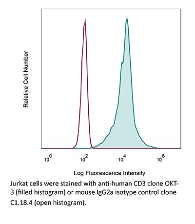
in vitro T cell stimulation/activation in vivo T cell depletion in humanized mice ex vivo T cell inhibition for xenografts Flow cytometry |
Formulation |
PBS, pH 7.0 Contains no stabilizers or preservatives |
Endotoxin |
<2EU/mg (<0.002EU/μg) Determined by LAL gel clotting assay |
Purity |
>95% Determined by SDS-PAGE |
Sterility | 0.2 µm filtration |
Production | Purified from cell culture supernatant in an animal-free facility |
Purification | Protein G |
RRID | AB_1107632 |
Molecular Weight | 150 kDa |
Storage | The antibody solution should be stored at the stock concentration at 4°C. Do not freeze. |
Additional Formats
Recommended Products
in vitro T cell stimulation/activation
Rochman, Y., et al. (2015). "Functional characterization of human T cell hyporesponsiveness induced by CTLA4-Ig" PLoS One 10(4): e0122198. PubMed
During activation, T cells integrate multiple signals from APCs and cytokine milieu. The blockade of these signals can have clinical benefits as exemplified by CTLA4-Ig, which blocks interaction of B7 co-stimulatory molecules on APCs with CD28 on T cells. Variants of CTLA4-Ig, abatacept and belatacept are FDA approved as immunosuppressive agents in arthritis and transplantation, yet murine studies suggested that CTLA4-Ig could be beneficial in a number of other diseases. However, detailed analysis of human CD4 cell hyporesponsivness induced by CTLA4-Ig has not been performed. Herein, we established a model to study the effect of CTLA4-Ig on the activation of human naive T cells in a human mixed lymphocytes system. Comparison of human CD4 cells activated in the presence or absence of CTLA4-Ig showed that co-stimulation blockade during TCR activation does not affect NFAT signaling but results in decreased activation of NF-kappaB and AP-1 transcription factors followed by a profound decrease in proliferation and cytokine production. The resulting T cells become hyporesponsive to secondary activation and, although capable of receiving TCR signals, fail to proliferate or produce cytokines, demonstrating properties of anergic cells. However, unlike some models of T cell anergy, these cells did not possess increased levels of the TCR signaling inhibitor CBLB. Rather, the CTLA4-Ig-induced hyporesponsiveness was associated with an elevated level of p27kip1 cyclin-dependent kinase inhibitor.
in vitro T cell stimulation/activation
Hill, E. V., et al. (2015). "Glycogen synthase kinase-3 controls IL-10 expression in CD4(+) effector T-cell subsets through epigenetic modification of the IL-10 promoter" Eur J Immunol 45(4): 1103-1115. PubMed
The serine/threonine kinase glycogen synthase kinase-3 (GSK3) plays an important role in balancing pro- and anti-inflammatory cytokines. We have examined the role of GSK3 in production of IL-10 by subsets of CD4(+) T helper cells. Treatment of naive murine CD4(+) T cells with GSK3 inhibitors did not affect their production of IL-10. However, treatment of Th1 and Th2 cells with GSK3 inhibitors dramatically increased production of IL-10. GSK3 inhibition also led to upregulation of IL-10 among Th1, Th2, and Th17 subsets isolated from human blood. The encephalitogenic potential of GSK3 inhibitor treated murine Th1 cells was significantly reduced in adoptive transfer experiments by an IL-10-dependent mechanism. Analysis of the murine IL-10 promoter in response to inhibition of GSK3 in Th1 cells showed modification to a transcriptionally active state indicated by changes in histone H3 acetylation and methylation. Additionally, GSK3 inhibition increased expression of the transcription factors c-Maf, Nfil3, and GATA3, correlating with the increase in IL-10. These findings are important in the context of autoimmune disease since they show that it is possible to reprogram disease-causing cells through GSK3 inhibition.
in vitro T cell stimulation/activation
Liu, H., et al. (2015). "The Immune Adaptor SLP-76 Binds to SUMO-RANGAP1 at Nuclear Pore Complex Filaments to Regulate Nuclear Import of Transcription Factors in T Cells" Mol Cell 59(5): 840-849. PubMed
While immune cell adaptors regulate proximal T cell signaling, direct regulation of the nuclear pore complex (NPC) has not been reported. NPC has cytoplasmic filaments composed of RanGAP1 and RanBP2 with the potential to interact with cytoplasmic mediators. Here, we show that the immune cell adaptor SLP-76 binds directly to SUMO-RanGAP1 of cytoplasmic fibrils of the NPC, and that this interaction is needed for optimal NFATc1 and NF-kappaB p65 nuclear entry in T cells. Transmission electron microscopy showed anti-SLP-76 cytoplasmic labeling of the majority of NPCs in anti-CD3 activated T cells. Further, SUMO-RanGAP1 bound to the N-terminal lysine 56 of SLP-76 where the interaction was needed for optimal RanGAP1-NPC localization and GAP exchange activity. While the SLP-76-RanGAP1 (K56E) mutant had no effect on proximal signaling, it impaired NF-ATc1 and p65/RelA nuclear entry and in vivo responses to OVA peptide. Overall, we have identified SLP-76 as a direct regulator of nuclear pore function in T cells.
in vitro T cell stimulation/activation
Sturner, K. H., et al. (2014). "A multiple sclerosis-associated variant of CBLB links genetic risk with type I IFN function" J Immunol 193(9): 4439-4447. PubMed
Multiple sclerosis (MS) is an autoimmune disease of the CNS, and autoreactive CD4(+) T cells are considered important for its pathogenesis. The etiology of MS involves a complex genetic trait and environmental triggers that include viral infections, particularly the EBV. Among the risk alleles that have repeatedly been identified by genome-wide association studies, three are located near the Casitas B-lineage lymphoma proto-oncogene b gene (CBLB). The CBLB protein (CBL-B) is a key regulator of peripheral immune tolerance by limiting T cell activation and expansion and hence T cell-mediated autoimmunity through its ubiquitin E3-ligase activity. In this study, we show that CBL-B expression is reduced in CD4(+) T cells from relapsing-remitting MS (RR-MS) patients during relapse. The MS risk-related single nucleotide polymorphism of CBLB rs12487066 is associated with diminished CBL-B expression levels and alters the effects of type I IFNs on human CD4(+) T cell proliferation. Mechanistically, the CBLB rs12487066 risk allele mediates increased binding of the transcription factor C/EBPbeta and reduced CBL-B expression in human CD4(+) T cells. Our data suggest a role of the CBLB rs12487066 variant in the interactions of a genetic risk factor and IFN function during viral infections in MS.
in vitro T cell stimulation/activation, Flow Cytometry
Willing, A., et al. (2014). "CD8(+) MAIT cells infiltrate into the CNS and alterations in their blood frequencies correlate with IL-18 serum levels in multiple sclerosis" Eur J Immunol 44(10): 3119-3128. PubMed
Recent findings indicate a pathogenic involvement of IL-17-producing CD8(+) T cells in multiple sclerosis (MS). IL-17 production has been attributed to a subset of CD8(+) T cells that belong to the mucosal-associated invariant T (MAIT) cell population. Here, we report a reduction of CD8(+) MAIT cells in the blood of MS patients compared with healthy individuals, which significantly correlated with IL-18 serum levels in MS patients. In vitro stimulation of peripheral blood mononuclear cells from healthy individuals and MS patients with IL-18 specifically activated CD8(+) MAIT cells. Moreover, IL-18 together with T-cell receptor stimulation induced, specifically on CD8(+) MAIT cells, an upregulation of the integrin very late antigen-4 that is essential for the infiltration of CD8(+) T cells into the CNS. Notably, we were able to identify CD8(+) MAIT cells in MS brain lesions by immunohistochemistry while they were almost absent in the cerebrospinal fluid (CSF). In summary, our findings indicate that an IL-18-driven activation of CD8(+) MAIT cells contributes to their CNS infiltration in MS, in turn leading to reduced CD8(+) MAIT-cell frequencies in the blood. Therefore, CD8(+) MAIT cells seem to play a role in the innate arm of immunopathology in MS.
in vitro T cell stimulation/activation
Esposito, L., et al. (2014). "Investigation of soluble and transmembrane CTLA-4 isoforms in serum and microvesicles" J Immunol 193(2): 889-900. PubMed
Expression of the CTLA-4 gene is absolutely required for immune homeostasis, but aspects of its molecular nature remain undefined. In particular, the characterization of the soluble CTLA-4 (sCTLA-4) protein isoform generated by an alternatively spliced mRNA of CTLA4 lacking transmembrane-encoding exon 3 has been hindered by the difficulty in distinguishing it from the transmembrane isoform of CTLA-4, Tm-CTLA-4. In the current study, sCTLA-4 has been analyzed using novel mAbs and polyclonal Abs specific for its unique C-terminal amino acid sequence. We demonstrate that the sCTLA-4 protein is secreted at low levels following the activation of primary human CD4(+) T cells and is increased only rarely in the serum of autoimmune patients. Unexpectedly, during our studies aimed to define the kinetics of sCTLA-4 produced by activated human CD4(+) T cells, we discovered that Tm-CTLA-4 is associated with microvesicles produced by the activated cells. The functional roles of sCTLA-4 and microvesicle-associated Tm-CTLA-4 warrant further investigation, especially as they relate to the multiple mechanisms of action described for the more commonly studied cell-associated Tm-CTLA-4.
in vivo T cell depletion in humanized mice, ex vivo T cell inhibtion for xenografts
Wunderlich, M., et al. (2014). "OKT3 prevents xenogeneic GVHD and allows reliable xenograft initiation from unfractionated human hematopoietic tissues" Blood 123(24): e134-144. PubMed
Immunodeficient mice are now readily engrafted with human hematopoietic cells. However, these mice are susceptible to graft-versus-host disease (GVHD) induced by the engraftment and rapid expansion of coinjected human T cells. Therefore, highly purified sample populations must be used, adding significant time, expense, and effort. Here, we have explored in vivo and in vitro methods utilizing anti-T-cell antibodies to circumvent this problem. Intraperitoneal injection of the antibody within 48 hours prevented GVHD. Alternatively, short-term in vitro incubation of cells with antibody immediately before transplant was equally effective. Although in vitro antithymocyte globulin treatment resulted in a dramatic loss of SCID-repopulating cells (SRCs), treatment with OKT3 or UCHT1 abrogated GVHD risk and preserved engraftment potential. Leukemia samples that presented with substantial human T-cell contamination were effectively rescued from GVHD. In addition, OKT3 treatment of unfractionated cord blood resulted in robust engraftment of primary and secondary mice that was indistinguishable from grafts obtained using purified CD34(+) cells. Limiting dilution analysis of unfractionated blood demonstrated a SRC frequency of 1 in 300 to 500 CD34(+) cells, similar to that of purified hematopoietic stem and progenitor cells. This protocol streamlines xenograft studies while significantly reducing the cost and time of the procedure.
in vitro T cell stimulation/activation
Lines, J. L., et al. (2014). "VISTA is an immune checkpoint molecule for human T cells" Cancer Res 74(7): 1924-1932. PubMed
V-domain Ig suppressor of T cell activation (VISTA) is a potent negative regulator of T-cell function that is expressed on hematopoietic cells. VISTA levels are heightened within the tumor microenvironment, in which its blockade can enhance antitumor immune responses in mice. In humans, blockade of the related programmed cell death 1 (PD-1) pathway has shown great potential in clinical immunotherapy trials. Here, we report the structure of human VISTA and examine its function in lymphocyte negative regulation in cancer. VISTA is expressed predominantly within the hematopoietic compartment with highest expression within the myeloid lineage. VISTA-Ig suppressed proliferation of T cells but not B cells and blunted the production of T-cell cytokines and activation markers. Our results establish VISTA as a negative checkpoint regulator that suppresses T-cell activation, induces Foxp3 expression, and is highly expressed within the tumor microenvironment. By analogy to PD-1 and PD-L1 blockade, VISTA blockade may offer an immunotherapeutic strategy for human cancer.


FcRn-silencing of IL-12Fc prevents toxicity of local IL-12 therapy and prolongs survival in experimental glioblastoma.
In Nature Communications on 22 May 2025 by Beffinger, M. M., Schellhammer, L., et al.
Glioblastoma remains a challenging indication for immunotherapy: the blood-brain barrier hampers accessibility for systemic treatments and the immunosuppressive microenvironment impedes immune attack. Intratumoral therapy with the proinflammatory cytokine interleukin-12 (IL-12) can revert immunosuppression but leakage into the circulation causes treatment-limiting toxicity. Here we engineer an IL-12Fc fusion cytokine with reduced binding to the neonatal Fc receptor FcRn. FcRn-silenced IL-12Fc avoids FcRn-mediated brain export, thus exhibits prolonged brain retention and reduced blood levels, which prevents toxicity. In murine glioblastoma, FcRn-silenced IL-12Fc induces more durable responses with negligible systemic cytokine exposure and boosts the efficacy of radio- and chemotherapy. It triggers anti-tumor responses independently of peripheral T cell influx or lymphopenia and leads to inflammatory polarization of the tumor microenvironment in patient-derived glioblastoma explants. FcRn-silencing of IL-12Fc may unlock the full potential of IL-12 for brain cancer therapy and could be further applied to containing the activity of other therapeutics targeting neurological diseases. Ā© 2025. The Author(s).
Unveiling cellular communications through rapid pan-membrane-protein labeling.
In Nature Communications on 15 April 2025 by Gunasekara, H., Cheng, Y. S., et al.
Dynamic protein distribution within and across the plasma membrane is pivotal in regulating cell communication. However, rapid, high-density labeling methods for multiplexed live imaging across diverse cell types remain scarce. Here, we demonstrate N-hydroxysuccinimide (NHS)-ester-based amine crosslinking of fluorescent dyes to uniformly label live mammalian cell surface proteins. Using model cell systems, we capture previously elusive membrane topology and cell-cell interactions. Live imaging shows transient membrane protein accumulation at cell-cell contacts and bidirectional migration patterns guided by membrane fibers in DC2.4 dendritic cells. Multiplexed superresolution imaging reveals the biogenesis of membrane tunneling nanotubes that facilitate intercellular transfer in DC2.4 cells, and caveolinĀ 1-dependent endocytosis of insulin receptors in HEK293T cells. 3D superresolution imaging reveals membrane topology remodeling in response to stimulation, generation of microvesicles, and phagocytic activities in Jurkat T cells. Furthermore, NHS-labeling remains stable in vivo, enabling visualization of intercellular transfer among splenocytes using a TĀ cell lymphoma mouse model. Ā© 2025. The Author(s).
- Immunology and Microbiology,
Polysialic acid is upregulated on activated immune cells and negatively regulates anticancer immune activity.
In Frontiers in Oncology on 4 April 2025 by Drummond-Guy, O., Daly, J., et al.
Suppression of anticancer immune function is a key driver of tumorigenesis. Identifying molecular pathways that inhibit anticancer immunity is critical for developing novel immunotherapeutics. One such molecule that has recently been identified is the carbohydrate polysialic acid (polySia), whose expression is dramatically upregulated on both cancer cells and immune cells in breast cancer patient tissues. The role of polySia in the anticancer immune response, however, remains incompletely understood. In this study, we profile polySia expression on both healthy primary immune cells and on infiltrating immune cells in the tumour microenvironment (TME). These studies reveal polySia expression on multiple immune cell subsets in patient breast tumors. We find that stimulation of primary T-cells and macrophages in vitro induces a significant upregulation of polySia expression. We subsequently show that polySia is appended to a range of different carrier proteins within these immune cells. Finally, we find that selective removal of polySia can significantly potentiate killing of breast cancer cells by innate immune cells. These studies implicate polySia as a significant negative regulator of anticancer immunity. Copyright Ā© 2025 Drummond-Guy, Daly, Wu, Stewart, Milne, Duff, Nelson, Williams and Wisnovsky.
- Immunology and Microbiology
Preclinical characterization of MTX-101: a novel bispecific CD8 Treg modulator that restores CD8 Treg functions to suppress pathogenic T cells in autoimmune diseases.
In Frontiers in Immunology on 19 November 2024 by Gardell, J. L., Maurer, M. E., et al.
Regulatory CD8 T cells (CD8 Treg) are responsible for the selective killing of self-reactive and pathogenic CD4 T cells. In autoimmune disease, CD8 Treg may accumulate in the peripheral blood but fail to control the expansion of pathogenic CD4 T cells that subsequently cause tissue destruction. This CD8 Treg dysfunction is due in part to the expression of inhibitory killer immunoglobulin-like receptors (KIR; KIR2DL isoforms [KIR2DL1, KIR2DL2, and KIR2DL3]); these molecules serve as autoimmune checkpoints and limit CD8 Treg activation. Here we describe the pre-clinical characterization of MTX-101, a bispecific antibody targeting inhibitory KIR and CD8. Using human peripheral blood mononuculear cells (PBMC) derived from healthy donors and autoimmune patients, humanized mouse models, and human derived tissue organoids, we evaluated the molecular mechanisms and functional effects of MTX-101. By binding to KIR, MTX-101 inhibited KIR signaling that can restore CD8 Treg ability to eliminate pathogenic CD4 T cells. MTX-101 bound and activated CD8 Treg in human peripheral blood mononuclear cells (PBMC), resulting in increased CD8 Treg cytolytic capacity, activation, and prevalence. Enhancing CD8 Treg function with MTX-101 reduced pathogenic CD4 T cell expansion and inflammation, without increasing pro-inflammatory cytokines or activating immune cells that express either target alone. MTX-101 reduced antigen induced epithelial cell death in disease affected tissues, including in tissue biopsies from individuals with autoimmune disease (i.e., celiac disease, Crohn's disease). The effects of MTX-101 were specific to autoreactive CD4 T cells and did not suppress responses to viral and bacterial antigens. In a human PBMC engrafted Graft versus Host Disease (GvHD) mouse model of acute inflammation, MTX-101 bound CD8 Treg and delayed onset of disease. MTX-101 induced dose dependent binding, increased prevalence and cytolytic capacity of CD8 Treg, as well as increased CD4 T cell death. MTX-101 selectively bound CD8 Treg without unwanted immune cell activation or increase of pro-inflammatory serum cytokines and exhibited an antibody-like half-life in pharmacokinetic and exploratory tolerability studies performed using IL-15 transgenic humanized mice with engrafted human lymphocytes, including CD8 Treg at physiologic ratios. Collectively, these data support the development of MTX-101 for the treatment of autoimmune diseases. Copyright Ā© 2024 Gardell, Maurer, Childs, Pham, Meengs, Julien, Tan, Boster, Quach, Therriault, Hermansky, Patton, Bowser, Chen, Morgan, Gilbertson, Bogatzki, Encarnacion, McMahan, Crane and Swiderek.
- Immunology and Microbiology
Progranulin protects against Clostridioides difficile infection by enhancing IL-22 production.
In Gut Microbes on 1 October 2024 by Huang, J., Liu, B., et al.
Enhanced mortality, relapse rates, and increased prevalence of Clostridioides difficile infection (CDI) emphasize the need for better therapies and management approaches. Modulating host immune response to ameliorate CDI-associated immunopathology may provide new advantages to currently inadequate antibiotic therapies. Here, we identified progranulin (PGRN) as an important immune target upregulated in response to CDI. PGRN-deficient mice displayed dramatically higher mortality and aggravated epithelial barrier disruption compared with wild type (WT) mice after CDI despite equivalent levels of bacterial burden or toxin in the large intestine. Mechanistically, PGRN protection was mediated by IL-22 production from CD4+ T helper cells, as demonstrated by a decrease in colonic IL-22-producing CD4+ T helper cells in the intestine of PGRN-deficient mice upon CDI and a boost of IL-22-producing CD4+ T helper cells activated by PGRN ex vivo. Clinical evidence suggests that CDI patients had significantly higher serum levels of PGRN compared with healthy controls, which was significantly and positively correlated with IL-22. Our findings thus indicate a critical role for PGRN-promoted CD4+ T cell IL-22 production in shaping gut immunity and reestablishing the intestinal barrier during CDI. As an alternative to pathogen-targeted therapy, this study may provide a new host-directed therapeutic strategy to attenuate severe, refractory CDI.
- Immunology and Microbiology
CD38 in SLE CD4 T cells promotes Ca2+ flux and suppresses interleukin-2 production by enhancing the expression of GM2 on the surface membrane.
In Nature Communications on 27 September 2024 by Katsuyama, E., Humbel, M., et al.
CD38 has emerged as a potential therapeutic target for patients with systemic lupus erythematosus (SLE) but it is not known whether CD38 alters CD4+ T cell function. Using primary human T cells and CD38-sufficient and CD38-deficient Jurkat T cells, we demonstrate that CD38 shifts the T cell lipid profile of gangliosides from GM3 to GM2 by upregulating B4GALNT1 in a Sirtuin 1-dependent manner. Enhanced expression of GM2 causes ER stress by enhancing Ca2+ flux through the PLCγ1-IP3 pathway. Interestingly, correction of the calcium overload by an IP3 receptor inhibitor, but not by a store-operated calcium entry (SOCE) inhibitor, improves IL-2 production by CD4+ T cells in SLE. This study demonstrates that CD38 affects calcium homeostasis in CD4+ T cells by controlling cell membrane lipid composition that results in suppressed IL-2 production. CD38 inhibition with biologics or small drugs should be expected to benefit patients with SLE. © 2024. The Author(s).
- Biochemistry and Molecular biology,
- Cancer Research,
- Cell Biology
Spermidine metabolism regulates leukemia stem and progenitor cell function through KAT7 expression in patient-derived mouse models.
In Science Translational Medicine on 25 September 2024 by Rondeau, V., Berman, J., et al.
Acute myeloid leukemia (AML) is a devastating disease initiated and maintained by a rare subset of cells called leukemia stem cells (LSCs). LSCs are responsible for driving disease relapse, making the development of new therapeutic strategies to target LSCs urgently needed. The use of mass spectrometry-based metabolomics profiling has enabled the discovery of unique and targetable metabolic properties in LSCs. However, we do not have a comprehensive understanding of metabolite differences between LSCs and their normal counterparts, hematopoietic stem and progenitor cells (HSPCs). In this study, we used an unbiased mass spectrometry-based metabolomics analysis to define differences in metabolites between primary human LSCs and HSPCs, which revealed that LSCs have a distinct metabolome. Spermidine was the most enriched metabolite in LSCs compared with HSPCs. Pharmacological reduction of spermidine concentrations decreased LSC function but spared normal HSPCs. Polyamine depletion also decreased leukemic burden in patient-derived xenografts. Mechanistically, spermidine depletion induced LSC myeloid differentiation by decreasing eIF5A-dependent protein synthesis, resulting in reduced expression of a select subset of proteins. KAT7, a histone acetyltransferase, was one of the top candidates identified to be down-regulated by spermidine depletion. Overexpression of KAT7 partially rescued polyamine depletion-induced decreased colony-forming ability, demonstrating that loss of KAT7 is an essential part of the mechanism by which spermidine depletion targets AML clonogenic potential. Together, we identified and mechanistically dissected a metabolic vulnerability of LSCs that has the potential to be rapidly translated into clinical trials to improve outcomes for patients with AML.
- Cancer Research,
- Immunology and Microbiology
Soluble Tim-3 serves as a tumor prognostic marker and therapeutic target for CD8+ T cell exhaustion and anti-PD-1 resistance.
In Cell Reports Medicine on 20 August 2024 by Chen, C., Zhao, F., et al.
Resistance to PD-1 blockade in onco-immunotherapy greatly limits its clinical application. TĀ cell immunoglobulin and mucin domain containing-3 (Tim-3), a promising immune checkpoint target, is cleaved by ADAM10/17 to produce its soluble form (sTim-3) in humans, potentially becoming involved in anti-PD-1 resistance. Herein, serum sTim-3 upregulation was observed in non-small cell lung cancer (NSCLC) and various digestive tumors. Notably, serum sTim-3 is further upregulated in non-responding patients undergoing anti-PD-1 therapy for NSCLC and anti-PD-1-resistant cholangiocarcinoma patients. Furthermore, sTim-3 overexpression facilitates tumor progression and confers anti-PD-1 resistance in multiple tumor mouse models. Mechanistically, sTim-3 induces terminal TĀ cell exhaustion and attenuates CD8+ TĀ cell response to PD-1 blockade through carcinoembryonic antigen-related cell adhesion molecule 1 (CEACAM-1). Moreover, the ADAM10 inhibitor GI254023X, which blocks sTim-3 production, reduces tumor progression in Tim-3 humanized mice and reverses anti-PD-1 resistance in human tumor-infiltrating lymphocytes (TILs). Overall, human sTim-3 holds great predictive and therapeutic potential in onco-immunotherapy. Copyright Ā© 2024 The Authors. Published by Elsevier Inc. All rights reserved.
- Cancer Research
Improvement of Tumor Neoantigen Detection by High-Field Asymmetric Waveform Ion Mobility Mass Spectrometry.
In Cancer Immunology Research on 1 August 2024 by Meng, W., Takeuchi, Y., et al.
Cancer neoantigens have been shown to elicit cancer-specific T-cell responses and have garnered much attention for their roles in both spontaneous and therapeutically induced antitumor responses. Mass spectrometry (MS) profiling of tumor immunopeptidomes has been used, in part, to identify MHC-bound mutant neoantigen ligands. However, under standard conditions, MS-based detection of such rare but clinically relevant neoantigens is relatively insensitive, requiring 300 million cells or more. Here, to quantitatively define the minimum detectable amounts of therapeutically relevant MHC-I and MHC-II neoantigen peptides, we analyzed different dilutions of immunopeptidomes isolated from the well-characterized T3 mouse methylcholanthrene (MCA)-induced cell line by MS. Using either data-dependent acquisition or parallel reaction monitoring (PRM), we established the minimum amount of material required to detect the major T3 neoantigens in the presence or absence of high field asymmetric waveform ion mobility spectrometry (FAIMS). This analysis yielded a 14-fold enhancement of sensitivity in detecting the major T3 MHC-I neoantigen (mLama4) with FAIMS-PRM compared with PRM without FAIMS, allowing ex vivo detection of this neoantigen from an individual 100 mg T3 tumor. These findings were then extended to two other independent MCA-sarcoma lines (1956 and F244). This study demonstrates that FAIMS substantially increases the sensitivity of MS-based characterization of validated neoantigens from tumors. ©2024 American Association for Cancer Research.
- COVID-19,
- Immunology and Microbiology
Resolution of SARS-CoV-2 infection in human lung tissues is driven by extravascular CD163+ monocytes
Preprint on BioRxiv : the Preprint Server for Biology on 8 March 2024 by Kenney, D., OāConnell, A. K., et al.
ABSTRACT The lung-resident immune mechanisms driving resolution of SARS-CoV-2 infection in humans remain elusive. Using mice co-engrafted with a genetically matched human immune system and fetal lung xenograft (fLX), we mapped the immunological events defining resolution of SARS-CoV-2 infection in human lung tissues. Viral infection is rapidly cleared from fLX following a peak of viral replication. Acute replication results in the emergence of cell subsets enriched in viral RNA, including extravascular inflammatory monocytes (iMO) and macrophage-like T-cells, which dissipate upon infection resolution. iMO display robust antiviral responses, are transcriptomically unique among myeloid lineages, and their emergence associates with the recruitment of circulating CD4+ monocytes. Consistently, mice depleted for human CD4+ cells but not CD3+ T-cells failed to robustly clear infectious viruses and displayed signatures of chronic infection. Our findings uncover the transient differentiation of extravascular iMO from CD4+ monocytes as a major hallmark of SARS-CoV-2 infection resolution and open avenues for unravelling viral and host adaptations defining persistently active SARS-CoV-2 infection.
- Immunology and Microbiology
Gut microbiota-derived LCA mediates the protective effect of PEDV infection in piglets.
In Microbiome on 5 February 2024 by Xing, J. H., Niu, T. M., et al.
The gut microbiota is a critical factor in the regulation of host health, but the relationship between the differential resistance of hosts to pathogens and the interaction of gut microbes is not yet clear. Herein, we investigated the potential correlation between the gut microbiota of piglets and their disease resistance using single-cell transcriptomics, 16S amplicon sequencing, metagenomics, and untargeted metabolomics. Porcine epidemic diarrhea virus (PEDV) infection leads to significant changes in the gut microbiota of piglets. Notably, Landrace pigs lose their resistance quickly after being infected with PEDV, but transplanting the fecal microbiota of Min pigs to Landrace pigs alleviated the infection status. Macrogenomic and animal protection models identified Lactobacillus reuteri and Lactobacillus amylovorus in the gut microbiota as playing an anti-infective role. Moreover, metabolomic screening of the secondary bile acids' deoxycholic acid (DCA) and lithocholic acid (LCA) correlated significantly with Lactobacillus reuteri and Lactobacillus amylovorus, but only LCA exerted a protective function in the animal model. In addition, LCA supplementation altered the distribution of intestinal T-cell populations and resulted in significantly enriched CD8+ CTLs, and in vivo and in vitro experiments showed that LCA increased SLA-I expression in porcine intestinal epithelial cells via FXR receptors, thereby recruiting CD8+ CTLs to exert antiviral effects. Overall, our findings indicate that the diversity of gut microbiota influences the development of the disease, and manipulating Lactobacillus reuteri and Lactobacillus amylovorus, as well as LCA, represents a promising strategy to improve PEDV infection in piglets. Video Abstract. Ā© 2024. The Author(s).
IL-21-armored B7H3 CAR-iNKT cells exert potent antitumor effects.
In IScience on 19 January 2024 by Liu, Y., Dang, Y., et al.
PubMed
CD1d-restricted invariant NKT (iNKT) cells play a critical role in tumor immunity. However, the scarcity and limited persistence restricts their development and clinical application. Here, we demonstrated that iNKT cells could be efficiently expanded using modified cytokines combination from peripheral blood mononuclear cells. Introduction of IL-21 significantly increased the frequency of CD62L-positive memory-like iNKT cells. iNKT cells armoring with B7H3-targeting second generation CAR and IL-21 showed potent tumor cell killing activity. Moreover, co-expression of IL-21 promoted the activation of Stat3 signaling and reduced the expression of exhaustion markers in CAR-iNKT cells inĀ vitro. Most importantly, IL-21-arming significantly prolonged B7H3 CAR-iNKT cell proliferation and survival inĀ vivo, thus improving their therapeutic efficacy in mouse renal cancer xerograph models without observed cytokine-related adverse events. In summary, these results suggest that B7H3 CAR-iNKT armored with IL-21 is a promising therapeutic strategy for cancer treatment. Ā© 2023 The Authors.
- Cancer Research
Facile generation of biepitopic antibodies with intrinsic agonism for activating receptors in the tumor necrosis factor superfamily
Preprint on BioRxiv : the Preprint Server for Biology on 12 December 2023 by Jhajj, H. S., Schardt, J. S., et al.
PubMed
Summary Agonist antibodies that activate cellular receptors are being pursued for therapeutic applications ranging from neurodegenerative diseases to cancer. For the tumor necrosis factor (TNF) receptor superfamily, higher-order clustering of three or more receptors is key to their potent activation. This can be achieved using antibodies that recognize two unique epitopes on the same receptor and mediate receptor superclustering. However, identifying compatible pairs of antibodies to generate biepitopic antibodies (also known as biparatopic antibodies) for activating TNF receptors typically requires animal immunization and is a laborious and unpredictable process. Here, we report a simple method for systematically identifying biepitopic antibodies that potently activate TNF receptors without the need for additional animal immunization. Our approach uses off-the-shelf, receptor-specific IgG antibodies, which lack intrinsic (Fc-gamma receptor-independent) agonist activity, to first block their corresponding epitopes. Next, we perform selections for single-chain antibodies from human nonimmune libraries that bind accessible epitopes on the same ectodomains using yeast surface display and fluorescence-activated cell sorting. The selected single-chain antibodies are finally fused to the light chains of IgGs to generate human tetravalent antibodies that engage two different receptor epitopes and mediate potent receptor activation. We highlight the broad utility of this approach by converting several existing clinical-stage antibodies against TNF receptors, including ivuxolimab and pogalizumab against OX40 and utomilumab against CD137, into biepitopic antibodies with highly potent agonist activity. We expect that this widely accessible methodology can be used to systematically generate biepitopic antibodies for activating other receptors in the TNF receptor superfamily and many other receptors whose activation is dependent on strong receptor clustering.
- Homo sapiens (Human),
- Immunology and Microbiology
Precise surface functionalization of PLGA particles for human T cell modulation.
In Nature Protocols on 1 November 2023 by Hadley, P., Chen, Y., et al.
PubMed
The biofunctionalization of synthetic materials has extensive utility for biomedical applications, but approaches to bioconjugation typically show insufficient efficiency and controllability. We recently developed an approach by building synthetic DNA scaffolds on biomaterial surfaces that enables the precise control of cargo density and ratio, thus improving the assembly and organization of functional cargos. We used this approach to show that the modulation and phenotypic adaptation of immune cells can be regulated using our precisely functionalized biomaterials. Here, we describe the three key procedures, including the fabrication of polymeric particles engrafted with short DNA scaffolds, the attachment of functional cargos with complementary DNA strands, and the surface assembly control and quantification. We also explain the critical checkpoints needed to ensure the overall quality and expected characteristics of the biological product. We provide additional experimental design considerations for modifying the approach by varying the material composition, size or cargo types. As an example, we cover the use of the protocol for human primary T cell activation and for the identification of parameters that affect ex vivo T cell manufacturing. The protocol requires users with diverse expertise ranging from synthetic materials to bioconjugation chemistry to immunology. The fabrication procedures and validation assays to design high-fidelity DNA-scaffolded biomaterials typically require 8 d. Ā© 2023. Springer Nature Limited.
- In Vitro,
- Homo sapiens (Human),
- Immunology and Microbiology,
- Pathology
Loss of CD4+ TĀ cell-intrinsic arginase 1 accelerates Th1 response kinetics and reduces lung pathology during influenza infection.
In Immunity on 12 September 2023 by West, E. E., Merle, N. S., et al.
PubMed
Arginase 1 (Arg1), the enzyme catalyzing the conversion of arginine to ornithine, is a hallmark of IL-10-producing immunoregulatory M2 macrophages. However, its expression in TĀ cells is disputed. Here, we demonstrate that induction of Arg1 expression is a key feature of lung CD4+ TĀ cells during mouse inĀ vivo influenza infection. Conditional ablation of Arg1 in CD4+ TĀ cells accelerated both virus-specific T helper 1 (Th1) effector responses and its resolution, resulting in efficient viral clearance and reduced lung pathology. Using unbiased transcriptomics and metabolomics, we found that Arg1-deficiency was distinct from Arg2-deficiency and caused altered glutamine metabolism. Rebalancing this perturbed glutamine flux normalized the cellular Th1 response. CD4+ TĀ cells from rare ARG1-deficient patients or CRISPR-Cas9-mediated ARG1-deletion in healthy donor cells phenocopied the murine cellular phenotype. Collectively, CD4+ TĀ cell-intrinsic Arg1 functions as an unexpected rheostat regulating the kinetics of the mammalian Th1 lifecycle with implications for Th1-associated tissue pathologies. Published by Elsevier Inc.
- Cell Biology,
- Immunology and Microbiology
LC3B conjugation machinery promotes autophagy-independent HIV-1 entry in CD4+ T lymphocytes
Preprint on BioRxiv : the Preprint Server for Biology on 11 July 2023 by Pradel, B., Deffieu, M. S., et al.
PubMed
HIV-1 entry into CD4+ T lymphocytes relies on the viral and cellular membranesā fusion, leading to viral capsid delivery in the cytoplasm of target cells. The conjugation of ATG8/LC3B protein, process referred to as ATG8ylation and mainly studied in the context of autophagy, occurs transiently in the early stages of the HIV-1 replication cycle in CD4+ T lymphocytes. Despite numerous studies investigating the interplays of HIV-1 with autophagy machinery, the impact of ATG8ylation in the early stages of HIV-1 infection remains unknown. Here we found that HIV-1 exposure leads to the rapid enrichment of LC3B towards the target cell plasma membrane, in close proximity with the incoming viral particles. Furthermore, we demonstrated that ATG8ylation is a key event that facilitates HIV-1 fusion with target CD4+ T cells. Interestingly, this effect is independent of the canonical autophagy pathway as ATG13 silencing does not prevent HIV-1 entry. Together, our results provide an unconventional role of LC3B conjugation subverted by HIV-1 to achieve a critical early step of its replication cycle. Teaser HIV-1 induces LC3B enrichment towards its target cell entry site and uses the conjugation of this protein to favor its entry step.
- FC/FACS,
- Homo sapiens (Human),
- Cancer Research,
- Immunology and Microbiology
STAT4, a potential predictor of prognosis, promotes CD8 Tācell infiltration in ovarian serous carcinoma by inducing CCL5 secretion.
In Oncology Reports on 1 July 2023 by Wang, W., Liu, S., et al.
PubMed
Ovarian serous carcinoma (OC) is a common cause of mortality among gynecological malignancies. Although tumorāinfiltrating CD8 T cells are associated with a favorable prognosis of OC, the underlying mechanisms are not clearly understood. The present study identified the key genes and potential molecular mechanisms associated with CD8 Tācell infiltration in OC. The score of CD8 T cells in The Cancer Genome Atlas dataset (376 samples from patients with OC) was estimated using the quanTIseq and MCPācounter algorithms. Thereafter, a proteināprotein interaction network of differentially expressed genes was constructed and the hub genes were identified using cytoHubba in Cytoscape. The results revealed that signal transducer and activator of transcription 4 (STAT4) was strongly correlated with CD8 Tācell infiltration in OC. Furthermore, the prognostic value of STAT4 in OC was verified by KaplanāMeier curve, and univariate and multivariate analyses. The biological functions of STAT4 were determined by Gene Ontology and Kyoto Encyclopedia of Genes and Genomes pathway analyses, which revealed that STAT4 is closely related to cytokines in OC. Moreover, Spearman correlation analysis suggested that STAT4 was most positively correlated with CC chemokine ligand 5 (CCL5). CCL5 was revealed to be critical for orchestrating Tācell infiltration in tumors. Moreover, immunohistochemistry and reverse transcriptionāquantitative PCR showed that STAT4, CCL5 and CD8A (a marker for CD8 T cells) were closely related in OC. Moreover, inĀ vitro analysis revealed that STAT4 knockdown led to a decrease in CCL5 expression and CD8 Tācell migration. Taken together, the present study suggested that STAT4 may regulate CD8 Tācell infiltration in OC tissues by inducing CCL5 secretion. Furthermore, STAT4 may be considered a promising prognostic biomarker for OC.
- Biochemistry and Molecular biology,
- Immunology and Microbiology
Surfaces for Study of Receptor Dynamics on T Cells.
In Methods in Molecular Biology (Clifton, N.J.) on 28 April 2023 by McColl, J. & Klenerman, D.
PubMed
Microscopy developments since the turn of the decade have seen an abundance of imaging modalities emerge that are revolutionizing the way we image the immune system. We are now able to image faster and utilize techniques that can image individual receptors, in real time, on live T cells. Total internal reflection fluorescence (TIRF) microscopy is one such technique, although it has one problem. The imaging must be carried out close to the glass interface. There are clearly issues with live cell imaging at glass surfaces as these are not biologically relevant. Manipulating the surface is key for maintaining biologically relevant imaging conditions. Here, we describe a simple approach to generate substrates for cell attachment and imaging of receptor dynamics and outline a guide for imaging and tracking T cell, surface receptors using TIRF microscopy. Ā© 2023. The Author(s), under exclusive license to Springer Science+Business Media, LLC, part of Springer Nature.
Selective inhibitors of JAK1 targeting an isoform-restricted allosteric cysteine.
In Nature Chemical Biology on 1 December 2022 by Kavanagh, M. E., Horning, B. D., et al.
The Janus tyrosine kinase (JAK) family of non-receptor tyrosine kinases includes four isoforms (JAK1, JAK2, JAK3, and TYK2) and is responsible for signal transduction downstream of diverse cytokine receptors. JAK inhibitors have emerged as important therapies for immun(onc)ological disorders, but their use is limited by undesirable side effects presumed to arise from poor isoform selectivity, a common challenge for inhibitors targeting the ATP-binding pocket of kinases. Here we describe the chemical proteomic discovery of a druggable allosteric cysteine present in the non-catalytic pseudokinase domain of JAK1 (C817) and TYK2 (C838), but absent from JAK2 or JAK3. Electrophilic compounds selectively engaging this site block JAK1-dependent trans-phosphorylation and cytokine signaling, while appearing to act largely as 'silent' ligands for TYK2. Importantly, the allosteric JAK1 inhibitors do not impair JAK2-dependent cytokine signaling and are inactive in cells expressing a C817A JAK1 mutant. Our findings thus reveal an allosteric approach for inhibiting JAK1 with unprecedented isoform selectivity. Ā© 2022. The Author(s), under exclusive licence to Springer Nature America, Inc.
- Cancer Research,
- Cell Biology,
- Stem Cells and Developmental Biology
Autophagy inhibition impairs leukemia stem cell function in FLT3-ITD AML but has antagonistic interactions with tyrosine kinase inhibition.
In Leukemia on 1 November 2022 by Qiu, S., Kumar, H., et al.
PubMed
The FLT3-ITD mutation is associated with poor prognosis in acute myeloid leukemia (AML). FLT3 tyrosine kinase inhibitors (TKIs) demonstrate clinical efficacy but fail to target leukemia stem cells (LSC) and do not generate sustained responses. Autophagy is an important cellular stress response contributing to hematopoietic stem cells (HSC) maintenance and promoting leukemia development. Here we investigated the role of autophagy in regulating FLT3-ITD AML stem cell function and response to TKI treatment. We show that autophagy inhibition reduced quiescence and depleted repopulating potential of FLT3-ITD AML LSC, associated with mitochondrial accumulation and increased oxidative phosphorylation. However, TKI treatment reduced mitochondrial respiration and unexpectedly antagonized the effects of autophagy inhibition on LSC attrition. We further show that TKI-mediated targeting of AML LSC and committed progenitors was p53-dependent, and that autophagy inhibition enhanced p53 activity and increased TKI-mediated targeting of AML progenitors, but decreased p53 activity in LSC and reduced TKI-mediated LSC inhibition. These results provide new insights into the role of autophagy in differentially regulating AML stem and progenitor cells, reveal unexpected antagonistic effects of combined oncogenic tyrosine kinase inhibition and autophagy inhibition in AML LSC, and suggest an alternative approach to target AML LSC quiescence and regenerative potential. Ā© 2022. The Author(s), under exclusive licence to Springer Nature Limited.