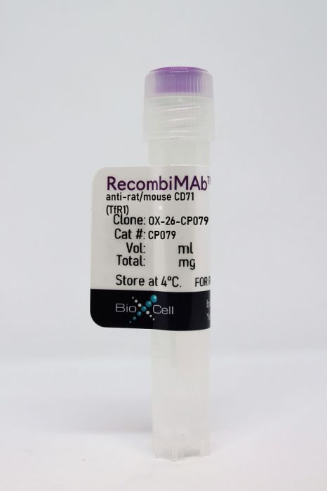
RecombiMAb anti-rat/mouse CD71 (TfR1)
Product Details
The OX-26-CP079 monoclonal antibody is a recombinant version of the original OX-26 antibody that contains the Fc-silencing mutations in the heavy chain rendering it unable to bind endogenous murine Fcγ receptors or C1q. The LALA-PG mutations prevent antibody-dependent, cell-mediated cytotoxicity (ADCC) and complement-dependent cytotoxicity (CDC). The LALA-PG variant has significantly reduced effector function, C1q binding and C3 fixation compared to other common silencing mutations such as the LALA and DANG variants while retaining favorable biophysical and manufacturing properties. Recent studies have demonstrated that administration of anti-TfR1 antibodies with Fc effector function in mice resulted in acute clinical signs of toxicity and a decrease in reticulocyte count. These negative effects were eliminated through Fc-silencing.The OX-26 monoclonal antibody reacts with rat and mouse CD71, also known as transferrin receptor protein 1 (TfR1). CD71 is a type II homodimeric transmembrane glycoprotein which is expressed on the surface of proliferating cells, reticulocytes, and erythroid precursors. CD71 plays a role in the control of cellular proliferation and is required for iron import from transferrin into cells by endocytosis. CD71 is overexpressed on many different types of cancer cells and expression level correlates with advanced stage and/or poorer prognosis in several cancers, including solid cancers. Elevated levels of CD71 expression on malignant cells, together with its extracellular accessibility, ability to internalize, and central role in cancer cell pathology make this receptor an attractive target for antibody-mediated therapy. In addition, cells of the vascular endothelium of brain capillaries that compose the blood-brain barrier (BBB) also express high levels of CD71 allowing for receptor-mediated transcytosis of large biomolecules into the brain. OX-26 has been used as a BBB transporter in rats and mice and is suitable for studying CD71 expression and iron uptake into different tissues. The OX-26 antibody binds to a distinct extracellular epitope on TfR that does not compete for binding with the natural ligand Tf or iron uptake. The OX-26 antibody is often used to transport conjugated drugs across the BBB in experimental models.
Specifications
| Isotype | Mouse IgG2a LALA-PG |
|---|---|
| Recommended Isotype Control(s) | RecombiMAb mouse IgG2a (LALA-PG) isotype control, anti-hen egg lysozyme |
| Recommended Dilution Buffer | InVivoPure pH 7.0 Dilution Buffer |
| Mutations | LALA-PG |
| Immunogen | PHA-activated PVG rat lymph node cells |
| Reported Applications |
Targeted drug delivery to the brain Immunohistochemistry Flow Cytometry *Reported for the original mouse IgG1 antibody. For information on in vivo applications, please contact technicalservice@bioxcell.com |
| Formulation |
PBS, pH 7.0 Contains no stabilizers or preservatives |
| Endotoxin |
<1EU/mg (<0.001EU/μg) Determined by LAL gel clotting assay |
| Aggregation |
<5% Determined by SEC |
| Purity |
>95% Determined by SDS-PAGE |
| Sterility | 0.2 µm filtration |
| Production | Purified from HEK293 cell supernatant in an animal-free facility |
| Purification | Protein G |
| Molecular Weight | 150 kDa |
| Murine Pathogen Tests |
Ectromelia/Mousepox Virus: Negative Hantavirus: Negative K Virus: Negative Lactate Dehydrogenase-Elevating Virus: Negative Lymphocytic Choriomeningitis virus: Negative Mouse Adenovirus: Negative Mouse Cytomegalovirus: Negative Mouse Hepatitis Virus: Negative Mouse Minute Virus: Negative Mouse Norovirus: Negative Mouse Parvovirus: Negative Mouse Rotavirus: Negative Mycoplasma Pulmonis: Negative Pneumonia Virus of Mice: Negative Polyoma Virus: Negative Reovirus Screen: Negative Sendai Virus: Negative Theiler’s Murine Encephalomyelitis: Negative |
| Storage | The antibody solution should be stored at the stock concentration at 4°C. Do not freeze. |
Additional Formats
Recommended Products
Immunohistochemistry, receptor blockade
DeGregorio-Rocasolano N, Martí-Sistac O, Ponce J, Castelló-Ruiz M, Millán M, Guirao V, García-Yébenes I, Salom JB, Ramos-Cabrer P, Alborch E, Lizasoain I, Castillo J, Dávalos A, Gasull T. (2018). "Iron-loaded transferrin (Tf) is detrimental whereas iron-free Tf confers protection against brain ischemia by modifying blood Tf saturation and subsequent neuronal damage" Redox Biol . PubMed
Despite transferrin being the main circulating carrier of iron in body fluids, and iron overload conditions being known to worsen stroke outcome through reactive oxygen species (ROS)-induced damage, the contribution of blood transferrin saturation (TSAT) to stroke brain damage is unknown. The objective of this study was to obtain evidence on whether TSAT determines the impact of experimental ischemic stroke on brain damage and whether iron-free transferrin (apotransferrin, ATf)-induced reduction of TSAT is neuroprotective. We found that experimental ischemic stroke promoted an early extravasation of circulating iron-loaded transferrin (holotransferrin, HTf) to the ischemic brain parenchyma. In vitro, HTf was found to boost ROS production and to be harmful to primary neuronal cultures exposed to oxygen and glucose deprivation. In stroked rats, whereas increasing TSAT with exogenous HTf was detrimental, administration of exogenous ATf and the subsequent reduction of TSAT was neuroprotective. Mechanistically, ATf did not prevent extravasation of HTf to the brain parenchyma in rats exposed to ischemic stroke. However, ATf in vitro reduced NMDA-induced neuronal uptake of HTf and also both the NMDA-mediated lipid peroxidation derived 4-HNE and the resulting neuronal death without altering Ca2+-calcineurin signaling downstream the NMDA receptor. Removal of transferrin from the culture media or blockade of transferrin receptors reduced neuronal death. Together, our data establish that blood TSAT exerts a critical role in experimental stroke-induced brain damage. In addition, our findings suggest that the protective effect of ATf at the neuronal level resides in preventing NMDA-induced HTf uptake and ROS production, which in turn reduces neuronal damage.
Haobam B, Nozawa T, Minowa-Nozawa A, Tanaka M, Oda S, Watanabe T, Aikawa C, Maruyama F, Nakagawa I. (2014). "Rab17-mediated recycling endosomes contribute to autophagosome formation in response to Group A Streptococcus invasion" Cell Microbiol 16(12):1806-21. PubMed
Autophagy plays a crucial role in host defence by facilitating the degradation of invading bacteria such as Group A Streptococcus (GAS). GAS-containing autophagosome-like vacuoles (GcAVs) form when GAS-targeting autophagic membranes entrap invading bacteria. However, the membrane origin and the precise molecular mechanism that underlies GcAV formation remain unclear. In this study, we found that Rab17 mediates the supply of membrane from recycling endosomes (REs) to GcAVs. We showed that GcAVs contain the RE marker transferrin receptor (TfR). Colocalization analyses demonstrated that Rab17 colocalized effectively with GcAV. Rab17 and TfR were visible as punctate structures attached to GcAVs and the Rab17-positive dots were recruited to the GAS-capturing membrane. Overexpression of Rab17 increased the TfR-positive GcAV content, whereas expression of the dominant-negative Rab17 form (Rab17 N132I) caused a decrease, thereby suggesting the involvement of Rab17 in RE-GcAV fusion. The efficiency of GcAV formation was lower in Rab17 N132I-overexpressing cells. Furthermore, knockdown of Rabex-5, the upstream activator of Rab17, reduced the GcAV formation efficiency. These results suggest that Rab17 and Rab17-mediated REs are involved in GcAV formation. This newly identified function of Rab17 in supplying membrane from REs to GcAVs demonstrates that RE functions as a primary membrane source during antibacterial autophagy.
Immunohistochemistry
Fabriek BO, Polfliet MM, Vloet RP, van der Schors RC, Ligtenberg AJ, Weaver LK, Geest C, Matsuno K, Moestrup SK, Dijkstra CD, van den Berg TK. (2007). "The macrophage CD163 surface glycoprotein is an erythroblast adhesion receptor" Blood 109(12):5223-9. PubMed
Erythropoiesis occurs in erythroblastic islands, where developing erythroblasts closely interact with macrophages. The adhesion molecules that govern macrophage-erythroblast contact have only been partially defined. Our previous work has implicated the rat ED2 antigen, which is highly expressed on the surface of macrophages in erythroblastic islands, in erythroblast binding. In particular, the monoclonal antibody ED2 was found to inhibit erythroblast binding to bone marrow macrophages. Here, we identify the ED2 antigen as the rat CD163 surface glycoprotein, a member of the group B scavenger receptor cysteine-rich (SRCR) family that has previously been shown to function as a receptor for hemoglobin-haptoglobin (Hb-Hp) complexes and is believed to contribute to the clearance of free hemoglobin. CD163 transfectants and recombinant protein containing the extracellular domain of CD163 supported the adhesion of erythroblastic cells. Furthermore, we identified a 13-amino acid motif (CD163p2) corresponding to a putative interaction site within the second scavenger receptor domain of CD163 that could mediate erythroblast binding. Finally, CD163p2 promoted erythroid expansion in vitro, suggesting that it enhanced erythroid proliferation and/or survival, but did not affect differentiation. These findings identify CD163 on macrophages as an adhesion receptor for erythroblasts in erythroblastic islands, and suggest a regulatory role for CD163 during erythropoiesis.
Jones AR, Shusta EV. (2007). "Blood-brain barrier transport of therapeutics via receptor-mediation" Pharm Res 24(9):1759-71. PubMed
Drug delivery to the brain is hindered by the presence of the blood-brain barrier (BBB). Although the BBB restricts the passage of many substances, it is actually selectively permeable to nutrients necessary for healthy brain function. To accomplish the task of nutrient transport, the brain endothelium is endowed with a diverse collection of molecular transport systems. One such class of transport system, known as a receptor-mediated transcytosis (RMT), employs the vesicular trafficking machinery of the endothelium to transport substrates between blood and brain. If appropriately targeted, RMT systems can also be used to shuttle a wide range of therapeutics into the brain in a noninvasive manner. Over the last decade, there have been significant developments in the arena of RMT-based brain drug transport, and this review will focus on those approaches that have been validated in an in vivo setting.
Flow Cytometry
Jiménez E, Sacedón R, Vicente A, Hernández-López C, Zapata AG, Varas A. (2002). "Rat peripheral CD4+CD8+ T lymphocytes are partially immunocompetent thymus-derived cells that undergo post-thymic maturation to become functionally mature CD4+ T lymphocytes" J Immunol 168(10):5005-13. PubMed
CD4+CD8+ double-positive (DP) T cells represent a minor subpopulation of T lymphocytes found in the periphery of adult rats. In this study, we show that peripheral DP T cells appear among the first T cells that colonize the peripheral lymphoid organs during fetal life, and represent approximately 40% of peripheral T cells during the perinatal period. Later their proportion decreases to reach the low values seen in adulthood. Most DP T cells are small size lymphocytes that do not exhibit an activated phenotype, and their proliferative rate is similar to that of the other peripheral T cell subpopulations. Only 30-40% of DP T cells expresses CD8beta chain, the remaining cells expressing CD8alphaalpha homodimers. However, both DP T cell subsets have an intrathymic origin since they appear in the recent thymic emigrant population after injection of FITC intrathymically. Functionally, although DP T cells are resistant to undergo apoptosis in response to glucocorticoids, they show poor proliferative responses upon CD3/TCR stimulation due to their inability to produce IL-2. A fraction of DP T cells are not actively synthesizing the CD8 coreceptor, and they gradually differentiate to the CD4 cell lineage in reaggregation cultures. Transfer of DP T lymphocytes into thymectomized SCID mice demonstrates that these cells undergo post-thymic maturation in the peripheral lymphoid organs and that their CD4 cell progeny is fully immunocompetent, as judged by its ability to survive and expand in peripheral lymphoid organs, to proliferate in response to CD3 ligation, and to produce IL-2 upon stimulation.


