RecombiMAb anti-mouse IL-10R (CD210)

Catalog #CP060
Clone:
1B1.3A-CP060
Reactivities:
Mouse

$541.00 - $7,325.00

$541.00 - $7,325.00

Choose an Option...
  • 25 mg - $7,325.00
  • 5 mg - $2,097.00
  • 1 mg - $541.00
  • Custom Amount (Quotes Only)
In stock
Only %1 left
Isotype:
Mouse IgG2a, kappa
(switched from Rat IgG1, kappa)

Product Details

The 1B1.3A-CP060 monoclonal antibody is a recombinant, chimeric version of the original 1B1.3A clone. The variable domain sequences are identical to the original 1B1.3A but the constant region sequences have been switched from Rat IgG1, Īŗ to mouse IgG2a, Īŗ for use in murine models. Species-matched chimeric antibodies exhibit regulated effector functions—including Fc receptor binding and complement activation—and cause less immunogenicity and formation of anti-drug antibodies (ADAs) than xenogenic antibodies in animal models. The highly controlled sequence and lack of genetic drift in recombinant antibodies provide more reliable and reproducible results compared to hybridoma derived antibodies.
The 1B1.3A monoclonal antibody reacts with mouse IL-10R (IL-10 receptor) also known as CD210. The IL-10R is a class II cytokine receptor and is expressed by a variety of cell types including thymocytes, T lymphocytes, B lymphocytes, NK cells, monocytes, and macrophages. Upon binding IL-10, IL-10R stimulation results in many pleiotropic, effects in immunoregulation and inflammation. IL-10R downregulates the expression of pro-inflammatory cytokines, MHC class II antigens, and co-stimulatory molecules on macrophages. It also enhances B lymphocyte survival, proliferation, and antibody production. IL-10R signaling can block NF-ĪŗB activity and is involved in the regulation of the JAK-STAT signaling pathway. The 1B1.3A antibody is a neutralizing antibody and has been shown to block the binding of human IL-10, which cross-reacts with the mouse IL-10R. However, this clone does not recognize the human IL-10R.

Specifications

Isotype Mouse IgG2a, Īŗ
Recommended Isotype Control(s) RecombiMAb mouse IgG2a isotype control, unknown specificity
Recommended Dilution Buffer InVivoPure pH 7.0 Dilution Buffer
Conjugation This product is unconjugated. Conjugation is available via our Antibody Conjugation Services.
Reported Applications Flow Cytometry
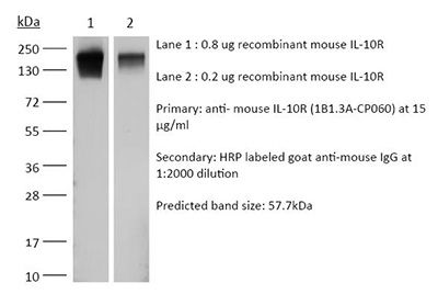
Western blot
in vitro blocking of IL-10R signaling
in vivo blocking of IL-10/IL-10R signaling
*Reported for the original rat IgG1 1B1.3A antibody
Formulation PBS, pH 7.0
Contains no stabilizers or preservatives
Endotoxin <1EU/mg (<0.001EU/μg)
Determined by LAL gel clotting assay
Aggregation <5%
Determined by SEC
Purity >95%
Determined by SDS-PAGE
Sterility 0.2 µm filtration
Production Purified from HEK293 cell supernatant in an animal-free facility
Purification Protein G
Molecular Weight 150 kDa
Murine Pathogen Tests Ectromelia/Mousepox Virus: Negative
Hantavirus: Negative
K Virus: Negative
Lactate Dehydrogenase-Elevating Virus: Negative
Lymphocytic Choriomeningitis virus: Negative
Mouse Adenovirus: Negative
Mouse Cytomegalovirus: Negative
Mouse Hepatitis Virus: Negative
Mouse Minute Virus: Negative
Mouse Norovirus: Negative
Mouse Parvovirus: Negative
Mouse Rotavirus: Negative
Mycoplasma Pulmonis: Negative
Pneumonia Virus of Mice: Negative
Polyoma Virus: Negative
Reovirus Screen: Negative
Sendai Virus: Negative
Theiler’s Murine Encephalomyelitis: Negative
Storage The antibody solution should be stored at the stock concentration at 4°C. Do not freeze.
in vivo blocking of IL-10/IL-10R signaling
Bravo M, Dileepan T, Dolan M, Hildebrand J, Wolford J, Hanson ID, Hamilton SE, Frosch AE, Burrack KS. (2024). "IL-15 Complex-Induced IL-10 Enhances Plasmodium-specific CD4+ T Follicular Helper Differentiation and Antibody Production" J Immunol 212(6):992-1001. PubMed

Malaria, which results from infection with Plasmodium parasites, remains a major public health problem. Although humans do not develop long-lived, sterilizing immunity, protection against symptomatic disease develops after repeated exposure to Plasmodium parasites and correlates with the acquisition of humoral immunity. Despite the established role Abs play in protection from malaria disease, dysregulated inflammation is thought to contribute to the suboptimal immune response to Plasmodium infection. Plasmodium berghei ANKA (PbA) infection results in a fatal severe malaria disease in mice. We previously demonstrated that treatment of mice with IL-15 complex (IL-15C; IL-15 bound to an IL-15Rα-Fc fusion protein) induces IL-10 expression in NK cells, which protects mice from PbA-induced death. Using a novel MHC class II tetramer to identify PbA-specific CD4+ T cells, in this study we demonstrate that IL-15C treatment enhances T follicular helper (Tfh) differentiation and modulates cytokine production by CD4+ T cells. Moreover, genetic deletion of NK cell-derived IL-10 or IL-10R expression on T cells prevents IL-15C-induced Tfh differentiation. Additionally, IL-15C treatment results in increased anti-PbA IgG Ab levels and improves survival following reinfection. Overall, these data demonstrate that IL-15C treatment, via its induction of IL-10 from NK cells, modulates the dysregulated inflammation during Plasmodium infection to promote Tfh differentiation and Ab generation, correlating with improved survival from reinfection. These findings will facilitate improved control of malaria infection and protection from disease by informing therapeutic strategies and vaccine design.

in vivo blocking of IL-10/IL-10R signaling
Xu M, Pokrovskii M, Ding Y, Yi R, Au C, Harrison OJ, Galan C, Belkaid Y, Bonneau R, Littman DR. (2018). "c-MAF-dependent regulatory T cells mediate immunological tolerance to a gut pathobiont" Nature 554(7692):373-377. PubMed

Both microbial and host genetic factors contribute to the pathogenesis of autoimmune diseases. There is accumulating evidence that microbial species that potentiate chronic inflammation, as in inflammatory bowel disease, often also colonize healthy individuals. These microorganisms, including the Helicobacter species, can induce pathogenic T cells and are collectively referred to as pathobionts. However, how such T cells are constrained in healthy individuals is not yet understood. Here we report that host tolerance to a potentially pathogenic bacterium, Helicobacter hepaticus, is mediated by the induction of RORγt+FOXP3+ regulatory T (iTreg) cells that selectively restrain pro-inflammatory T helper 17 (TH17) cells and whose function is dependent on the transcription factor c-MAF. Whereas colonization of wild-type mice by H. hepaticus promoted differentiation of RORγt-expressing microorganism-specific iTreg cells in the large intestine, in disease-susceptible IL-10-deficient mice, there was instead expansion of colitogenic TH17 cells. Inactivation of c-MAF in the Treg cell compartment impaired differentiation and function, including IL-10 production, of bacteria-specific iTreg cells, and resulted in the accumulation of H. hepaticus-specific inflammatory TH17 cells and spontaneous colitis. By contrast, RORγt inactivation in Treg cells had only a minor effect on the bacteria-specific Treg and TH17 cell balance, and did not result in inflammation. Our results suggest that pathobiont-dependent inflammatory bowel disease is driven by microbiota-reactive T cells that have escaped this c-MAF-dependent mechanism of iTreg-TH17 homeostasis.

Burrack KS, Huggins MA, Taras E, Dougherty P, Henzler CM, Yang R, Alter S, Jeng EK, Wong HC, Felices M, Cichocki F, Miller JS, Hart GT, Johnson AJ, Jameson SC, Hamilton SE. (2018). "Interleukin-15 Complex Treatment Protects Mice from Cerebral Malaria by Inducing Interleukin-10-Producing Natural Killer Cells" Immunity 48(4):760-772.e4. PubMed

Cerebral malaria is a deadly complication of Plasmodium infection and involves blood brain barrier (BBB) disruption following infiltration of white blood cells. During experimental cerebral malaria (ECM), mice inoculated with Plasmodium berghei ANKA-infected red blood cells develop a fatal CM-like disease caused by CD8+ T cell-mediated pathology. We found that treatment with interleukin-15 complex (IL-15C) prevented ECM, whereas IL-2C treatment had no effect. IL-15C-expanded natural killer (NK) cells were necessary and sufficient for protection against ECM. IL-15C treatment also decreased CD8+ T cell activation in the brain and prevented BBB breakdown without influencing parasite load. IL-15C induced NK cells to express IL-10, which was required for IL-15C-mediated protection against ECM. Finally, we show that ALT-803, a modified human IL-15C, mediates similar induction of IL-10 in NK cells and protection against ECM. These data identify a regulatory role for cytokine-stimulated NK cells in the prevention of a pathogenic immune response.

Lin CC, Bradstreet TR, Schwarzkopf EA, Sim J, Carrero JA, Chou C, Cook LE, Egawa T, Taneja R, Murphy TL, Russell JH, Edelson BT. (2014). "Bhlhe40 controls cytokine production by T cells and is essential for pathogenicity in autoimmune neuroinflammation" Nat Commun . PubMed

TH1 and TH17 cells mediate neuroinflammation in experimental autoimmune encephalomyelitis (EAE), a mouse model of multiple sclerosis. Pathogenic TH cells in EAE must produce the pro-inflammatory cytokine granulocyte-macrophage colony stimulating factor (GM-CSF). TH cell pathogenicity in EAE is also regulated by cell-intrinsic production of the immunosuppressive cytokine interleukin 10 (IL-10). Here we demonstrate that mice deficient for the basic helix-loop-helix (bHLH) transcription factor Bhlhe40 (Bhlhe40(-/-)) are resistant to the induction of EAE. Bhlhe40 is required in vivo in a T cell-intrinsic manner, where it positively regulates the production of GM-CSF and negatively regulates the production of IL-10. In vitro, GM-CSF secretion is selectively abrogated in polarized Bhlhe40(-/-) TH1 and TH17 cells, and these cells show increased production of IL-10. Blockade of IL-10 receptor in Bhlhe40(-/-) mice renders them susceptible to EAE. These findings identify Bhlhe40 as a critical regulator of autoreactive T-cell pathogenicity.