InVivoMAb anti-mouse IL-10R (CD210)

Catalog #BE0050
Product Citations:
67
Clone:
1B1.3A
Reactivities:
Mouse

$164.00 - $4,280.00

Choose an Option...
  • 100 mg - $4,280.00
  • 50 mg - $3,024.00
  • 25 mg - $2,009.00
  • 5 mg - $600.00
  • 1 mg - $164.00
  • Custom Amount (Quotes Only)
In stock
Only %1 left

Product Details

The 1B1.3A monoclonal antibody reacts with mouse IL-10R (IL-10 receptor) also known as CD210. The IL-10R is a class II cytokine receptor and is expressed by a variety of cell types including thymocytes, T lymphocytes, B lymphocytes, NK cells, monocytes, and macrophages. Upon binding IL-10, IL-10R stimulation results in many pleiotropic, effects in immunoregulation and inflammation. IL-10R downregulates the expression of pro-inflammatory cytokines, MHC class II antigens, and co-stimulatory molecules on macrophages. It also enhances B lymphocyte survival, proliferation, and antibody production. IL-10R signaling can block NF-ĪŗB activity, and is involved in the regulation of the JAK-STAT signaling pathway. The 1B1.3A antibody is a neutralizing antibody and has been shown to block the binding of human IL-10, which cross-reacts with the mouse IL-10R. However, this clone does not recognize the human IL-10R.

Specifications

Isotype Rat IgG1,Ā Īŗ
Recommended Isotype Control(s) InVivoMAb rat IgG1 isotype control, anti-horseradish peroxidase
Recommended Dilution Buffer InVivoPure pH 6.5T Dilution Buffer
Conjugation This product is unconjugated. Conjugation is available via our Antibody Conjugation Services.
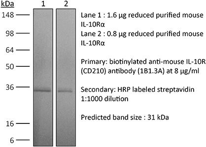
Immunogen Recombinant ligand-binding domain of mouse IL-10R
Reported Applications in vivo blocking of IL-10/IL-10R signaling
in vitro blocking of IL-10R signaling
Flow cytometry
Western Blot
Formulation PBS, pH 6.5
0.01% Tween


Contains no stabilizers or preservatives
Endotoxin <2EU/mg (<0.002EU/Ī¼g)
Determined by LAL gel clotting assay
Purity >95%
Determined by SDS-PAGE
Sterility 0.2 Āµm filtration
Production Purified from cell culture supernatant in an animal-free facility
Purification Protein G
RRID AB_1107611
Molecular Weight 150 kDa
Storage The antibody solution should be stored at the stock concentration at 4Ā°C. Do not freeze.

Additional Formats

in vivo blocking of IL-10/IL-10R signaling
Xu, M., et al. (2018). "c-MAF-dependent regulatory T cells mediate immunological tolerance to a gut pathobiont" Nature 554(7692): 373-377. PubMed

Both microbial and host genetic factors contribute to the pathogenesis of autoimmune diseases. There is accumulating evidence that microbial species that potentiate chronic inflammation, as in inflammatory bowel disease, often also colonize healthy individuals. These microorganisms, including the Helicobacter species, can induce pathogenic T cells and are collectively referred to as pathobionts. However, how such T cells are constrained in healthy individuals is not yet understood. Here we report that host tolerance to a potentially pathogenic bacterium, Helicobacter hepaticus, is mediated by the induction of RORgammat(+)FOXP3(+) regulatory T (iTreg) cells that selectively restrain pro-inflammatory T helper 17 (TH17) cells and whose function is dependent on the transcription factor c-MAF. Whereas colonization of wild-type mice by H. hepaticus promoted differentiation of RORgammat-expressing microorganism-specific iTreg cells in the large intestine, in disease-susceptible IL-10-deficient mice, there was instead expansion of colitogenic TH17 cells. Inactivation of c-MAF in the Treg cell compartment impaired differentiation and function, including IL-10 production, of bacteria-specific iTreg cells, and resulted in the accumulation of H. hepaticus-specific inflammatory TH17 cells and spontaneous colitis. By contrast, RORgammat inactivation in Treg cells had only a minor effect on the bacteria-specific Treg and TH17 cell balance, and did not result in inflammation. Our results suggest that pathobiont-dependent inflammatory bowel disease is driven by microbiota-reactive T cells that have escaped this c-MAF-dependent mechanism of iTreg-TH17 homeostasis.

in vivo blocking of IL-10/IL-10R signaling
Burrack, K. S., et al. (2018). "Interleukin-15 Complex Treatment Protects Mice from Cerebral Malaria by Inducing Interleukin-10-Producing Natural Killer Cells" Immunity 48(4): 760-772 e764. PubMed

Cerebral malaria is a deadly complication of Plasmodium infection and involves blood brain barrier (BBB) disruption following infiltration of white blood cells. During experimental cerebral malaria (ECM), mice inoculated with Plasmodium berghei ANKA-infected red blood cells develop a fatal CM-like disease caused by CD8(+) T cell-mediated pathology. We found that treatment with interleukin-15 complex (IL-15C) prevented ECM, whereas IL-2C treatment had no effect. IL-15C-expanded natural killer (NK) cells were necessary and sufficient for protection against ECM. IL-15C treatment also decreased CD8(+) T cell activation in the brain and prevented BBB breakdown without influencing parasite load. IL-15C induced NK cells to express IL-10, which was required for IL-15C-mediated protection against ECM. Finally, we show that ALT-803, a modified human IL-15C, mediates similar induction of IL-10 in NK cells and protection against ECM. These data identify a regulatory role for cytokine-stimulated NK cells in the prevention of a pathogenic immune response.

in vivo blocking of IL-10/IL-10R signaling
Sun, M., et al. (2018). "Microbiota-derived short-chain fatty acids promote Th1 cell IL-10 production to maintain intestinal homeostasis" Nat Commun 9(1): 3555. PubMed

T-cells are crucial in maintanence of intestinal homeostasis, however, it is still unclear how microbiota metabolites regulate T-effector cells. Here we show gut microbiota-derived short-chain fatty acids (SCFAs) promote microbiota antigen-specific Th1 cell IL-10 production, mediated by G-protein coupled receptors 43 (GPR43). Microbiota antigen-specific Gpr43(-/-) CBir1 transgenic (Tg) Th1 cells, specific for microbiota antigen CBir1 flagellin, induce more severe colitis compared with wide type (WT) CBir1 Tg Th1 cells in Rag(-/-) recipient mice. Treatment with SCFAs limits colitis induction by promoting IL-10 production, and administration of anti-IL-10R antibody promotes colitis development. Mechanistically, SCFAs activate Th1 cell STAT3 and mTOR, and consequently upregulate transcription factor B lymphocyte-induced maturation protein 1 (Blimp-1), which mediates SCFA-induction of IL-10. SCFA-treated Blimp1(-/-) Th1 cells produce less IL-10 and induce more severe colitis compared to SCFA-treated WT Th1 cells. Our studies, thus, provide insight into how microbiota metabolites regulate Th1 cell functions to maintain intestinal homeostasis.

in vivo blocking of IL-10/IL-10R signaling
Christensen, A. D., et al. (2015). "Depletion of regulatory T cells in a hapten-induced inflammation model results in prolonged and increased inflammation driven by T cells" Clin Exp Immunol 179(3): 485-499. PubMed

Regulatory T cells (Tregs ) are known to play an immunosuppressive role in the response of contact hypersensitivity (CHS), but neither the dynamics of Tregs during the CHS response nor the exaggerated inflammatory response after depletion of Tregs has been characterized in detail. In this study we show that the number of Tregs in the challenged tissue peak at the same time as the ear-swelling reaches its maximum on day 1 after challenge, whereas the number of Tregs in the draining lymph nodes peaks at day 2. As expected, depletion of Tregs by injection of a monoclonal antibody to CD25 prior to sensitization led to a prolonged and sustained inflammatory response which was dependent upon CD8 T cells, and co-stimulatory blockade with cytotoxic T lymphocyte antigen-4-immunoglobulin (CTLA-4-Ig) suppressed the exaggerated inflammation. In contrast, blockade of the interleukin (IL)-10-receptor (IL-10R) did not further increase the exaggerated inflammatory response in the Treg -depleted mice. In the absence of Tregs , the response changed from a mainly acute reaction with heavy infiltration of neutrophils to a sustained response with more chronic characteristics (fewer neutrophils and dominated by macrophages). Furthermore, depletion of Tregs enhanced the release of cytokines and chemokines locally in the inflamed ear and augmented serum levels of the systemic inflammatory mediators serum amyloid (SAP) and haptoglobin early in the response.

in vivo blocking of IL-10/IL-10R signaling
Liu, G., et al. (2015). "IL-27 Signaling Is Crucial for Survival of Mice Infected with African Trypanosomes via Preventing Lethal Effects of CD4+ T Cells and IFN-gamma" PLoS Pathog 11(7): e1005065. PubMed

African trypanosomes are extracellular protozoan parasites causing a chronic debilitating disease associated with a persistent inflammatory response. Maintaining the balance of the inflammatory response via downregulation of activation of M1-type myeloid cells was previously shown to be crucial to allow prolonged survival. Here we demonstrate that infection with African trypanosomes of IL-27 receptor-deficient (IL-27R-/-) mice results in severe liver immunopathology and dramatically reduced survival as compared to wild-type mice. This coincides with the development of an exacerbated Th1-mediated immune response with overactivation of CD4+ T cells and strongly enhanced production of inflammatory cytokines including IFN-gamma. What is important is that IL-10 production was not impaired in infected IL-27R-/- mice. Depletion of CD4+ T cells in infected IL-27R-/- mice resulted in a dramatically reduced production of IFN-gamma, preventing the early mortality of infected IL-27R-/- mice. This was accompanied by a significantly reduced inflammatory response and a major amelioration of liver pathology. These results could be mimicked by treating IL-27R-/- mice with a neutralizing anti-IFN-gamma antibody. Thus, our data identify IL-27 signaling as a novel pathway to prevent early mortality via inhibiting hyperactivation of CD4+ Th1 cells and their excessive secretion of IFN-gamma during infection with African trypanosomes. These data are the first to demonstrate the essential role of IL-27 signaling in regulating immune responses to extracellular protozoan infections.

in vivo blocking of IL-10/IL-10R signaling
Lin, C. C., et al. (2014). "Bhlhe40 controls cytokine production by T cells and is essential for pathogenicity in autoimmune neuroinflammation" Nat Commun 5: 3551. PubMed

TH1 and TH17 cells mediate neuroinflammation in experimental autoimmune encephalomyelitis (EAE), a mouse model of multiple sclerosis. Pathogenic TH cells in EAE must produce the pro-inflammatory cytokine granulocyte-macrophage colony stimulating factor (GM-CSF). TH cell pathogenicity in EAE is also regulated by cell-intrinsic production of the immunosuppressive cytokine interleukin 10 (IL-10). Here we demonstrate that mice deficient for the basic helix-loop-helix (bHLH) transcription factor Bhlhe40 (Bhlhe40(-/-)) are resistant to the induction of EAE. Bhlhe40 is required in vivo in a T cell-intrinsic manner, where it positively regulates the production of GM-CSF and negatively regulates the production of IL-10. In vitro, GM-CSF secretion is selectively abrogated in polarized Bhlhe40(-/-) TH1 and TH17 cells, and these cells show increased production of IL-10. Blockade of IL-10 receptor in Bhlhe40(-/-) mice renders them susceptible to EAE. These findings identify Bhlhe40 as a critical regulator of autoreactive T-cell pathogenicity.

in vitro blocking of IL-10R signaling
Verhagen, J. and D. C. Wraith. (2014). "Blockade of LFA-1 augments in vitro differentiation of antigen-induced Foxp3(+) Treg cells" J Immunol Methods 414: 58-64. PubMed

Adoptive transfer of antigen-specific, in vitro-induced Foxp3(+) Treg (iTreg) cells protects against autoimmune disease. To generate antigen-specific iTreg cells at high purity, however, remains a challenge. Whereas polyclonal T cell stimulation with anti-CD3 and anti-CD28 antibody yields Foxp3(+) iTreg cells at a purity of 90-95%, antigen-induced iTreg cells typically do not exceed a purity of 65-75%, even in a TCR-transgenic model. In a similar vein to thymic Treg cell selection, iTreg cell differentiation is influenced not only by antigen recognition and the availability of TGF-beta but also by co-factors including costimulation and adhesion molecules. In this study, we demonstrate that blockade of the T cell integrin Leukocyte Function-associated Antigen-1 (LFA-1) during antigen-mediated iTreg cell differentiation augments Foxp3 induction, leading to approximately 90% purity of Foxp3(+) iTreg cells. This increased efficacy not only boosts the yield of Foxp3(+) iTreg cells, it also reduces contamination with activated effector T cells, thus improving the safety of adoptive transfer immunotherapy.

in vivo blocking of IL-10/IL-10R signaling
Masson, F., et al. (2014). "Id2 represses E2A-mediated activation of IL-10 expression in T cells" Blood 123(22): 3420-3428. PubMed

Interleukin-10 (IL-10) is a key immunoregulatory cytokine that functions to prevent inflammatory and autoimmune diseases. Despite the critical role for IL-10 produced by effector CD8(+) T cells during pathogen infection and autoimmunity, the mechanisms regulating its production are poorly understood. We show that loss of the inhibitor of DNA binding 2 (Id2) in T cells resulted in aberrant IL-10 expression in vitro and in vivo during influenza virus infection and in a model of acute graft-versus-host disease (GVHD). Furthermore, IL-10 overproduction substantially reduced the immunopathology associated with GVHD. We demonstrate that Id2 acts to repress the E2A-mediated trans-activation of the Il10 locus. Collectively, our findings uncover a key regulatory role of Id2 during effector T cell differentiation necessary to limit IL-10 production by activated T cells and minimize their suppressive activity during the effector phase of disease control.

in vivo blocking of IL-10/IL-10R signaling
Dolina, J. S., et al. (2014). "Liver-primed CD8+ T cells suppress antiviral adaptive immunity through galectin-9-independent T-cell immunoglobulin and mucin 3 engagement of high-mobility group box 1 in mice" Hepatology 59(4): 1351-1365. PubMed

The liver is a tolerogenic environment exploited by persistent infections, such as hepatitis B (HBV) and C (HCV) viruses. In a murine model of intravenous hepatotropic adenovirus infection, liver-primed antiviral CD8(+) T cells fail to produce proinflammatory cytokines and do not display cytolytic activity characteristic of effector CD8(+) T cells generated by infection at an extrahepatic, that is, subcutaneous, site. Importantly, liver-generated CD8(+) T cells also appear to have a T-regulatory (Treg) cell function exemplified by their ability to limit proliferation of antigen-specific T-effector (Teff ) cells in vitro and in vivo via T-cell immunoglobulin and mucin 3 (Tim-3) expressed by the CD8(+) Treg cells. Regulatory activity did not require recognition of the canonical Tim-3 ligand, galectin-9, but was dependent on CD8(+) Treg cell-surface Tim-3 binding to the alarmin, high-mobility group box 1 (HMGB-1). CONCLUSION: Virus-specific Tim-3(+) CD8(+) T cells operating through HMGB-1 recognition in the setting of acute and chronic viral infections of the liver may act to dampen hepatic T-cell responses in the liver microenvironment and, as a consequence, limit immune-mediated tissue injury or promote the establishment of persistent infections.

in vivo blocking of IL-10/IL-10R signaling
Ruffell, B., et al. (2014). "Macrophage IL-10 blocks CD8+ T cell-dependent responses to chemotherapy by suppressing IL-12 expression in intratumoral dendritic cells" Cancer Cell 26(5): 623-637. PubMed

Blockade of colony-stimulating factor-1 (CSF-1) limits macrophage infiltration and improves response of mammary carcinomas to chemotherapy. Herein we identify interleukin (IL)-10 expression by macrophages as the critical mediator of this phenotype. Infiltrating macrophages were the primary source of IL-10 within tumors, and therapeutic blockade of IL-10 receptor (IL-10R) was equivalent to CSF-1 neutralization in enhancing primary tumor response to paclitaxel and carboplatin. Improved response to chemotherapy was CD8(+) T cell-dependent, but IL-10 did not directly suppress CD8(+) T cells or alter macrophage polarization. Instead, IL-10R blockade increased intratumoral dendritic cell expression of IL-12, which was necessary for improved outcomes. In human breast cancer, expression of IL12A and cytotoxic effector molecules were predictive of pathological complete response rates to paclitaxel.

in vivo blocking of IL-10/IL-10R signaling
Richter, K. and A. Oxenius. (2013). "Non-neutralizing antibodies protect from chronic LCMV infection independently of activating FcgammaR or complement" Eur J Immunol 43(9): 2349-2360. PubMed

Chronic viral infections lead to CD8(+) T cell exhaustion, characterized by impaired cytokine secretion. The presence of the immune-regulatory cytokine IL-10 promotes chronic lymphocytic choriomeningitis virus (LCMV) Clone 13 infection in mice, whereas the absence of IL-10/IL-10R signaling early during infection results in viral clearance and higher percentages and numbers of antiviral, cytokine-producing T cells. However, it is currently unclear which cell populations and effector molecules are crucial to protect against chronic infection. In this study, we demonstrate that antiviral, LCMV-binding, non-neutralizing antibodies are needed, in addition to CD4(+) and CD8(+) T cells, to clear a high-dose LCMV infection in mice, in the absence of IL-10. The interaction between CD4(+) T cells and B cells in B-cell follicles via CD40/CD40L, in addition to class switch and/or somatic hypermutation, is crucial for viral control in the absence of IL-10. Interestingly, transfer of LCMV-binding non-neutralizing antibodies protected recipients from chronic infection. In addition, viral clearance in the absence of IL-10R signaling was independent of activating Fcgamma receptors and complement. These data highlight that non-neutralizing antibodies effectively contribute to the control of LCMV infection when present prior to infection, suggesting that the induction of neutralizing antibodies is not implicitly necessary for the generation of successful vaccines.

in vivo blocking of IL-10/IL-10R signaling
Mishra, P. K., et al. (2013). "Prevention of type 1 diabetes through infection with an intestinal nematode parasite requires IL-10 in the absence of a Th2-type response" Mucosal Immunol 6(2): 297-308. PubMed

Helminth infection can prevent type 1 diabetes (T1D); however, the regulatory mechanisms inhibiting disease remain largely undefined. In these studies, nonobese diabetic (NOD) IL-4(-/-) mice were infected with the strictly enteric nematode parasite, Heligmosomoides polygyrus. Short-term infection, 5-7 weeks of age, inhibited T1D onset, as late as 40 weeks of age. CD4(+) T-cell STAT6 phosphorylation was inhibited, while suppressed signal transducer and activator of transcription 1 phosphorylation was sustained, as were increases in FOXP3(-), CD4(+) T-cell interleukin (IL)-10 production. Blockade of IL-10 signaling in NOD-IL-4(-/-), but not in NOD, mice during this short interval abrogated protective effects resulting in pancreatic beta-cell destruction and ultimately T1D. Transfer of CD4(+) T cells from H. polygyrus (Hp)-inoculated NOD IL-4(-/-) mice to NOD mice blocked the onset of T1D. These studies indicate that Hp infection induces non-T-regulatory cells to produce IL-10 independently of STAT6 signaling and that in this Th2-deficient environment IL-10 is essential for T1D inhibition.

in vitro blocking of IL-10R signaling, Flow Cytometry
Hu, Z., et al. (2013). "Regulatory CD8+ T cells associated with erosion of immune surveillance in persistent virus infection suppress in vitro and have a reversible proliferative defect" J Immunol 191(1): 312-322. PubMed

CD4(+) T cell help is critical for CD8(+) T cell memory and immune surveillance against persistent virus infections. Our recent data have showed the lack of CD4(+) T cells leads to the generation of an IL-10-producing CD8(+) T cell population during persistent murine gamma-herpesvirus-68 (MHV-68) infection. IL-10 from these cells is partly responsible for erosion in immune surveillance, leading to spontaneous virus reactivation in lungs. In this study, we further characterized the generation, phenotype, and function of these IL-10-producing CD8(+) T cells by comparing with a newly identified IL-10-producing CD8(+) T cell population present during the acute stage of the infection. The IL-10-producing CD8(+) populations in acute and chronic stages differed in their requirement for CD4(+) T cell help, the dependence on IL-2/CD25 and CD40-CD40L pathways, and the ability to proliferate in vitro in response to anti-CD3 stimulation. IL-10-producing CD8(+) T cells in the chronic stage showed a distinct immunophenotypic profile, sharing partial overlap with the markers of previously reported regulatory CD8(+) T cells, and suppressed the proliferation of naive CD8(+) T cells. Notably, they retained the ability to produce effector cytokines and cytotoxic activity. In addition, the proliferative defect of the cells could be restored by addition of exogenous IL-2 or blockade of IL-10. These data suggest that the IL-10-producing CD8(+) T cells arising in chronic MHV-68 infection in the absence of CD4(+) T cell help belong to a subset of CD8(+) regulatory T cells.

in vivo blocking of IL-10/IL-10R signaling
Wilson, M. S., et al. (2011). "IL-10 blocks the development of resistance to re-infection with Schistosoma mansoni" PLoS Pathog 7(8): e1002171. PubMed

Despite effective chemotherapy to treat schistosome infections, re-infection rates are extremely high. Resistance to reinfection can develop, however it typically takes several years following numerous rounds of treatment and re-infection, and often develops in only a small cohort of individuals. Using a well-established and highly permissive mouse model, we investigated whether immunoregulatory mechanisms influence the development of resistance. Following Praziquantel (PZQ) treatment of S. mansoni infected mice we observed a significant and mixed anti-worm response, characterized by Th1, Th2 and Th17 responses. Despite the elevated anti-worm response in PBMCā€™s, liver, spleen and mesenteric lymph nodes, this did not confer any protection from a secondary challenge infection. Because a significant increase in IL-10-producing CD4(+)CD44(+)CD25(+)GITR(+) lymphocytes was observed, we hypothesised that IL-10 was obstructing the development of resistance. Blockade of IL-10 combined with PZQ treatment afforded a greater than 50% reduction in parasite establishment during reinfection, compared to PZQ treatment alone, indicating that IL-10 obstructs the development of acquired resistance. Markedly enhanced Th1, Th2 and Th17 responses, worm-specific IgG1, IgG2b and IgE and circulating eosinophils characterized the protection. This study demonstrates that blocking IL-10 signalling during PZQ treatment can facilitate the development of protective immunity and provide a highly effective strategy to protect against reinfection with S. mansoni.

in vivo blocking of IL-10/IL-10R signaling
Kastenmuller, W., et al. (2011). "Regulatory T cells selectively control CD8+ T cell effector pool size via IL-2 restriction" J Immunol 187(6): 3186-3197. PubMed

Regulatory T cells (Treg) are key players in maintaining immune homeostasis but have also been shown to regulate immune responses against infectious pathogens. Therefore, Treg are a promising target for modulating immune responses to vaccines to improve their efficacy. Using a viral vector system, we found that Treg act on the developing immune response early postinfection by reducing the extent of dendritic cell costimulatory molecule expression. Due to this change and the lower IL-2 production that results, a substantial fraction of CD8(+) effector T cells lose CD25 expression several days after activation. Surprisingly, such Treg-dependent limitations in IL-2 signaling by Ag-activated CD8(+) T cells prevent effector differentiation without interfering with memory cell formation. In this way, Treg fine-tune the numbers of effector T cells generated while preserving the capacity for a rapid recall response upon pathogen re-exposure. This selective effect of Treg on a subpopulation of CD8(+) T cells indicates that although manipulation of the Treg compartment might not be optimal for prophylactic vaccinations, it can be potentially exploited to optimize vaccine efficacy for therapeutic interventions.

    • Immunology and Microbiology
    • ,
    Deletion of Endogenous Neuregulin-4 Limits Adaptive Immunity During Interleukin-10 Receptor-Neutralizing Colitis.

    In Inflammatory Bowel Diseases on 2 November 2023 by Bernard, J. K., Bucar, E. B., et al.

    PubMed

    Growth factors are essential for maintenance of intestinal health. We previously showed that exogenous neuregulin-4 (NRG4) promotes colonocyte survival during cytokine challenge and is protective against acute models of intestinal inflammation. However, the function(s) of endogenous NRG4 are not well understood. Using NRG4-/- mice, we tested the role of endogenous NRG4 in models of colitis skewed toward either adaptive (interleukin-10 receptor [IL-10R] neutralization) or innate (dextran sulfate sodium [DSS]) immune responses. NRG4-/- and wild-type cage mate mice were subjected to chronic IL-10R neutralization colitis and acute DSS colitis. Disease was assessed by histological examination, inflammatory cytokine levels, fecal lipocalin-2 levels, and single cell mass cytometry immune cell profiling. Homeostatic gene alterations were evaluated by RNA sequencing analysis from colonic homogenates, with real-time quantitative polymerase chain reaction confirmation in both tissue and isolated epithelium. During IL-10R neutralization colitis, NRG4-/- mice had reduced colonic inflammatory cytokine expression, histological damage, and colonic CD8+ T cell numbers vs wild-type cage mates. Conversely, in DSS colitis, NRG4-/- mice had elevated cytokine expression, fecal lipocalin-2 levels, and impaired weight recovery. RNA sequencing showed a loss of St3gal4, a sialyltransferase involved in immune cell trafficking, in NRG4-null colons, which was verified in both tissue and isolated epithelium. The regulation of St3gal4 by NRG4 was confirmed with ex vivo epithelial colon organoid cultures from NRG4-/- mice and by induction of St3gal4 in vivo following NRG4 treatment. NRG4 regulates colonic epithelial ST3GAL4 and thus may allow for robust recruitment of CD8+ T cells during adaptive immune responses in colitis. On the other hand, NRG4 loss exacerbates injury driven by innate immune responses. Ā© The Author(s) 2023. Published by Oxford University Press on behalf of Crohnā€™s & Colitis Foundation. All rights reserved. For permissions, please e-mail: journals.permissions@oup.com.

    Distinct stimulus-dependent neutrophil dynamics revealed by real-time imaging of intestinal mucosa after acute injury.

    In PNAS Nexus on 1 November 2022 by Azcutia, V., Kelm, M., et al.

    PubMed

    Clinical symptoms in many inflammatory diseases of the intestine are directly related to neutrophil (PMN) migration across colonic mucosa and into the intestinal lumen, yet in-vivo studies detailing this process are lacking. Using real-time intravital microscopy and a new distal colon loop model, we report distinct PMN migratory dynamics in response to several models of acute colonic injury. PMNs exhibited rapid swarming responses after mechanically induced intestinal wounds. Similar numbers of PMNs infiltrated colonic mucosa after wounding in germ-free mice, suggesting microbiota-independent mechanisms. By contrast, acute mucosal injury secondary to either a treatment of mice with dextran sodium sulfate or an IL-10 receptor blockade model of colitis resulted in lamina propria infiltration with PMNs that were largely immotile. Biopsy wounding of colonic mucosa in DSS-treated mice did not result in enhanced PMN swarming however, intraluminal application of the neutrophil chemoattractant LTB4 under such conditions resulted in enhanced transepithelial migration of PMNs. Analyses of PMNs that had migrated into the colonic lumen revealed that the majority of PMNs were directly recruited from the circulation and not from the immotile pool in the mucosa. Decreased PMN motility parallels upregulation of the receptor CXCR4 and apoptosis. Similarly, increased expression of CXCR4 on human PMNs was observed in colonic biopsies from people with active ulcerative colitis. This new approach adds an important tool to investigate mechanisms regulating PMN migration across mucosa within the distal intestine and will provide new insights for developing future anti-inflammatory and pro-repair therapies. Ā© The Author(s) 2022. Published by Oxford University Press on behalf of National Academy of Sciences.

    • Immunology and Microbiology
    • ,
    • Mus musculus (House mouse)
    CD11c+ myeloid cell exosomes reduce intestinal inflammation during colitis.

    In JCI Insight on 10 October 2022 by Bauer, K. M., Nelson, M. C., et al.

    PubMed

    Intercellular communication is critical for homeostasis in mammalian systems, including the gastrointestinal (GI) tract. Exosomes are nanoscale lipid extracellular vesicles that mediate communication between many cell types. Notably, the roles of immune cell exosomes in regulating GI homeostasis and inflammation are largely uncharacterized. By generating mouse strains deficient in cell-specific exosome production, we demonstrate deletion of the small GTPase Rab27A in CD11c+ cells exacerbated murine colitis, which was reversible through administration of DC-derived exosomes. Profiling RNAs within colon exosomes revealed a distinct subset of miRNAs carried by colon- and DC-derived exosomes. Among antiinflammatory exosomal miRNAs, miR-146a was transferred from gut immune cells to myeloid and T cells through a Rab27-dependent mechanism, targeting Traf6, IRAK-1, and NLRP3 in macrophages. Further, we have identified a potentially novel mode of exosome-mediated DC and macrophage crosstalk that is capable of skewing gut macrophages toward an antiinflammatory phenotype. Assessing clinical samples, RAB27A, select miRNAs, and RNA-binding proteins that load exosomal miRNAs were dysregulated in ulcerative colitis patient samples, consistent with our preclinical mouse model findings. Together, our work reveals an exosome-mediated regulatory mechanism underlying gut inflammation and paves the way for potential use of miRNA-containing exosomes as a novel therapeutic for inflammatory bowel disease.

    • In Vivo
    • ,
    • Mus musculus (House mouse)
    Unravelling the sex-specific diversity and functions of adrenal gland macrophages.

    In Cell Reports on 14 June 2022 by Dolfi, B., Gallerand, A., et al.

    PubMed

    Despite the ubiquitous function of macrophages across the body, the diversity, origin, and function of adrenal gland macrophages remain largely unknown. We define the heterogeneity of adrenal gland immune cells using single-cell RNA sequencing and use genetic models to explore the developmental mechanisms yielding macrophage diversity. We define populations of monocyte-derived and embryonically seeded adrenal gland macrophages and identify a female-specific subset with low major histocompatibility complex (MHC) class II expression. In adulthood, monocyte recruitment dominates adrenal gland macrophage maintenance in female mice. Adrenal gland macrophage sub-tissular distribution follows a sex-dimorphic pattern, with MHC class IIlow macrophages located at the cortico-medullary junction. Macrophage sex dimorphism depends on the presence of the cortical X-zone. Adrenal gland macrophage depletion results in altered tissue homeostasis, modulated lipid metabolism, and decreased local aldosterone production during stress exposure. Overall, these data reveal the heterogeneity of adrenal gland macrophages and point toward sex-restricted distribution and functions of these cells.Copyright Ā© 2022 The Author(s). Published by Elsevier Inc. All rights reserved.

    • Cancer Research
    • ,
    • Immunology and Microbiology
    Targeting SNORA38B attenuates tumorigenesis and sensitizes immune checkpoint blockade in non-small cell lung cancer by remodeling the tumor microenvironment via regulation of GAB2/AKT/mTOR signaling pathway.

    In Journal for Immunotherapy of Cancer on 1 May 2022 by Zhuo, Y., Li, S., et al.

    PubMed

    Non-coding RNAs (ncRNAs), including small nucleolar RNAs (snoRNAs), are widely involved in the physiological and pathological processes of human beings. While up to date, although considerable progress has been achieved in ncRNA-related pathogenesis of non-small cell lung cancer (NSCLC), the underlying mechanisms and biological significance of snoRNAs in NSCLC still need to be further clarified. Quantitative real-time polymerase chain reaction or RNAscope was performed to verify the expression of Small Nucleolar RNA, H/ACA Box 38B (SNORA38B) in NSCLC cell lines or clinical samples. BALB/c nude mice xenograft model or C57BL/6J mice syngeneic tumor model were estimated to detect the effects of SNORA38B in tumor growth or tumor immune microenvironment in vivo. Cytometry by time of flight, enzyme-linked immunosorbent assay and flow cytometry assay were conducted to clarify the effects and mechanisms of SNORA38B-mediated tumor immunosuppressive microenvironment. The binding activity between SNORA38B and E2F transcription factor 1(E2F1) was detected by RNA immunoprecipitation and RNA pull-down assays. Then, bioinformatics analysis and chromatin immunoprecipitation were utilized to demonstrate the regulation of GRB2-associated-binding protein 2 (GAB2) by E2F1. Moreover, the combinatorial treatment of SNORA38B locked nucleic acid (LNA) and immune checkpoint blockade (ICB) was used to treat murine Lewis lung carcinoma-derived tumor burden C57BL/6J mice to clarify the effectiveness of targeting SNORA38B in NSCLC immunotherapy. SNORA38B was found highly expressed in NSCLC tissues and cell lines, and associated with worse prognosis. Further results showed that SNORA38B functioned as an oncogene via facilitating cell proliferation, migration, invasion, and inhibiting cell apoptosis in vitro and promoting tumorigenesis of NSCLC cells in vivo. SNORA38B could also recruit the CD4+FOXP3+ regulatory T cells by triggering tumor cells to secrete interleukin 10, which in turn reduced the infiltration of CD3+CD8+ T cells in NSCLC tumor microenvironment (TME), favoring tumor progression and poorer immune efficacy. Mechanistically, SNORA38B mainly distributed in the nucleus, and promoted NSCLC progression by regulating GAB2 transcription to activate protein kinase B (AKT)/mammalian target of rapamycin (mTOR) pathway through directly binding with E2F1. Moreover, we found that SNORA38B LNAs were able to ameliorate CD3+CD8+ T cell infiltration in TME, which sensitized NSCLC to the treatment of ICB. In conclusion, our data demonstrated that SNORA38B functioned as an oncogene in NSCLC both in vitro and in vivo at least in part by regulating the GAB2/AKT/mTOR pathway via directly binding to E2F1. SNORA38B could also sensitize NSCLC to immunotherapy, which may be a critical therapeutic target for NSCLC. Ā© Author(s) (or their employer(s)) 2022. Re-use permitted under CC BY-NC. No commercial re-use. See rights and permissions. Published by BMJ.

    • Immunology and Microbiology
    Gut Microbiota Dysbiosis Strengthens Kupffer Cell-mediated Hepatitis B Virus Persistence through Inducing Endotoxemia in Mice.

    In Journal of Clinical and Translational Hepatology on 28 February 2022 by Zhou, W., Luo, J., et al.

    PubMed

    Change of gut microbiota composition is associated with the outcome of hepatitis B virus (HBV) infection, yet the related mechanisms are not fully characterized. The objective of this study was to investigate the immune mechanism associated with HBV persistence induced by gut microbiota dysbiosis. C57BL/6 mice were sterilized for gut-microbiota by using an antibiotic (ABX) mixture protocol, and were monitored for their serum endotoxin (lipopolysaccharide [LPS]) levels. An HBV-replicating mouse model was established by performing HBV-expressing plasmid pAAV/HBV1.2 hydrodynamic injection (HDI) with or without LPS, and was monitored for serum hepatitis B surface antigen, hepatitis B e antigen, HBV DNA, and cytokine levels. Kupffer cells (KCs) were purified from antibiotic-treated mice and HBV-replicating mice and analyzed for IL-10 production and T cell suppression ability. ABX treatment resulted in increased serum LPS levels in mice. The KCs separated from both ABX-treated and LPS-treated HBV-replicating mice showed significantly increased IL-10 production and enhanced ability to suppress IFN-Ī³ production of TCR-activated T cells than the KCs separated from their counterpart controls. HDI of pAAV/HBV1.2 in combination with LPS in mice led to a delayed HBV clearance and early elevation of serum IL-10 levels compared to pAAV/HBV1.2 HDI alone. Moreover, IL-10 function blockade or KC depletion led to accelerated HBV clearance in LPS-treated HBV-replicating mice. Our results suggest that dysbiosis of the gut microbiota in mice leads to endotoxemia, which induces KC IL-10 production and strengthens KC-mediated T cell suppression, and thus facilitates HBV persistence. Ā© 2022 Authors.

    • Immunology and Microbiology
    JAK1 signaling in dendritic cells promotes peripheral tolerance in autoimmunity through PD-L1-mediated regulatory TĀ cell induction.

    In Cell Reports on 22 February 2022 by Vogel, A., Martin, K., et al.

    PubMed

    Dendritic cells (DCs) induce peripheral TĀ cell tolerance, but cell-intrinsic signaling cascades governing their stable tolerogenesis remain poorly defined. Janus Kinase 1 (JAK1) transduces cytokine-receptor signaling, and JAK inhibitors (Jakinibs), including JAK1-specific filgotinib, break inflammatory cycles in autoimmunity. Here, we report in heterogeneous DC populations of multiple secondary lymphoid organs that JAK1 promotes peripheral TĀ cell tolerance during experimental autoimmune encephalomyelitis (EAE). Mice harboring DC-specific JAK1 deletion exhibit elevated peripheral CD4+ TĀ cell expansion, less regulatory TĀ cells (Tregs), and worse EAE outcomes, whereas adoptive DC transfer ameliorates EAE pathogenesis by inducing peripheral Tregs, programmed cell death ligand 1 (PD-L1) dependently. This tolerogenic program is substantially reduced upon the transfer of JAK1-deficient DCs. DC-intrinsic IFN-Ī³-JAK1-STAT1 signaling induces PD-L1, which is required for DCs to convert CD4+ TĀ cells into Tregs inĀ vitro and attenuated upon JAK1 deficiency and filgotinib treatment. Thus, DC-intrinsic JAK1 promotes peripheral tolerance, suggesting potential unwarranted DC-mediated effects of Jakinibs in autoimmune diseases. Copyright Ā© 2022 The Author(s). Published by Elsevier Inc. All rights reserved.

    • In Vivo
    • ,
    • Mus musculus (House mouse)
    • ,
    • Immunology and Microbiology
    Regulation of Peripheral Inflammation by a Non-Viable, Non-Colonizing Strain of Commensal Bacteria.

    In Frontiers in Immunology on 22 February 2022 by Ramani, K., Cormack, T., et al.

    PubMed

    The gastrointestinal tract represents one of the largest body surfaces that is exposed to the outside world. It is the only mucosal surface that is required to simultaneously recognize and defend against pathogens, while allowing nutrients containing foreign antigens to be tolerated and absorbed. It differentiates between these foreign substances through a complex system of pattern recognition receptors expressed on the surface of the intestinal epithelial cells as well as the underlying immune cells. These immune cells actively sample and evaluate microbes and other particles that pass through the lumen of the gut. This local sensing system is part of a broader distributed signaling system that is connected to the rest of the body through the enteric nervous system, the immune system, and the metabolic system. While local tissue homeostasis is maintained by commensal bacteria that colonize the gut, colonization itself may not be required for the activation of distributed signaling networks that can result in modulation of peripheral inflammation. Herein, we describe the ability of a gut-restricted strain of commensal bacteria to drive systemic anti-inflammatory effects in a manner that does not rely upon its ability to colonize the gastrointestinal tract or alter the mucosal microbiome. Orally administered EDP1867, a gamma-irradiated strain of Veillonella parvula, rapidly transits through the murine gut without colonization or alteration of the background microbiome flora.Ā In murine models of inflammatory diseaseĀ includingĀ delayed-type hypersensitivity (DTH),Ā atopic dermatitis,Ā psoriasis, and experimental autoimmune encephalomyelitis (EAE), treatment with EDP1867 resultedĀ in significant reduction in inflammation and immunopathology.Ā ExĀ vivoĀ cytokine analyses revealedĀ that EDP1867 treatment diminishedĀ production of pro-inflammatory cytokines involved in inflammatory cascades. Furthermore, blockade of lymphocyte migration to the gut-associated lymphoid tissues impaired the ability of EDP1867 to resolve peripheral inflammation, supporting the hypothesis that circulating immune cells are responsible for promulgating the signals from the gut to peripheral tissues. Finally, we show that adoptively transferred TĀ cells from EDP1867-treated mice inhibit inflammation induced in recipient mice. These results demonstrate that an orally-delivered, non-viable strain of commensal bacteria can mediate potent anti-inflammatory effects in peripheral tissues through transient occupancy of the gastrointestinal tract, and support the development of non-living bacterial strains for therapeutic applications. Copyright Ā© 2022 Ramani, Cormack, Cartwright, Alami, Parameswaran, Abdou, Wang, Hilliard-Barth, Argueta, Raghunathan, Caffry, Davitt, Romano, Ng, Kravitz, Rommel, Sizova, Kiran, Pradeep, Ponichtera, Ganguly, Bodmer and Itano.

    • Immunology and Microbiology
    Psychological stress impairs IL22-driven protective gut mucosal immunity against colonising pathobionts.

    In Nature Communications on 18 November 2021 by Shaler, C. R., Parco, A. A., et al.

    PubMed

    Crohn's disease is an inflammatory disease of the gastrointestinal tract characterized by an aberrant response to microbial and environmental triggers. This includes an altered microbiome dominated by Enterobacteriaceae and in particular adherent-invasive E. coli (AIEC). Clinical evidence implicates periods of psychological stress in Crohn's disease exacerbation, and disturbances in the gut microbiome might contribute to theĀ pathogenic mechanism. Here we show that stress-exposed mice develop ileal dysbiosis, dominated by the expansion of Enterobacteriaceae. In an AIEC colonisation model, stress-induced glucocorticoids promote apoptosis of CD45+CD90+ cells that normally produce IL-22, a cytokine that is essential for the maintenance of ileal mucosal barrier integrity. Blockade of glucocorticoid signaling or administration of recombinant IL-22 restores mucosal immunity, prevents ileal dysbiosis, and blocks AIEC expansion. We conclude that psychological stress impairs IL-22-driven protective immunity in the gut, which creates a favorable niche for the expansion of pathobionts that have been implicated in Crohn's disease. Importantly, this work also shows that immunomodulation can counteract the negative effects of psychological stress on gut immunity and hence disease-associated dysbiosis. Ā© 2021. The Author(s).

    • In Vivo
    • ,
    • Mus musculus (House mouse)
    • ,
    • Immunology and Microbiology
    Commensal Cryptosporidium colonization elicits a cDC1-dependent Th1 response that promotes intestinal homeostasis and limits other infections.

    In Immunity on 9 November 2021 by Russler-Germain, E. V., Jung, J., et al.

    PubMed

    Cryptosporidium can cause severe diarrhea and morbidity, but many infections are asymptomatic. Here, we studied the immune response to a commensal strain of Cryptosporidium tyzzeri (Ct-STL) serendipitously discovered when conventional type 1 dendritic cell (cDC1)-deficient mice developed cryptosporidiosis. Ct-STL was vertically transmitted without negative health effects in wild-type mice. Yet, Ct-STL provoked profound changes in the intestinal immune system, including induction of an IFN-Ī³-producing Th1 response. TCR sequencing coupled with inĀ vitro and inĀ vivo analysis of common Th1 TCRs revealed that Ct-STL elicited a dominant antigen-specific Th1 response. In contrast, deficiency in cDC1s skewed the Ct-STL CD4 TĀ cell response toward Th17 and regulatory TĀ cells. Although Ct-STL predominantly colonized the small intestine, colon Th1 responses were enhanced and associated with protection against Citrobacter rodentium infection and exacerbation of dextran sodium sulfate and anti-IL10R-triggered colitis. Thus, Ct-STL represents a commensal pathobiont that elicits Th1-mediated intestinal homeostasis that may reflect asymptomatic human Cryptosporidium infection. Copyright Ā© 2021 Elsevier Inc. All rights reserved.

    • Immunology and Microbiology
    IL-10 receptor blockade delivered simultaneous with BCG vaccination sustains long term protection against Mycobacterium tuberculosis infection in mice

    Preprint on BioRxiv : the Preprint Server for Biology on 6 September 2021 by Dwivedi, V., Gautam, S., et al.

    PubMed

    Mycobacterium bovis bacillus Calmette-GuĆ©rin (BCG) immunization still remains the best vaccination strategy available to control the development of active tuberculosis (TB). Protection afforded by BCG vaccination gradually wanes over time and while booster strategies have promise, they remain under development. An alternative approach is to improve BCG efficacy through host-directed therapy. Building upon prior knowledge that blockade of interleukin-10 receptor 1 (IL-10R1) during early Mycobacterium tuberculosis (M.tb) infection improves and extends control of M.tb infection in mice, we employed a combined anti-IL-10R1/BCG vaccine strategy. A subcutaneous, single vaccination of BCG/Ī±IL10-R1 increased the numbers of CD4+ and CD8+ central memory T cells, and reduced TH1 and TH17 cytokine levels in the lung for up to 7 weeks post vaccination. Subsequent M.tb challenge in mice showed both an early (4 week) and sustained long-term (47 week) control of infection, which was associated with increased survival. In contrast, protection of BCG/saline vaccinated mice waned 8 weeks post M.tb infection. Our findings demonstrate that a single and simultaneous vaccination with BCG/Ī±IL10-R1 sustains long-term protection, identifying a promising approach to enhance and extend the current BCG mediated protection against TB.

    • Endocrinology and Physiology
    • ,
    • Immunology and Microbiology
    • ,
    • Stem Cells and Developmental Biology
    • ,
    • Mus musculus (House mouse)
    Persistence of Lgr5+ colonic epithelial stem cells in mouse models of inflammatory bowel disease.

    In American Journal of Physiology - Gastrointestinal and Liver Physiology on 1 September 2021 by Girish, N., Liu, C. Y., et al.

    PubMed

    Intestinal mucosal healing is the primary therapeutic goal of medical treatments for inflammatory bowel disease (IBD). Epithelial stem cells are key players in the healing process. Lgr5+ stem cells maintain cellular turnover during homeostasis in the colonic crypt. However, they are lost and dispensable for repair in a wide variety of injury models, including dextran sulfate sodium (DSS) colitis, radiation, helminth infection, and T-cell activation. The direct loss of Lgr5+ cells activates a plasticity response in the epithelium in which other cell types can serve as stem cells. Whether this paradigm applies to mouse models of IBD remains unknown. In contrast to previously tested models, IBD models involve an inflammatory response rooted in the loss of immunologic tolerance to intestinal luminal contents including the microbiome. Here, we show the persistence of Lgr5+ cells in oxazolone, 2,4,6-trinitrobenzene sulfonic acid (TNBS), and Il10-/-, and Il10-/- Tnfr1-/- IBD models. This contrasts with results obtained from DSS-induced injury. Through high-throughput expression profiling, we find that these colitis models were associated with distinct patterns of cytokine expression. Direct exposure of colonic epithelial organoids to DSS, oxazolone, or TNBS resulted in increased apoptosis and loss of Lgr5+ cells. Targeted ablation of Lgr5+ cells resulted in severe exacerbation of chronic, antibody-induced IL-10-deficient colitis, but had only modest effects in TNBS-induced colitis. These results show that distinct mouse models of IBD-like colitis induce different patterns of Lgr5+ stem cell retention and function.NEW NOTEWORTHY Acute intestinal injury and epithelial repair are associated with the loss of fast-cycling Lgr5+ stem cells and plasticity in the activation of formerly quiescent cell populations. In contrast, here we show in murine inflammatory bowel disease the persistence of the Lgr5+ stem cell population and its essential role in restricting the severity of chronic colitis. This demonstrates a diversity of stem cell responses to colitis.

    • In Vivo
    • ,
    • Mus musculus (House mouse)
    • ,
    • Immunology and Microbiology
    Dual targeting of lymphocyte homing and retention through Ī±4Ī²7 and Ī±EĪ²7 inhibition in inflammatory bowel disease.

    In Cell Reports Medicine on 17 August 2021 by Dai, B., Hackney, J. A., et al.

    PubMed

    Anti-integrins are therapeutically effective for inflammatory bowel disease, yet the relative contribution of Ī±4Ī²7 and Ī±EĪ²7 to gut lymphocyte trafficking is not fully elucidated. Here, we evaluate the effect of Ī±4Ī²7 and Ī±EĪ²7 blockade using a combination of murine models of gut trafficking and longitudinal gene expression analysis in etrolizumab-treated patients with Crohn's disease (CD). Dual blockade of Ī±4Ī²7 and Ī±EĪ²7 reduces CD8+ TĀ cell accumulation in the gut to a greater extent than blockade of either integrin alone. Anti-Ī±EĪ²7 reduces epithelial:T cell interactions and promotes egress of activated TĀ cells from the mucosa into lymphatics. Inflammatory gene expression is greater in human intestinal Ī±EĪ²7+ TĀ cells. Etrolizumab-treated patients with CD display a treatment-specific reduction in inflammatory and cytotoxic intraepithelial lymphocytes (IEL) genes. Concurrent blockade of Ī±4Ī²7 and Ī±EĪ²7 promotes reduction of cytotoxic IELs and inflammatory TĀ cells in the gut mucosa through a stepwise inhibition of intestinal tissue entry and retention.Ā© 2021 The Authors.

    • Mus musculus (House mouse)
    • ,
    • Immunology and Microbiology
    A PGE2-MEF2A axis enables context-dependent control of inflammatory gene expression.

    In Immunity on 10 August 2021 by Cilenti, F., Barbiera, G., et al.

    PubMed

    Tight control of inflammatory gene expression by antagonistic environmental cues is key to ensure immune protection while preventing tissue damage. Prostaglandin E2 (PGE2) modulates macrophage activation during homeostasis and disease, but the underlying mechanisms remain incompletely characterized. Here we dissected the genomic properties of lipopolysaccharide (LPS)-induced genes whose expression is antagonized by PGE2. The latter molecule targeted a set of inflammatory gene enhancers that, already in unstimulated macrophages, displayed poorly permissive chromatin organization and were marked by the transcription factor myocyte enhancer factor 2A (MEF2A). Deletion of MEF2A phenocopied PGE2 treatment and abolished type I interferon (IFN I) induction upon exposure to innate immune stimuli. Mechanistically, PGE2 interfered with LPS-mediated activation of ERK5, a known transcriptional partner of MEF2. This study highlights principles of plasticity and adaptation in cells exposed to a complex environment and uncovers a transcriptional circuit for IFN I induction with relevance for infectious diseases or cancer. Copyright Ā© 2021 The Author(s). Published by Elsevier Inc. All rights reserved.

    • Mus musculus (House mouse)
    TGFĪ²2 and TGFĪ²3 isoforms drive fibrotic disease pathogenesis.

    In Science Translational Medicine on 4 August 2021 by Sun, T., Huang, Z., et al.

    PubMed

    Transforming growth factor-Ī² (TGFĪ²) is a key driver of fibrogenesis. Three TGFĪ² isoforms (TGFĪ²1, TGFĪ²2, and TGFĪ²3) in mammals have distinct functions in embryonic development; however, the postnatal pathological roles and activation mechanisms of TGFĪ²2 and TGFĪ²3 have not been well characterized. Here, we show that the latent forms of TGFĪ²2 and TGFĪ²3 can be activated by integrin-independent mechanisms and have lower activation thresholds compared to TGFĪ²1. Unlike TGFB1, TGFB2 and TGFB3 expression is increased in human lung and liver fibrotic tissues compared to healthy control tissues. Thus, TGFĪ²2 and TGFĪ²3 may play a pathological role in fibrosis. Inducible conditional knockout mice and anti-TGFĪ² isoform-selective antibodies demonstrated that TGFĪ²2 and TGFĪ²3 are independently involved in mouse fibrosis models in vivo, and selective TGFĪ²2 and TGFĪ²3 inhibition does not lead to the increased inflammation observed with pan-TGFĪ² isoform inhibition. A cocrystal structure of a TGFĪ²2-anti-TGFĪ²2/3 antibody complex reveals an allosteric isoform-selective inhibitory mechanism. Therefore, inhibiting TGFĪ²2 and/or TGFĪ²3 while sparing TGFĪ²1 may alleviate fibrosis without toxicity concerns associated with pan-TGFĪ² blockade. Copyright Ā© 2021 The Authors, some rights reserved; exclusive licensee American Association for the Advancement of Science. No claim to original U.S. Government Works.

    • Mus musculus (House mouse)
    IL-10 Receptor Neutralization-Induced Colitis in Mice: A Comprehensive Guide.

    In Current Protocols on 1 August 2021 by Saha, P., Golonka, R. M., et al.

    PubMed

    Interleukin-10 (IL-10) and its receptor (IL-10R) have been foremost targets to understand inflammatory bowel disease (IBD) pathogenesis. For the past several decades, IL-10-deficient (Il10-/- ) mice were considered one of the best models to study immune-mediated colitis. Several physiologic limitations with this model, e.g., delayed and varied disease onset, have hindered investigators in testing new clinical therapies for IBD. In this article, we provide comprehensive guidance for using anti-IL-10R monoclonal antibody (Ī±IL-10R mAb) neutralization as a superior alternative model to study IBD. This article describes the feasibility of using Ī±IL-10R mAb to induce chronic colitis (within 4 weeks), perform time-dependent mechanistic studies, and assess the efficacy of IBD therapeutics. This article also delineates protocols for in-house assays to critically assess colitis and associated inflammatory parameters. Overall, we underscore Ī±IL-10R mAb neutralization as a relevant immune-mediated murine colitis model to study human Crohn's disease. Ā© 2021 Wiley Periodicals LLC. Basic Protocol 1: Induction of chronic colitis in mice via Ī±IL-10R mAb neutralization Basic Protocol 2: Biochemical evaluation of Ī±IL-10R mAb neutralization-induced chronic colitis Support Protocol 1: Stool analysis and scoring Support Protocol 2: Swiss roll method. Ā© 2021 Wiley Periodicals LLC.

    • Biochemistry and Molecular biology
    • ,
    • Mus musculus (House mouse)
    Food colorants metabolized by commensal bacteria promote colitis in mice with dysregulated expression of interleukin-23.

    In Cell Metabolism on 6 July 2021 by He, Z., Chen, L., et al.

    PubMed

    Both genetic predisposition and environmental factors appear to play a role in inflammatory bowel disease (IBD) development. Genetic studies in humans have linked the interleukin (IL)-23 signaling pathway with IBD, but the environmental factors contributing to disease have remained elusive. Here, we show that the azo dyes Red 40 and Yellow 6, the most abundant food colorants in the world, can trigger an IBD-like colitis in mice conditionally expressing IL-23, or in two additional animal models in which IL-23 expression was augmented. Increased IL-23 expression led to generation of activated CD4+ TĀ cells that expressed interferon-Ī³ and transferred disease to mice exposed to Red 40. Colitis induction was dependent on the commensal microbiota promoting the azo reduction of Red 40 and generation of a metabolite, 1-amino-2-naphthol-6-sulfonate sodium salt. Together these findings suggest that specific food colorants represent novel risk factors for development of colitis in mice with increased IL-23 signaling. Copyright Ā© 2021 Elsevier Inc. All rights reserved.

    • Cardiovascular biology
    • ,
    • Immunology and Microbiology
    Viral coinfection promotes tuberculosis immunopathogenesis by type I IFN signaling-dependent impediment of Th1 pulmonary influx

    Preprint on Research Square on 23 June 2021 by Shin, S. J., Kang, T. G., et al.

    PubMed

    Tuberculosis (TB), caused by Mycobacterium tuberculosis (Mtb), is often exacerbated upon coinfection, but the underlying immunological mechanisms remain unclear. Here, to elucidate these mechanisms, we used a Mtb and lymphocytic choriomeningitis virus coinfection model. Viral coinfection significantly suppressed Mtb-specific IFN-Ī³ production, with elevated bacterial loads and hyperinflammation in the lungs. Type I IFN signaling blockade rescued the Mtb-specific IFN-Ī³ response and ameliorated lung immunopathology. Single-cell sequencing, tissue immunofluorescence staining, and adoptive transfer experiments revealed that type I IFN signaling produced in response to viral infection inhibited CXCL9/10 production in myeloid cells, resulting in impaired pulmonary migration of Mtb-specific CD4 + T cells from lymph nodes. Thus, virus coinfection-induced type I IFN signaling prior to the pulmonary localization of Mtb-specific Th1 cells exacerbates TB immunopathogenesis by impeding the Mtb-specific Th1 cell influx. Our study highlights another novel negative role of viral coinfection and/or type I IFNs in delaying Mtb-specific Th1 responses in the lung.

    • Biochemistry and Molecular biology
    • ,
    • Cell Biology
    Aging mitigates the severity of obesity-associated metabolic sequelae in a gender independent manner.

    In Nutrition Diabetes on 7 June 2021 by Moreno-Fernandez, M. E., Sharma, V., et al.

    PubMed

    Understanding gender-associated bias in aging and obesity-driven metabolic derangements has been hindered by the inability to model severe obesity in female mice. Here, using chow- or high fat diet (HFD)-feeding regimens at standard (TS) and thermoneutral (TN) housing temperatures, the latter to model obesity in female mice, we examined the impact of gender and aging on obesity-associated metabolic derangements and immune responsiveness. Analysis included quantification of: (i) weight gain and adiposity; (ii) the development and severity of glucose dysmetabolism and non-alcoholic fatty liver disease (NAFLD); and (iii) induction of inflammatory pathways related to metabolic dysfunction. We show that under chow diet feeding regimen, aging was accompanied by increased body weight and white adipose tissue (WAT) expansion in a gender independent manner. HFD feeding regimen in aged, compared to young, male mice at TS, resulted in attenuated glucose dysmetabolism and hepatic steatosis. However, under TS housing conditions only aged, but not young, HFD fed female mice developed obesity. At TN however, both young and aged HFD fed female mice developed severe obesity. Independent of gender or housing conditions, aging attenuated the severity of metabolic derangements in HFD-fed obese mice. Tempered severity of metabolic derangements in aged mice was associated with increased splenic frequency of regulatory T (Treg) cells, Type I regulatory (Tr1)-like cells and circulating IL-10 levels and decreased vigor of HFD-driven induction of inflammatory pathways in adipose and liver tissues. Our findings suggest that aging-associated altered immunological profile and inflammatory vigor may play a dominant role in the attenuation of obesogenic diet-driven metabolic dysfunction.

    • Immunology and Microbiology
    Cardiolipin-mediated PPARĪ³ S112 phosphorylation impairs IL-10 production and inflammation resolution during bacterial pneumonia.

    In Cell Reports on 9 February 2021 by Garg, M., Johri, S., et al.

    PubMed

    Bacterial pneumonia is a global healthcare burden, and unwarranted inflammation is suggested as an important cause of mortality. Optimum levels of the anti-inflammatory cytokine IL-10 are essential to reduce inflammation and improve survival in pneumonia. Elevated levels of the mitochondrial-DAMP cardiolipin (CL), reported in tracheal aspirates of pneumonia patients, have been shown to block IL-10 production from lung MDSCs. Although CL-mediated K107 SUMOylation of PPARĪ³ has been suggested to impair this IL-10 production, the mechanism remains elusive. We identify PIAS2 to be the specific E3-SUMOligase responsible for this SUMOylation. Moreover, we identify a concomitant CL-mediated PPARĪ³ S112 phosphorylation, mediated by JNK-MAPK, to be essential for PIAS2 recruitment. Furthermore, using a clinically tested peptide inhibitor targeting JNK-MAPK, we blocked these post-translational modifications (PTMs) of PPARĪ³ and rescued IL-10 expression, improving survival in murine pneumonia models. Thus, we explore the mechanism of mito-DAMP-mediated impaired lung inflammation resolution and propose a therapeutic strategy targeting PPARĪ³ PTMs. Copyright Ā© 2021 The Authors. Published by Elsevier Inc. All rights reserved.

1 2 3 4