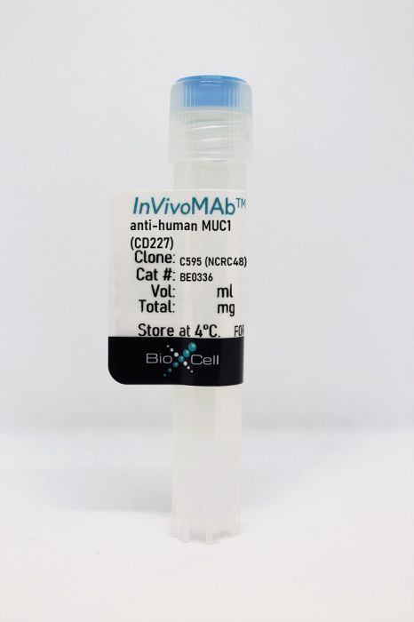
InVivoMAb anti-human MUC1 (CD227)
Product Details
The C595 (also known as NCRC48) monoclonal antibody reacts with human mucin 1 (MUC1), a 500-1000 kDa transmembrane glycoprotein with a large mucin-like extracellular domain. The epitope of C595 is the tetrameric motif (RPAP) within the protein core of MUC1. MUC1 is highly polymorphic and is expressed on most mucosal epithelial cells, including mammary gland and some hematopoietic cells. MUC1 is heavily glycosylated and plays a crucial role in the lubrication and protection of normal epithelial cells. MUC1 is abnormally expressed in a wide variety of malignancies, including colon, breast, ovarian, lung and pancreatic cancers. MUC1 promotes cancer cell growth and metastases through multiple mechanisms. The C595 antibody has been shown to suppress ovarian tumor xenograft growth in mice.Specifications
| Isotype | Mouse IgG3, κ |
|---|---|
| Recommended Isotype Control(s) | InVivoMAb polyclonal mouse IgG |
| Recommended Dilution Buffer | InVivoPure pH 7.0 Dilution Buffer |
| Conjugation | This product is unconjugated. Conjugation is available via our Antibody Conjugation Services. |
| Immunogen | Purified human MUC1 |
| Reported Applications |
in vivo administration in mouse xenograft models Immunohistochemistry (paraffin) Immunofluorescence in vitro cell cytotoxicity assay Western blot |
| Formulation |
PBS, pH 7.0 Contains no stabilizers or preservatives |
| Endotoxin |
<2EU/mg (<0.002EU/μg) Determined by LAL gel clotting assay |
| Purity |
>95% Determined by SDS-PAGE |
| Sterility | 0.2 µm filtration |
| Production | Purified from cell culture supernatant in an animal-free facility |
| Purification | Protein A |
| RRID | AB_2894756 |
| Molecular Weight | 150 kDa |
| Storage | The antibody solution should be stored at the stock concentration at 4°C. Do not freeze. |
Recommended Products
in vivo administration in mouse xenograft models
Wang, L., et al. (2011). "Anti-MUC1 monoclonal antibody (C595) and docetaxel markedly reduce tumor burden and ascites, and prolong survival in an in vivo ovarian cancer model" PLoS One 6(9): e24405. PubMed
MUC1 is associated with cellular transformation and tumorigenicity and is considered as an important tumor-associated antigen (TAA) for cancer therapy. We previously reported that anti-MUC1 monoclonal antibody C595 (MAb C595) plus docetaxel (DTX) increased efficacy of DTX alone and caused cultured human epithelial ovarian cancer (EOC) cells to undergo apoptosis. To further study the mechanisms of this combination-mediated apoptosis, we investigated the effectiveness of this combination therapy in vivo in an intraperitoneal (i.p.) EOC mouse model. OVCAR-3 cells were implanted intraperitoneally in female athymic nude mice and allowed to grow tumor and ascites. Mice were then treated with single MAb C595, DTX, combination test (MAb C595 and DTX), combination control (negative MAb IgG(3) and DTX) or vehicle control i.p. for 3 weeks. Treated mice were killed 4 weeks post-treatment. Ascites volume, tumor weight, CA125 levels from ascites and survival of animals were assessed. The expression of MUC1, CD31, Ki-67, TUNEL and apoptotic proteins in tumor xenografts was evaluated by immunohistochemistry. MAb C595 alone inhibited i.p. tumor growth and ascites production in a dose-dependent manner but did not obviously prevent tumor development. However, combination test significantly reduced ascites volume, tumor growth and metastases, CA125 levels in ascites and improved survival of treated mice compared with single agent-treated mice, combination control or vehicle control-treated mice (P<0.05). The data was in a good agreement with that from cultured cells in vitro. The mechanisms behind the observed effects could be through targeting MUC1 antigens, inhibition of tumor angiogenesis, and induction of apoptosis. Our results suggest that this combination approach can effectively reduce tumor burden and ascites, prolong survival of animals through induction of tumor apoptosis and necrosis, and may provide a potential therapy for advanced metastatic EOC.
Western Blot
Storr, S. J., et al. (2008). "The O-linked glycosylation of secretory/shed MUC1 from an advanced breast cancer patient’s serum" Glycobiology 18(6): 456-462. PubMed
MUC1 is a high molecular weight glycoprotein that is overexpressed in breast cancer. Aberrant O-linked glycosylation of MUC1 in cancer has been implicated in disease progression. We investigated the O-linked glycosylation of MUC1 purified from the serum of an advanced breast cancer patient. O-Glycans were released by hydrazinolysis and analyzed by liquid chromatography-electrospray ionization-mass spectrometry and by high performance liquid chromatography coupled with sequential exoglycosidase digestions. Core 1 type glycans (83%) dominated the profile which also confirmed high levels of sialylation: 80% of the glycans were mono-, di- or trisialylated. Core 2 type structures contributed approximately 17% of the assigned glycans and the oncofoetal Thomsen-Friedenreich (TF) antigen (Galbeta1-3GalNAc) accounted for 14% of the total glycans. Interestingly, two core 1 type glycans were identified that had sialic acid alpha2-8 linked to sialylated core 1 type structures (9% of the total glycan pool). This is the first O-glycan analysis of MUC1 from the serum of a breast cancer patient; the results suggest that amongst the cell lines commonly used to express recombinant MUC1 the T47D cell line processes glycans that are most similar to patient-derived material.
in vitro cell cytotoxicity assay, Immunofluorescence, Immunohistochemistry (paraffin)
Qu, C. F., et al. (2004). "MUC1 expression in primary and metastatic pancreatic cancer cells for in vitro treatment by (213)Bi-C595 radioimmunoconjugate" Br J Cancer 91(12): 2086-2093. PubMed
Control of micrometastatic pancreatic cancer remains a major objective in pancreatic cancer treatment. The overexpression of MUC1 mucin plays an important role in cancer metastasis. The aim of this study was to detect the expression of MUC1 in human primary tumour tissues and three pancreatic cancer cell lines (CAPAN-1, CFPAC-1 and PANC-1), and target MUC1-positive cancer cells in vitro using (213)Bi-C595 alpha-immunoconjugate (AIC). The expression of MUC1 on pancreatic tumour tissues and cancer cell lines was performed by immunohistochemistry and further confirmed by confocal microscope and flow cytometry analysis on the cell surface. Cytotoxicity of (213)Bi-C595 was tested by MTS assay. Apoptosis was documented using TUNEL assay. Overexpression of MUC1 was found in approximately 90% of tested tumour samples and the three pancreatic cancer cell lines. (213)Bi-C595 is specifically cytotoxic to pancreatic cancer cells in a concentration-dependent fashion. These results suggest that overexpression of MUC1 in pancreatic cancer is a useful target, and that the novel (213)Bi-C595 AIC selectively targets pancreatic cancer cells in vitro. (213)Bi-C595 may be a useful agent for the treatment of micrometastases or minimal residual disease (MRD) in pancreatic cancer patients with overexpression of MUC1 antigen.


