InVivoPlus anti-mouse/human/rat/monkey/hamster/canine/bovine TGF-β
Product Description
Specifications
| Isotype | Mouse IgG1, κ |
|---|---|
| Recommended Isotype Control(s) | InVivoPlus mouse IgG1 isotype control, unknown specificity |
| Recommended Dilution Buffer | InVivoPure pH 7.0 Dilution Buffer |
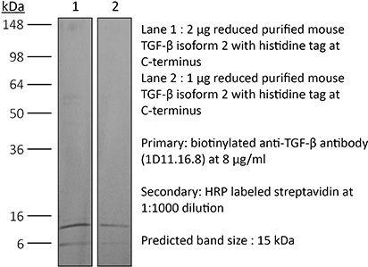
| Conjugation | This product is unconjugated. Conjugation is available via our Antibody Conjugation Services. |
| Immunogen | Bovine TGFβ isoform 2 |
| Reported Applications |
in vivo TGFβ neutralization in vitro TGFβ neutralization Western blot |
| Formulation |
PBS, pH 7.0 Contains no stabilizers or preservatives |
| Endotoxin* |
≤0.5EU/mg (≤0.0005EU/μg) Determined by LAL assay |
| Aggregation* |
<5% Determined by SEC |
| Purity |
≥95% Determined by SDS-PAGE |
| Sterility | 0.2 µm filtration |
| Production | Purified from cell culture supernatant in an animal-free facility |
| Purification | Protein G |
| RRID | AB_1107757 |
| Molecular Weight | 150 kDa |
| Murine Pathogen Tests* |
Ectromelia/Mousepox Virus: Negative Hantavirus: Negative K Virus: Negative Lactate Dehydrogenase-Elevating Virus: Negative Lymphocytic Choriomeningitis virus: Negative Mouse Adenovirus: Negative Mouse Cytomegalovirus: Negative Mouse Hepatitis Virus: Negative Mouse Minute Virus: Negative Mouse Norovirus: Negative Mouse Parvovirus: Negative Mouse Rotavirus: Negative Mycoplasma Pulmonis: Negative Pneumonia Virus of Mice: Negative Polyoma Virus: Negative Reovirus Screen: Negative Sendai Virus: Negative Theiler’s Murine Encephalomyelitis: Negative |
| Storage | The antibody solution should be stored at the stock concentration at 4°C. Do not freeze. |
| Need a Custom Formulation? | See All Antibody Customization Options |
Application References
-
Leon, B., et al (2014). "FoxP3+ regulatory T cells promote influenza-specific Tfh responses by controlling IL-2 availability" Nat Commun 5: 3495.
PubMed
Here, we test the role of FoxP3(+) regulatory T cells (Tregs) in controlling T follicular helper (Tfh) and germinal centre (GC) B-cell responses to influenza. In contrast to the idea that Tregs suppress T-cell responses, we find that Treg depletion severely reduces the Tfh cell response to influenza virus. Furthermore, Treg depletion prevents the accumulation of influenza-specific GCs. These effects are not due to alterations in TGFbeta availability or a precursor-progeny relationship between Tregs and Tfh cells, but are instead mediated by increased availability of IL-2, which suppresses the differentiation of Tfh cells and as a consequence, compromises the GC B response. Thus, Tregs promote influenza-specific GC responses by preventing excessive IL-2 signalling, which suppresses Tfh cell differentiation.
-
Choi, Y. S., et al (2015). "LEF-1 and TCF-1 orchestrate TFH differentiation by regulating differentiation circuits upstream of the transcriptional repressor Bcl6" Nat Immunol 16(9): 980-990.
PubMed
Follicular helper T cells (TFH cells) are specialized effector CD4(+) T cells that help B cells develop germinal centers (GCs) and memory. However, the transcription factors that regulate the differentiation of TFH cells remain incompletely understood. Here we report that selective loss of Lef1 or Tcf7 (which encode the transcription factor LEF-1 or TCF-1, respectively) resulted in TFH cell defects, while deletion of both Lef1 and Tcf7 severely impaired the differentiation of TFH cells and the formation of GCs. Forced expression of LEF-1 enhanced TFH differentiation. LEF-1 and TCF-1 coordinated such differentiation by two general mechanisms. First, they established the responsiveness of naive CD4(+) T cells to TFH cell signals. Second, they promoted early TFH differentiation via the multipronged approach of sustaining expression of the cytokine receptors IL-6Ralpha and gp130, enhancing expression of the costimulatory receptor ICOS and promoting expression of the transcriptional repressor Bcl6.
-
Bodogai, M., et al (2015). "Immunosuppressive and Prometastatic Functions of Myeloid-Derived Suppressive Cells Rely upon Education from Tumor-Associated B Cells" Cancer Res 75(17): 3456-3465.
PubMed
Myeloid-derived suppressive cells (MDSC) have been reported to promote metastasis, but the loss of cancer-induced B cells/B regulatory cells (tBreg) can block metastasis despite MDSC expansion in cancer. Here, using multiple murine tumor models and human MDSC, we show that MDSC populations that expand in cancer have only partially primed regulatory function and limited prometastatic activity unless they are fully educated by tBregs. Cancer-induced tBregs directly activate the regulatory function of both the monocyte and granulocyte subpopulations of MDSC, relying, in part, on TgfbetaR1/TgfbetaR2 signaling. MDSC fully educated in this manner exhibit an increased production of reactive oxygen species and NO and more efficiently suppress CD4(+) and CD8(+) T cells, thereby promoting tumor growth and metastasis. Thus, loss of tBregs or TgfbetaR deficiency in MDSC is sufficient to disable their suppressive function and to block metastasis. Overall, our data indicate that cancer-induced B cells/B regulatory cells are important regulators of the immunosuppressive and prometastatic functions of MDSC.
-
Clemente-Casares, X., et al (2016). "Expanding antigen-specific regulatory networks to treat autoimmunity" Nature 530(7591): 434-440.
PubMed
Regulatory T cells hold promise as targets for therapeutic intervention in autoimmunity, but approaches capable of expanding antigen-specific regulatory T cells in vivo are currently not available. Here we show that systemic delivery of nanoparticles coated with autoimmune-disease-relevant peptides bound to major histocompatibility complex class II (pMHCII) molecules triggers the generation and expansion of antigen-specific regulatory CD4(+) T cell type 1 (TR1)-like cells in different mouse models, including mice humanized with lymphocytes from patients, leading to resolution of established autoimmune phenomena. Ten pMHCII-based nanomedicines show similar biological effects, regardless of genetic background, prevalence of the cognate T-cell population or MHC restriction. These nanomedicines promote the differentiation of disease-primed autoreactive T cells into TR1-like cells, which in turn suppress autoantigen-loaded antigen-presenting cells and drive the differentiation of cognate B cells into disease-suppressing regulatory B cells, without compromising systemic immunity. pMHCII-based nanomedicines thus represent a new class of drugs, potentially useful for treating a broad spectrum of autoimmune conditions in a disease-specific manner.
Product Citations
-
Erythropoietin receptor on cDC1s dictates immune tolerance.
In Nature on 1 February 2026 by Zhang, X., McGinnis, C. S., et al.
PubMed
Type 1 conventional dendritic cells (cDC1s) are unique in their efferocytosis1 and cross-presenting abilities2, resulting in antigen-specific T cell immunity3 or tolerance4-8. However, the mechanisms that underlie cDC1 tolerogenic function remain largely unknown. Here we show that the erythropoietin receptor (EPOR) acts as a critical switch that determines the tolerogenic function of cDC1s and the threshold of antigen-specific T cell responses. In total lymphoid irradiation-induced allograft tolerance9,10, cDC1s upregulate EPOR expression, and conditional knockout of EPOR in cDC1s diminishes antigen-specific induction and expansion of FOXP3+ regulatory T (Treg) cells, resulting in allograft rejection. Mechanistically, EPOR promotes efferocytosis-induced tolerogenic maturation7,11 of splenic cDC1s towards late-stage CCR7+ cDC1s characterized by increased expression of the integrin β8 gene12 (Itgb8), and conditional knockout of Itgb8 in cDC1s impairs tolerance induced by total lymphoid irradiation plus anti-thymocyte serum. Migratory cDC1s in peripheral lymph nodes preferentially express EPOR, and their FOXP3+ Treg cell-inducing capacity is enhanced by erythropoietin. Reciprocally, loss of EPOR enables immunogenic maturation of peripheral lymph node migratory and splenic CCR7+ cDC1s by upregulating genes involved in MHC class II- and class I-mediated antigen presentation, cross-presentation and costimulation. EPOR deficiency in cDC1s reduces tumour growth by enhancing anti-tumour T cell immunity, particularly increasing the generation of precursor exhausted tumour antigen-specific CD8+ T cells13 in tumour-draining lymph nodes and supporting their maintenance within tumours, while concurrently reducing intratumoural Treg cells. Targeting EPOR on cDC1s to induce or inhibit T cell immune tolerance could have potential for treating a variety of diseases.
-
Bidirectional CRISPR screens decode a GLIS3-dependent fibrotic cell circuit.
In Nature on 1 February 2026 by Pokatayev, V., Jaiswal, A., et al.
PubMed
The stromal cell compartment plays a central part in the maintenance of tissue homeostasis by coordinating with the immune system throughout inception, amplification and resolution of inflammation1. Chronic inflammation can impede the phased regulation of tissue restitution, resulting in the scarring complication of fibrosis. In inflammatory bowel disease, stromal fibroblasts have been implicated in treatment-refractory disease and fibrosis2,3; however, their mechanisms of activation have remained undefined. Through integrative single-cell and spatial profiling of intestinal tissues from patients with inflammatory bowel disease, we uncovered a pathological cell nexus centred on inflammation-associated fibroblasts. These fibroblasts were induced by proinflammatory macrophages (FCN1+IL1B+) and, in turn, produced profibrotic cytokine IL-11. We investigated the inflammation-associated fibroblast activation program at a mechanistic level using genome-wide CRISPR knockout and activation screens and identified the transcription factor GLIS3 as a key regulator of a gene regulatory network governing expression of inflammatory and fibrotic genes. We further demonstrated that the magnitude of the GLIS3 gene expression program in intestinal biopsies could be used to stratify patients with ulcerative colitis by disease severity, and that fibroblast-specific deletion of Glis3 in mice alleviated pathological features of chronic colitis. Taken together, our findings identify a critical immune-stromal cell circuit that functions as a central node in the inflammation-fibrosis cycle.
-
Targeting TGF-β signaling, oxidative stress, and cellular senescence rescues osteoporosis in gerodermia osteodysplastica.
In Aging Cell on 1 December 2024 by Chan, W. L., Bucher, C. H., et al.
PubMed
GORAB is a key regulator of Golgi vesicle transport and protein glycanation. Loss of GORAB function in gerodermia osteodysplastica (GO) causes shortening of glycosaminoglycan chains, leading to extracellular matrix disorganization that results in wrinkled skin, osteoporosis and elevated TGF-β signaling. In this study, we investigated the role of TGF-β-signaling, oxidative stress, and resulting cellular senescence in the osteoporosis phenotype of GO. Treatment of GorabPrx1 conditional knockouts with the TGF-β neutralizing antibody 1D11 rescued the trabecular bone loss, indicating that TGF-β overactivation causes osteoporosis in GO. Using an inducible knockout system, we demonstrated that TGF-β dysregulation was not a cell-intrinsic effect of GORAB inactivation, but a consequence of a disorganized extracellular matrix. Enhanced TGF-β signaling caused elevated Nox4 expression in GorabPrx1 mutants and in GO patients' fibroblasts, resulting in overproduction of mitochondrial superoxide. The resulting oxidative stress was detected in GORAB null cells and also in wildtype bystander cells. The same effect was observed in zebrafish after TALEN-mediated gorab inactivation, indicating that the pathway is evolutionarily conserved. Treating GorabPrx1 mutants with the antioxidant N-acetylcysteine ameliorated the osteoporosis phenotype. TGF-β induced oxidative stress coincided with accumulation of DNA damage and elevated expression of senescence markers. Inactivation of Cdkn2a in the GorabPrx1 rescued the osteoporosis phenotype. Reduced colony formation and altered subpopulations of bone marrow stromal cells were normalized upon inactivation of Cdkn2a, thus further demonstrating the relevance of cellular senescence in the pathogenesis. Our results shed light on the causative role of a TGF-β-Nox4-senescence axis and therapeutic strategies for GO.
-
Natural killer cell function is regulated by TGF-β signaling in pregnancy and tumor progression
In bioRxiv on 22 September 2024 by Yalin, A., Wang, S. Y., et al.