
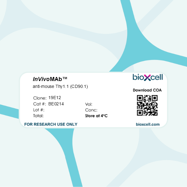
InVivoMAb anti-mouse Thy1.1 (CD90.1)
Product Description
Specifications
| Isotype | Mouse IgG2a, κ |
|---|---|
| Recommended Isotype Control(s) | InVivoMAb mouse IgG2a isotype control, unknown specificity |
| Recommended Dilution Buffer | InVivoPure pH 7.0 Dilution Buffer |
| Conjugation | This product is unconjugated. Conjugation is available via our Antibody Conjugation Services. |
| Immunogen | AKR mouse SL3 leukemia cells |
| Reported Applications | in vivo T cell depletion |
| Formulation |
PBS, pH 7.0 Contains no stabilizers or preservatives |
| Endotoxin |
≤1EU/mg (≤0.001EU/μg) Determined by LAL assay |
| Purity |
≥95% Determined by SDS-PAGE |
| Sterility | 0.2 µm filtration |
| Production | Purified from cell culture supernatant in an animal-free facility |
| Purification | Protein G |
| RRID | AB_2687700 |
| Molecular Weight | 150 kDa |
| Storage | The antibody solution should be stored at the stock concentration at 4°C. Do not freeze. |
| Need a Custom Formulation? | See All Antibody Customization Options |
Application References
-
Kim, J., et al (2015). "Memory programming in CD8(+) T-cell differentiation is intrinsic and is not determined by CD4 help" Nat Commun 6: 7994.
PubMed
CD8(+) T cells activated without CD4(+) T-cell help are impaired in memory expansion. To understand the underlying cellular mechanism, here we track the dynamics of helper-deficient CD8(+) T-cell response to a minor histocompatibility antigen by phenotypic and in vivo imaging analyses. Helper-deficient CD8(+) T cells show reduced burst expansion, rapid peripheral egress, delayed antigen clearance and continuous activation, and are eventually exhausted. Contrary to the general consensus that CD4 help encodes memory programmes in CD8(+) T cells and helper-deficient CD8(+) T cells are abortive, these cells can differentiate into effectors and memory precursors. Importantly, accelerating antigen clearance or simply increasing the burst effector size enables generation of memory cells by CD8(+) T cells, regardless of CD4 help. These results suggest that the memory programme is CD8(+) T-cell-intrinsic, and provide insight into the role of CD4 help in CD8(+) T-cell responses.
-
Campisi, L., et al (2016). "Apoptosis in response to microbial infection induces autoreactive TH17 cells" Nat Immunol. doi : 10.1038/ni.3512.
PubMed
Microbial infections often precede the onset of autoimmunity. How infections trigger autoimmunity remains poorly understood. We investigated the possibility that infection might create conditions that allow the stimulatory presentation of self peptides themselves and that this might suffice to elicit autoreactive T cell responses that lead to autoimmunity. Self-reactive CD4+ T cells are major drivers of autoimmune disease, but their activation is normally prevented through regulatory mechanisms that limit the immunostimulatory presentation of self antigens. Here we found that the apoptosis of infected host cells enabled the presentation of self antigens by major histocompatibility complex class II molecules in an inflammatory context. This was sufficient for the generation of an autoreactive TH17 subset of helper T cells, prominently associated with autoimmune disease. Once induced, the self-reactive TH17 cells promoted auto-inflammation and autoantibody generation. Our findings have implications for how infections precipitate autoimmunity.
-
Badell, I. R., et al (2015). "Pathogen Stimulation History Impacts Donor-Specific CD8 T Cell Susceptibility to Costimulation/Integrin Blockade-Based Therapy" Am J Transplant. doi : 10.1111/ajt.13399.
PubMed
Recent studies have shown that the quantity of donor-reactive memory T cells is an important factor in determining the relative heterologous immunity barrier posed during transplantation. Here, we hypothesized that the quality of T cell memory also potently influences the response to costimulation blockade-based immunosuppression. Using a murine skin graft model of CD8+ memory T cell-mediated costimulation blockade resistance, we elicited donor-reactive memory T cells using three distinct types of pathogen infections. Strikingly, we observed differential efficacy of a costimulation and integrin blockade regimen based on the type of pathogen used to elicit the donor-reactive memory T cell response. Intriguingly, the most immunosuppression-sensitive memory T cell populations were composed primarily of central memory cells that possessed greater recall potential, exhibited a less differentiated phenotype, and contained more multi-cytokine producers. These data, therefore, demonstrate that the memory T cell barrier is dependent on the specific type of pathogen infection via which the donor-reactive memory T cells are elicited, and suggest that the immune stimulation history of a given transplant patient may profoundly influence the relative barrier posed by heterologous immunity during transplantation.
-
Liu, B., et al (2015). "Collaborative interactions between type 2 innate lymphoid cells and antigen-specific CD4+ Th2 cells exacerbate murine allergic airway diseases with prominent eosinophilia" J Immunol 194(8): 3583-3593.
PubMed
Type-2 innate lymphoid cells (ILC2s) and the acquired CD4(+) Th2 and Th17 cells contribute to the pathogenesis of experimental asthma; however, their roles in Ag-driven exacerbation of chronic murine allergic airway diseases remain elusive. In this study, we report that repeated intranasal rechallenges with only OVA Ag were sufficient to trigger airway hyperresponsiveness, prominent eosinophilic inflammation, and significantly increased serum OVA-specific IgG1 and IgE in rested mice that previously developed murine allergic airway diseases. The recall response to repeated OVA inoculation preferentially triggered a further increase of lung OVA-specific CD4(+) Th2 cells, whereas CD4(+) Th17 and ILC2 cell numbers remained constant. Furthermore, the acquired CD4(+) Th17 cells in Stat6(-/-)/IL-17-GFP mice, or innate ILC2s in CD4(+) T cell-ablated mice, failed to mount an allergic recall response to OVA Ag. After repeated OVA rechallenge or CD4(+) T cell ablation, the increase or loss of CD4(+) Th2 cells resulted in an enhanced or reduced IL-13 production by lung ILC2s in response to IL-25 and IL-33 stimulation, respectively. In return, ILC2s enhanced Ag-mediated proliferation of cocultured CD4(+) Th2 cells and their cytokine production, and promoted eosinophilic airway inflammation and goblet cell hyperplasia driven by adoptively transferred Ag-specific CD4(+) Th2 cells. Thus, these results suggest that an allergic recall response to recurring Ag exposures preferentially triggers an increase of Ag-specific CD4(+) Th2 cells, which facilitates the collaborative interactions between acquired CD4(+) Th2 cells and innate ILC2s to drive the exacerbation of a murine allergic airway diseases with an eosinophilic phenotype.
Product Citations
-
Systemic pre-conditioning favors effector over exhausted CD8 T-cell subsets following Sup2-IL33 armored CAR T-cell therapy.
In J Immunother Cancer on 3 June 2026 by Ferry, S. L., Abdullah, L., et al.
PubMed
Systemic pre-conditioning prior to chimeric antigen receptor (CAR) T-cell infusion enhances engraftment and increases infiltration and activity of adoptively transferred cells within the tumor. Despite promising responses in some patients, additional strategies are needed to enhance overall efficacy of CAR T cells in patients with solid tumors. Armoring CAR T cells to express immunomodulatory cytokines can enhance treatment efficacy. In this study, we assess how systemic pre-conditioning impacts CAR T cells alone and in combination with interleukin (IL)-2 superkine (Sup2) and IL-33 cytokine armoring in the context of B16F10 melanoma.
-
Expression of an interleukin-2 partial agonist enhances regulatory T cell persistence and efficacy in mouse autoimmune models.
In Nat Commun on 27 May 2025 by Robert, J., Feuillolay, M., et al.
PubMed
Regulatory T (Treg)-based cell therapy holds promise for autoimmune and inflammatory diseases, yet challenges remain regarding the functional stability and persistence of transferred Tregs. Here we engineer Tregs to express a partial agonist form of IL-2 (IL-2pa) to enhance persistence while avoiding toxicity from excessive signaling. Mouse Tregs expressing wild-type IL-2 (Tregs-IL2wt) have only a transient growth advantage, limited by toxicity from likely excessive signaling. By contrast, mouse Tregs-IL2pa exhibit sustained expansion, long-term survival in immunocompetent mice for over a year, and bystander expansion of endogenous Tregs. Tregs-IL2pa maintain a stable activated phenotype, Treg-specific demethylation, and a diverse TCR repertoire. In vivo, prophylactic transfer of Tregs-IL2pa ameliorates multi-organ autoimmunity in a Treg depletion-induced mouse autoimmune model. Lastly, compared with control Treg, human Tregs-IL2pa show enhanced survival in the IL-2-depleted environment of immune-deficient mice and improved control of xenogeneic graft-versus-host disease. Our results thus show that IL-2pa self-sufficiency enhances the stability, durability and efficacy of Treg therapies in preclinical settings.
-
Repetitive antigen stimulation in the periphery dictates the composition and recall responses of brain-resident memory CD8+ T cells.
In Cell Rep on 25 February 2025 by Mix, M. R., van de Wall, S., et al.
PubMed
The human brain harbors virus-specific, tissue-resident memory (TRM) CD8+ T cells. However, the impact of repeated peripheral viral infection on the generation, phenotype, localization, and recall responses of brain TRM remains elusive. Here, utilizing two murine models of peripheral viral infection, we demonstrate that circulating memory CD8+ T cells with previous antigen exposure exhibit a markedly reduced capacity to form brain TRM compared to naive CD8+ T cells. Repetitively stimulated brain TRM also demonstrate differential inhibitory receptor expression, preserved functionality, and divergent localization patterns compared to primary memory counterparts. Despite these differences, repetitively stimulated brain TRM provide similar protection against intracranial infection as primary populations with superior recall-based recruitment of peripheral lymphocytes. As CD8+ T cells may distinctly seed the brain with each repeated infection of the same host, these findings point to heterogeneity in the brain TRM pool that is dictated by prior peripheral antigen stimulation history.
-
Dynamic landscapes and protective immunity coordinated by influenza-specific lung-resident memory CD8+ T cells revealed by intravital imaging.
In Immunity on 13 August 2024 by van de Wall, S., Anthony, S. M., et al.
PubMed
Lung-tissue-resident memory (TRM) CD8+ T cells are critical for heterosubtypic immunity against influenza virus (IAV) reinfection. How TRM cells surveil the lung, respond to infection, and interact with other cells remains unresolved. Here, we used IAV infection of mice in combination with intravital and static imaging to define the spatiotemporal dynamics of lung TRM cells before and after recall infection. CD69+CD103+ TRM cells preferentially localized to lung sites of prior IAV infection, where they exhibited patrolling behavior. After rechallenge, lung TRM cells formed tight clusters in an antigen-dependent manner. Transcriptomic analysis of IAV-specific TRM cells revealed the expression of several factors that regulate myeloid cell biology. In vivo rechallenge experiments demonstrated that protection elicited by TRM cells is orchestrated in part by interferon (IFN)-γ-mediated recruitment of inflammatory monocytes into the lungs. Overall, these data illustrate the dynamic landscapes of CD103+ lung TRM cells that mediate early protective immunity against IAV infection.