InVivoMAb anti-mouse IL-6
Product Description
Specifications
| Isotype | Rat IgG1, κ |
|---|---|
| Recommended Isotype Control(s) | InVivoMAb rat IgG1 isotype control, anti-horseradish peroxidase |
| Recommended Dilution Buffer | InVivoPure pH 7.0 Dilution Buffer |
| Conjugation | This product is unconjugated. Conjugation is available via our Antibody Conjugation Services. |
| Immunogen | Recombinant mouse IL-6 |
| Reported Applications |
in vivo IL-6 neutralization in vitro IL-6 neutralization |
| Formulation |
PBS, pH 7.0 Contains no stabilizers or preservatives |
| Endotoxin |
≤1EU/mg (≤0.001EU/μg) Determined by LAL assay |
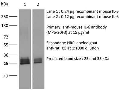
| Purity |
≥95% Determined by SDS-PAGE |
| Sterility | 0.2 µm filtration |
| Production | Purified from cell culture supernatant in an animal-free facility |
| Purification | Protein G |
| RRID | AB_1107709 |
| Molecular Weight | 150 kDa |
| Storage | The antibody solution should be stored at the stock concentration at 4°C. Do not freeze. |
| Need a Custom Formulation? | See All Antibody Customization Options |
Application References
-
Khmaladze, I., et al (2014). "Mannan induces ROS-regulated, IL-17A-dependent psoriasis arthritis-like disease in mice" Proc Natl Acad Sci U S A 111(35): E3669-3678.
PubMed
Psoriasis (Ps) and psoriasis arthritis (PsA) are poorly understood common diseases, induced by unknown environmental factors, affecting skin and articular joints. A single i.p. exposure to mannan from Saccharomyces cerevisiae induced an acute inflammation in inbred mouse strains resembling human Ps and PsA-like disease, whereas multiple injections induced a relapsing disease. Exacerbation of disease severity was observed in mice deficient for generation of reactive oxygen species (ROS). Interestingly, restoration of ROS production, specifically in macrophages, ameliorated both skin and joint disease. Neutralization of IL-17A, mainly produced by gammadelta T cells, completely blocked disease symptoms. Furthermore, mice depleted of granulocytes were resistant to disease development. In contrast, certain acute inflammatory mediators (C5, Fcgamma receptor III, mast cells, and histamine) and adaptive immune players (alphabeta T and B cells) were redundant in disease induction. Hence, we propose that mannan-induced activation of macrophages leads to TNF-alpha secretion and stimulation of local gammadelta T cells secreting IL-17A. The combined action of activated macrophages and IL-17A produced in situ drives neutrophil infiltration in the epidermis and dermis of the skin, leading to disease manifestations. Thus, our finding suggests a new mechanism triggered by exposure to exogenous microbial components, such as mannan, that can induce and exacerbate Ps and PsA.
-
Berger, H., et al (2013). "SOCS3 transactivation by PPARgamma prevents IL-17-driven cancer growth" Cancer Res 73(12): 3578-3590.
PubMed
Activation of the transcription factor PPARgamma by the n-3 fatty acid docosahexaenoic acid (DHA) is implicated in controlling proinflammatory cytokine secretion, but the intracellular signaling pathways engaged by PPARgamma are incompletely characterized. Here, we identify the adapter-encoding gene SOCS3 as a critical transcriptional target of PPARgamma. SOCS3 promoter binding and gene transactivation by PPARgamma was associated with a repression in differentiation of proinflammatory T-helper (TH)17 cells. Accordingly, TH17 cells induced in vitro displayed increased SOCS3 expression and diminished capacity to produce interleukin (IL)-17 following activation of PPARgamma by DHA. Furthermore, naive CD4 T cells derived from mice fed a DHA-enriched diet displayed less capability to differentiate into TH17 cells. In two different mouse models of cancer, DHA prevented tumor outgrowth and angiogenesis in an IL-17-dependent manner. Altogether, our results uncover a novel molecular pathway by which PPARgamma-induced SOCS3 expression prevents IL-17-mediated cancer growth.
-
Benevides, L., et al (2015). "IL17 Promotes Mammary Tumor Progression by Changing the Behavior of Tumor Cells and Eliciting Tumorigenic Neutrophils Recruitment" Cancer Res 75(18): 3788-3799.
PubMed
The aggressiveness of invasive ductal carcinoma (IDC) of the breast is associated with increased IL17 levels. Studying the role of IL17 in invasive breast tumor pathogenesis, we found that metastatic primary tumor-infiltrating T lymphocytes produced elevated levels of IL17, whereas IL17 neutralization inhibited tumor growth and prevented the migration of neutrophils and tumor cells to secondary disease sites. Tumorigenic neutrophils promote disease progression, producing CXCL1, MMP9, VEGF, and TNFalpha, and their depletion suppressed tumor growth. IL17A also induced IL6 and CCL20 production in metastatic tumor cells, favoring the recruitment and differentiation of Th17. In addition, IL17A changed the gene-expression profile and the behavior of nonmetastatic tumor cells, causing tumor growth in vivo, confirming the protumor role of IL17. Furthermore, high IL17 expression was associated with lower disease-free survival and worse prognosis in IDC patients. Thus, IL17 blockade represents an attractive approach for the control of invasive breast tumors. Cancer Res; 75(18); 3788-99. (c)2015 AACR.
-
Kugler, D. G., et al (2013). "CD4+ T cells are trigger and target of the glucocorticoid response that prevents lethal immunopathology in toxoplasma infection" J Exp Med 210(10): 1919-1927.
PubMed
Synthetic glucocorticoids (GCs) are commonly used in the treatment of inflammatory diseases, but the role of endogenous GCs in the regulation of host-protective immune responses is poorly understood. Here we show that GCs are induced during acute Toxoplasma gondii infection and directly control the T cell response to the parasite. When infected with toxoplasma, mice that selectively lack GC receptor (GR) expression in T cells (GR(lck-Cre)) rapidly succumb to infection despite displaying parasite burdens indistinguishable from control animals and unaltered levels of the innate cytokines IL-12 and IL-27. Mortality in the GR(lck-Cre) mice was associated with immunopathology and hyperactive Th1 cell function as revealed by enhanced IFN-gamma and TNF production in vivo. Unexpectedly, these CD4(+) T lymphocytes also overexpressed IL-10. Importantly, CD4(+) T cell depletion in wild-type or GR(lck-Cre) mice led to ablation of the GC response to infection. Moreover, in toxoplasma-infected RAG(-/-) animals, adoptive transfer of CD4(+) T lymphocytes was required for GC induction. These findings establish a novel IL-10-independent immunomodulatory circuit in which CD4(+) T cells trigger a GC response that in turn dampens their own effector function. In the case of T. gondii infection, this self-regulatory pathway is critical for preventing collateral tissue damage and promoting host survival.
Product Citations
-
Hypoxia-Induced Fibroblast IL-6 Promotes Immunosuppressive Macrophage Phenotypes in Pancreatic Cancer.
In Cells on 13 April 2026 by Hannifin, S., Mello, A. M., et al.
PubMed
Pancreatic ductal adenocarcinoma (PDAC) is a lethal malignancy characterized by a dense fibroinflammatory stroma and profound hypoxia. Using pancreatic stellate cell-tumor organoid coculture models and single-cell RNA sequencing analyses, we uncover that hypoxia-driven fibroblast reprogramming promotes immunosuppressive macrophage phenotypes in PDAC. Mechanistically, hypoxia acts through tumor-fibroblast crosstalk to increase IL-6 expression in fibroblasts; in turn, fibroblast-derived IL-6 induces expression of arginase 1 (ARG1), a key mediator of immunosuppression, in macrophages via activation of the JAK/STAT signaling pathway. Consistent with these findings, macrophages enriched for hypoxia signatures are strongly associated with elevated immunosuppression programs and IL6/JAK/STAT3 signaling signatures in PDAC. Our study reveals a paracrine mechanism by which hypoxia coordinates tumor cell, fibroblast, and macrophage interactions to promote immune suppression in PDAC.
-
Three-dimensional bioprinted hiHeps hepatorganoids with enhanced hepatic functions for the treatment of liver failure and promotion of liver regeneration.
In Bioact Mater on 1 April 2026 by Ye, Z., Yan, J., et al.
PubMed
The intricate architecture of the liver, combined with its limited regenerative ability in severe injury, has spurred the development of innovative approaches for hepatic repair and functional restoration. Three-dimensional (3D) bioprinting provides a unique platform to reconstruct biomimetic liver tissues through spatially orchestrated cellular and extracellular matrix integration. Here, we developed 3D bioprinted hepatorganoids derived from human induced hepatocytes (hiHeps), which faithfully recapitulate the native lobular zonation crucial for spatially segregated metabolic functions in vivo. 3D bioprinted hiHeps hepatorganoids (3DP-HHO) exhibited markedly enhanced metabolic performance, including improved glucose and lipid regulation and elevated albumin synthesis, highlighting their potential as advanced liver models. The hepatorganoids demonstrated robust regenerative potential, which reversed chronic liver fibrosis (CLF) by resolving pathological collagen deposition, rescued acute liver failure (ALF) through rapid functional compensation, and accelerated liver regeneration in partial hepatectomy models by stimulating endogenous hepatocyte proliferation. Preclinical validation of post-hepatectomy liver failure (PHLF) model revealed that the implantation of 3DP-HHO significantly improved survival outcomes and promoted liver regeneration, compared to controls. In the future, by integrating patient-specific cells with regulable 3D microenvironments, our platform will achieve superior functional integration and regenerative efficacy over conventional approaches. This work establishes a paradigm for bioengineered liver grafts that actively drive tissue repair and regeneration. As a scalable and physiologically relevant approach, these bioprinted hepatic units pioneer a transformative strategy in regenerative hepatology, addressing critical challenges in treating liver failure and post-resection recovery while illuminating microenvironmental factors essential for organ-level regeneration.
-
Tumor-secreted BDNF selectively depletes macrophage NAD+ via CD73 upregulation to suppress antitumor immunity
In Research Square on 6 March 2026 by Huang, M., Wang, T., et al.
-
Tet2 deficiency-induced expansion of monocyte-derived macrophages promotes liver fibrosis.
In J Exp Med on 2 February 2026 by Feng, J., Wu, B., et al.
PubMed
Clonal hematopoiesis driven by Tet2 deficiency in myeloid cells (TetΔMye) is prevalent in elderly individuals; however, the role of Tet2ΔMye in liver fibrosis pathogenesis remains elusive. In this study, we demonstrated that Tet2-deficient monocyte-derived macrophages (MDMs) promoted cellular expansion and elevated C-C motif chemokine ligand 2/8 (Ccl2/8) secretion by stabilizing their mRNAs through 5hmC-mediated alterations in RNA-protein interactions. These chemokines engaged with the upregulated C-C motif chemokine receptor (Ccr2/3) on Tet2-/- monocytes, forming a positive feedback loop that amplified pro-inflammatory MDMs (pMDMs) accumulation in liver. Tet2-/- pMDMs activated hepatic stellate cells through IL-6, driving extracellular matrix deposition and fibrotic progression. Pharmacological inhibition of Ccl2/Ccl8 with Bindarit attenuated MDMs accumulation and liver fibrosis, whereas combined therapy with Bindarit and IL-6 neutralization synergistically suppressed liver fibrosis in Tet2ΔMye mice and aged chimeric models recapitulating Tet2ΔMye-related myeloid hematopoiesis. These findings present the mechanism that Tet2ΔMye aggravates liver fibrosis and highlight MDMs depletion plus IL-6 neutralization as a promising therapy for liver fibrosis in patients with Tet2ΔMye-related myeloid hematopoiesis.