
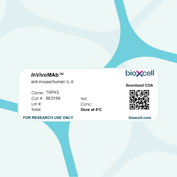
InVivoMAb anti-mouse/human IL-5
in vivo eosinophil depletion
Product Description
Specifications
| Isotype | Rat IgG1, κ |
|---|---|
| Recommended Isotype Control(s) | InVivoMAb rat IgG1 isotype control, anti-horseradish peroxidase |
| Recommended Dilution Buffer | InVivoPure pH 7.0 Dilution Buffer |
| Conjugation | This product is unconjugated. Conjugation is available via our Antibody Conjugation Services. |
| Immunogen | Mouse partially-purified T cell clone supernatant |
| Reported Applications |
in vivo IL-5 neutralization in vivo eosinophil depletion |
| Formulation |
PBS, pH 7.0 Contains no stabilizers or preservatives |
| Endotoxin |
≤1EU/mg (≤0.001EU/μg) Determined by LAL assay |
| Purity |
≥95% Determined by SDS-PAGE |
| Sterility | 0.2 µm filtration |
| Production | Purified from cell culture supernatant in an animal-free facility |
| Purification | Protein G |
| RRID | AB_10950522 |
| Molecular Weight | 150 kDa |
| Storage | The antibody solution should be stored at the stock concentration at 4°C. Do not freeze. |
| Need a Custom Formulation? | See All Antibody Customization Options |
Application References
-
Moynihan, K. D., et al (2016). "Eradication of large established tumors in mice by combination immunotherapy that engages innate and adaptive immune responses" Nat Med. doi : 10.1038/nm.4200.
PubMed
Checkpoint blockade with antibodies specific for cytotoxic T lymphocyte-associated protein (CTLA)-4 or programmed cell death 1 (PDCD1; also known as PD-1) elicits durable tumor regression in metastatic cancer, but these dramatic responses are confined to a minority of patients. This suboptimal outcome is probably due in part to the complex network of immunosuppressive pathways present in advanced tumors, which are unlikely to be overcome by intervention at a single signaling checkpoint. Here we describe a combination immunotherapy that recruits a variety of innate and adaptive immune cells to eliminate large tumor burdens in syngeneic tumor models and a genetically engineered mouse model of melanoma; to our knowledge tumors of this size have not previously been curable by treatments relying on endogenous immunity. Maximal antitumor efficacy required four components: a tumor-antigen-targeting antibody, a recombinant interleukin-2 with an extended half-life, anti-PD-1 and a powerful T cell vaccine. Depletion experiments revealed that CD8+ T cells, cross-presenting dendritic cells and several other innate immune cell subsets were required for tumor regression. Effective treatment induced infiltration of immune cells and production of inflammatory cytokines in the tumor, enhanced antibody-mediated tumor antigen uptake and promoted antigen spreading. These results demonstrate the capacity of an elicited endogenous immune response to destroy large, established tumors and elucidate essential characteristics of combination immunotherapies that are capable of curing a majority of tumors in experimental settings typically viewed as intractable.
-
Griseri, T., et al (2015). "Granulocyte Macrophage Colony-Stimulating Factor-Activated Eosinophils Promote Interleukin-23 Driven Chronic Colitis" Immunity 43(1): 187-199.
PubMed
The role of intestinal eosinophils in immune homeostasis is enigmatic and the molecular signals that drive them from protective to tissue damaging are unknown. Most commonly associated with Th2 cell-mediated diseases, we describe a role for eosinophils as crucial effectors of the interleukin-23 (IL-23)-granulocyte macrophage colony-stimulating factor (GM-CSF) axis in colitis. Chronic intestinal inflammation was characterized by increased bone marrow eosinopoiesis and accumulation of activated intestinal eosinophils. IL-5 blockade or eosinophil depletion ameliorated colitis, implicating eosinophils in disease pathogenesis. GM-CSF was a potent activator of eosinophil effector functions and intestinal accumulation, and GM-CSF blockade inhibited chronic colitis. By contrast neutrophil accumulation was GM-CSF independent and dispensable for colitis. In addition to TNF secretion, release of eosinophil peroxidase promoted colitis identifying direct tissue-toxic mechanisms. Thus, eosinophils are key perpetrators of chronic inflammation and tissue damage in IL-23-mediated immune diseases and it suggests the GM-CSF-eosinophil axis as an attractive therapeutic target.
-
Sanfilippo, A. M., et al (2015). "Allergic Lung Inflammation Reduces Tissue Invasion and Enhances Survival from Pulmonary Pneumococcal Infection in Mice, Which Correlates with Increased Expression of Transforming Growth Factor beta1 and SiglecF(low)
PubMed
Asthma is generally thought to confer an increased risk for invasive pneumococcal disease (IPD) in humans. However, recent reports suggest that mortality rates from IPD are unaffected in patients with asthma and that chronic obstructive pulmonary disease (COPD), a condition similar to asthma, protects against the development of complicated pneumonia. To clarify the effects of asthma on the subsequent susceptibility to pneumococcal infection, ovalbumin (OVA)-induced allergic lung inflammation (ALI) was induced in mice followed by intranasal infection with A66.1 serotype 3 Streptococcus pneumoniae. Surprisingly, mice with ALI were significantly more resistant to lethal infection than non-ALI mice. The heightened resistance observed following ALI correlated with enhanced early clearance of pneumococci from the lung, decreased bacterial invasion from the airway into the lung tissue, a blunted inflammatory cytokine and neutrophil response to infection, and enhanced expression of transforming growth factor beta1 (TGF-beta1). Neutrophil depletion prior to infection had no effect on enhanced early bacterial clearance or resistance to IPD in mice with ALI. Although eosinophils recruited into the lung during ALI appeared to be capable of phagocytizing bacteria, neutralization of interleukin-5 (IL-5) to inhibit eosinophil recruitment likewise had no effect on early clearance or survival following infection. However, enhanced resistance was associated with an increase in levels of clodronate-sensitive, phagocytic SiglecF(low) alveolar macrophages within the airways following ALI. These findings suggest that, while the risk of developing IPD may actually be decreased in patients with acute asthma, additional clinical data are needed to better understand the risk of IPD in patients with different asthma phenotypes.
-
Shi, Y., et al (2014). "Pristane-induced granulocyte recruitment promotes phenotypic conversion of macrophages and protects against diffuse pulmonary hemorrhage in Mac-1 deficiency" J Immunol 193(10): 5129-5139.
PubMed
Diffuse pulmonary hemorrhage (DPH) is an uncommon but critical complication of systemic lupus erythematosus. Peritoneal administration of 2,6,10,14-tetramethylpentadecane (pristane) can recapitulate a lupus-like syndrome in mice, which can develop into DPH within a few weeks, especially in C57BL/6 mice. Mac-1 (CD11b/CD18), a leukocyte adhesion molecule, is known to play a role in inflammation by regulating migration of leukocytes into injured tissue. In this study, we aimed to clarify the role of Mac-1 in pristane-induced DPH, using Mac-1(-/-) and wild-type (WT) mice on a C57BL/6 background. After pristane injection, Mac-1(-/-) mice showed reduced prevalence of DPH and attenuated peritonitis compared with WT mice. Analysis of the peritoneal lavage on days 5 and 10 after pristane treatment revealed increased numbers of eosinophils and alternatively activated macrophages, but decreased numbers of neutrophils and classically activated macrophages in Mac-1(-/-) mice compared with WT. Enhanced production of IL-4 and IL-13, both key mediators of macrophage polarization toward the mannose receptor(+) (MMR(+)) phenotype, was observed in the peritoneal cavity of Mac-1(-/-) mice. Depletion of neutrophils and eosinophils or adoptive transfer of classically activated macrophages resulted in the exacerbation of pristane-mediated DPH in both WT and Mac-1(-/-) mice. Moreover, peritoneal transfer of F4/80(high)MMR(+) alternatively activated macrophages successfully reduced the prevalence of DPH in WT mice. Collectively, Mac-1 promoted acute inflammatory responses in the peritoneal cavity and the lungs by downregulating granulocyte migration and subsequent phenotypic conversion of macrophages in a pristane-induced systemic lupus erythematosus model.
Product Citations
-
Metabolically reprogrammed eosinophils impair T cell immunity and cause chronic skin infection.
In EMBO Mol Med on 1 April 2026 by Barinberg, D., Sebald, H., et al.
PubMed
Eosinophils exhibit antimicrobial, cytotoxic and immunoregulatory effects, but our knowledge of their transcriptional and functional heterogeneity is still limited, especially in non-intestinal tissues. Here, we used a mouse model of chronic cutaneous inflammation elicited by the protozoan pathogen Leishmania mexicana to investigate the function and transcriptional dynamics of skin eosinophils. Infection of C57BL/6 mice triggered local and systemic eosinophilia that was driven by type 2 innate lymphoid cells and interleukin-5. Genetic and pharmacological eosinophil depletion led to an enhanced Th1 response, polarization towards M1-like macrophages and resolution of clinical disease, despite an unexpected simultaneous upregulation of IL-4. Single-cell transcriptomics revealed a skin-imprinted trajectory of inflammatory eosinophils that strongly expressed the glucose transporter Slc2a3 (GLUT3) These eosinophils impeded the function of Th1 cells by forming a competitive metabolic niche through preferential glucose uptake. Our findings uncover an inflammatory, metabolically reprogrammed eosinophil population that promotes chronic skin inflammation by limiting protective T cell responses.
-
Umbilical cord-derived mesenchymal stromal cells prime monocytes to enhance eosinophils-mediated defense against Klebsiella pneumoniae
In Research Square on 24 March 2026 by Zou, Q., Ming, Y., et al.
-
Depletion of eosinophils during sensitization but not challenge phase in mice blocks the development of food allergy early in life.
In J Immunol on 1 April 2025 by Gao, H., Kosins, A. E., et al.
PubMed
Food allergy can be life threatening and often develops early in life, especially in infants and children with atopic dermatitis. Food allergy is induced in neonatal mice with skin barrier mutations (Flaky Tail, FT+/- mice with filaggrin and mattrin gene mutations) by epicutaneous sensitization with co-exposures to the food allergen peanut extract (PNE), the environmental allergen Alternaria alternata (Alt), and detergent (4% SDS); oral PNE-challenge induces anaphylaxis. Sensitization in these neonates also induces eosinophil infiltration into the skin and elevates skin expression of eotaxins (CCL11 and CCL24). However, roles for eosinophils in food allergy are not known. In this study, the iPhil+/- FT+/- pups, which have an inducible eosinophil-deficiency upon injection of diphtheria toxin (DTX), were sensitized and then received PNE by gavage to assess anaphylaxis. DTX depletion of eosinophils, during sensitization and oral PNE-challenge, blocked the recruitment and activation of mast cells, blocked the Alt+PNE-induced increase in plasma IL-33 and OSM, attenuated serum PNE-specific IgE/IgG1/IgG2b, and blocked oral-PNE-induced anaphylaxis. Anti-IL-5 depletion of eosinophils during sensitization/challenge also blocked anaphylaxis. When eosinophils were depleted during allergen-skin-sensitization and restored before oral PNE-challenge, anaphylaxis was blocked. In contrast, when eosinophils were present during allergen-skin-sensitization but then depleted during oral PNE-challenge, anaphylaxis was not blocked. Together, these data indicate that although eosinophils are not necessary during oral food allergen-induced anaphylaxis, eosinophils have a critical role during the development of food allergy early in life by regulating the sensitization-induced increase in mast cell numbers and food allergen-specific IgE.
-
Antibiotic use during influenza infection augments lung eosinophils that impair immunity against secondary bacterial pneumonia.
In The Journal of Clical vestigation on 10 September 2024 by Sanches Santos Rizzo Zuttion, M., Parimon, T., et al.
A leading cause of mortality after influenza infection is the development of a secondary bacterial pneumonia. In the absence of a bacterial superinfection, prescribing antibacterial therapies is not indicated but has become a common clinical practice for those presenting with a respiratory viral illness. In a murine model, we found that antibiotic use during influenza infection impaired the lung innate immunologic defenses toward a secondary challenge with methicillin-resistant Staphylococcus aureus (MRSA). Antibiotics augment lung eosinophils, which have inhibitory effects on macrophage function through the release of major basic protein. Moreover, we demonstrated that antibiotic treatment during influenza infection caused a fungal dysbiosis that drove lung eosinophilia and impaired MRSA clearance. Finally, we evaluated 3 cohorts of hospitalized patients and found that eosinophils positively correlated with antibiotic use, systemic inflammation, and worsened outcomes. Altogether, our work demonstrates a detrimental effect of antibiotic treatment during influenza infection that has harmful immunologic consequences via recruitment of eosinophils to the lungs, thereby increasing the risk of developing a secondary bacterial infection.