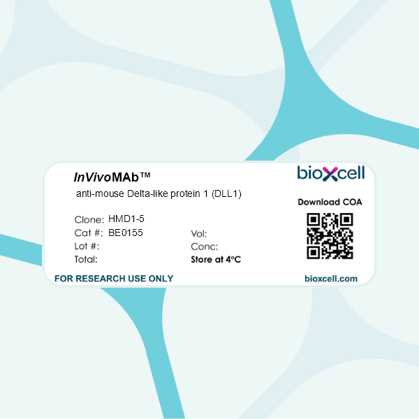
InVivoMAb anti-mouse Delta-like protein 1 (DLL1)
Product Description
Specifications
| Isotype | Armenian Hamster IgG, κ |
|---|---|
| Recommended Isotype Control(s) | InVivoMAb polyclonal Armenian hamster IgG |
| Recommended Dilution Buffer | InVivoPure pH 7.0 Dilution Buffer |
| Conjugation | This product is unconjugated. Conjugation is available via our Antibody Conjugation Services. |
| Immunogen | Mouse DLL1 |
| Reported Applications |
in vivo DLL1 neutralization Flow cytometry |
| Formulation |
PBS, pH 7.0 Contains no stabilizers or preservatives |
| Endotoxin |
≤1EU/mg (≤0.001EU/μg) Determined by LAL assay |
| Purity |
≥95% Determined by SDS-PAGE |
| Sterility | 0.2 µm filtration |
| Production | Purified from cell culture supernatant in an animal-free facility |
| Purification | Protein G |
| RRID | AB_10950546 |
| Molecular Weight | 150 kDa |
| Storage | The antibody solution should be stored at the stock concentration at 4°C. Do not freeze. |
| Need a Custom Formulation? | See All Antibody Customization Options |
Application References
-
Sakai, M., et al (2019). "Liver-Derived Signals Sequentially Reprogram Myeloid Enhancers to Initiate and Maintain Kupffer Cell Identity" Immunity 51(4): 655-670.e658.
PubMed
Tissue environment plays a powerful role in establishing and maintaining the distinct phenotypes of resident macrophages, but the underlying molecular mechanisms remain poorly understood. Here, we characterized transcriptomic and epigenetic changes in repopulating liver macrophages following acute Kupffer cell depletion as a means to infer signaling pathways and transcription factors that promote Kupffer cell differentiation. We obtained evidence that combinatorial interactions of the Notch ligand DLL4 and transforming growth factor-b (TGF-β) family ligands produced by sinusoidal endothelial cells and endogenous LXR ligands were required for the induction and maintenance of Kupffer cell identity. DLL4 regulation of the Notch transcriptional effector RBPJ activated poised enhancers to rapidly induce LXRα and other Kupffer cell lineage-determining factors. These factors in turn reprogrammed the repopulating liver macrophage enhancer landscape to converge on that of the original resident Kupffer cells. Collectively, these findings provide a framework for understanding how macrophage progenitor cells acquire tissue-specific phenotypes.
-
Riella, L. V., et al (2011). "Blockade of Notch ligand delta1 promotes allograft survival by inhibiting alloreactive Th1 cells and cytotoxic T cell generation" J Immunol 187(9): 4629-4638.
PubMed
The Notch signaling pathway has been recently shown to contribute to T cell differentiation in vitro. However, the in vivo function of Notch signaling in transplantation remains unknown. In this study, we investigated the importance of Delta1 in regulating the alloimmune response in vivo. Delta1 expression was upregulated on dendritic cells and monocytes/macrophages upon transplantation in a BALB/c into B6 vascularized cardiac transplant model. Whereas administration of anti-Delta1 mAb only slightly delayed survival of cardiac allografts in this fully MHC-mismatched model, it significantly prolonged graft survival in combination with single-dose CTLA4-Ig or in CD28 knockout recipients. The prolongation of allograft survival was associated with Th2 polarization and a decrease in Th1 and granzyme B-producing cytotoxic T cells. The survival benefit of Delta1 blockade was abrogated after IL-4 neutralization and in STAT6KO recipients, but was maintained in STAT4KO recipients, reinforcing the key role of Th2 cell development in its graft-prolonging effects. To our knowledge, these data demonstrate for the first time an important role of Delta1 in alloimmunity, identifying Delta1 ligand as a potential novel target for immunomodulation in transplantation.
Product Citations
-
Jagged2 targeting in lung cancer activates anti-tumor immunity via Notch-induced functional reprogramming of tumor-associated macrophages.
In Immunity on 14 May 2024 by Mandula, J. K., Sierra-Mondragon, R. A., et al.
PubMed
Signaling through Notch receptors intrinsically regulates tumor cell development and growth. Here, we studied the role of the Notch ligand Jagged2 on immune evasion in non-small cell lung cancer (NSCLC). Higher expression of JAG2 in NSCLC negatively correlated with survival. In NSCLC pre-clinical models, deletion of Jag2, but not Jag1, in cancer cells attenuated tumor growth and activated protective anti-tumor T cell responses. Jag2-/- lung tumors exhibited higher frequencies of macrophages that expressed immunostimulatory mediators and triggered T cell-dependent anti-tumor immunity. Mechanistically, Jag2 ablation promoted Nr4a-mediated induction of Notch ligands DLL1/4 on cancer cells. DLL1/4-initiated Notch1/2 signaling in macrophages induced the expression of transcription factor IRF4 and macrophage immunostimulatory functionality. IRF4 expression was required for the anti-tumor effects of Jag2 deletion in lung tumors. Antibody targeting of Jagged2 inhibited tumor growth and activated IRF4-driven macrophage-mediated anti-tumor immunity. Thus, Jagged2 orchestrates immunosuppressive systems in NSCLC that can be overcome to incite macrophage-mediated anti-tumor immunity.
-
Group 3 Innate Lymphoid Cells Exacerbate Lupus Nephritis by Promoting B Cell Activation in Kidney Ectopic Lymphoid Structures.
In Adv Sci (Weinh) on 1 December 2023 by Li, F., Liang, Z., et al.
PubMed
Group 3 innate lymphoid cells (ILC3s) represent a new population in immune regulation, yet their role in lupus nephritis (LN) remains elusive. In the present work, systemic increases in ILC3s, particularly in the kidney, are observed to correlate strongly with disease severity in both human and murine LN. Using MRL/lpr lupus mice and a nephrotoxic serum-induced LN model, this study demonstrates that ILC3s accumulated in the kidney migrate predominantly from the intestine. Furthermore, intestinal ILC3s accelerate LN progression, manifested by exacerbated autoimmunity and kidney injuries. In LN kidneys, ILC3s are located adjacent to B cells within ectopic lymphoid structures (ELS), directly activating B cell differentiation into plasma cells and antibody production in a Delta-like1 (DLL1)/Notch-dependent manner. Blocking DLL1 attenuates ILC3s' effects and protects against LN. Altogether, these findings reveal a novel pathogenic role of ILC3s in B cell activation, renal ELS formation and autoimmune injuries during LN, shedding light on the therapeutic value of targeting ILC3s for LN.
-
Reciprocal communication between astrocytes and endothelial cells is required for astrocytic glutamate transporter 1 (GLT-1) expression.
In Neurochem Int on 1 October 2020 by Martínez-Lozada, Z., Robinson, M. B., et al.
PubMed
Astrocytes have diverse functions that are supported by their anatomic localization between neurons and blood vessels. One of these functions is the clearance of extracellular glutamate. Astrocytes clear glutamate using two Na+-dependent glutamate transporters, GLT-1 (also called EAAT2) and GLAST (also called EAAT1). GLT-1 expression increases during synaptogenesis and is a marker of astrocyte maturation. Over 20 years ago, several groups demonstrated that astrocytes in culture express little or no GLT-1 and that neurons induce expression. We recently demonstrated that co-culturing endothelia with mouse astrocytes also induced expression of GLT-1 and GLAST. These increases were blocked by an inhibitor of γ-secretase. This and other observations are consistent with the hypothesis that Notch signaling is required, but the ligands involved were not identified. In the present study, we used rat astrocyte cultures to further define the mechanisms by which endothelia induce expression of GLT-1 and GLAST. We found that co-cultures of astrocytes and endothelia express higher levels of GLT-1 and GLAST protein and mRNA. That endothelia activate Hes5, a transcription factor target of Notch, in astrocytes. Using recombinant Notch ligands, anti-Notch ligand neutralizing antibodies, and shRNAs, we provide evidence that both Dll1 and Dll4 contribute to endothelia-dependent regulation of GLT-1. We also provide evidence that astrocytes secrete a factor(s) that induces expression of Dll4 in endothelia and that this effect is required for Notch-dependent induction of GLT-1. Together these studies indicate that reciprocal communication between astrocytes and endothelia is required for appropriate astrocyte maturation and that endothelia likely deploy additional non-Notch signals to induce GLT-1.
-
Liver-Derived Signals Sequentially Reprogram Myeloid Enhancers to Initiate and Maintain Kupffer Cell Identity.
In Immunity on 15 October 2019 by Sakai, M., Troutman, T. D., et al.
PubMed
Tissue environment plays a powerful role in establishing and maintaining the distinct phenotypes of resident macrophages, but the underlying molecular mechanisms remain poorly understood. Here, we characterized transcriptomic and epigenetic changes in repopulating liver macrophages following acute Kupffer cell depletion as a means to infer signaling pathways and transcription factors that promote Kupffer cell differentiation. We obtained evidence that combinatorial interactions of the Notch ligand DLL4 and transforming growth factor-b (TGF-β) family ligands produced by sinusoidal endothelial cells and endogenous LXR ligands were required for the induction and maintenance of Kupffer cell identity. DLL4 regulation of the Notch transcriptional effector RBPJ activated poised enhancers to rapidly induce LXRα and other Kupffer cell lineage-determining factors. These factors in turn reprogrammed the repopulating liver macrophage enhancer landscape to converge on that of the original resident Kupffer cells. Collectively, these findings provide a framework for understanding how macrophage progenitor cells acquire tissue-specific phenotypes.