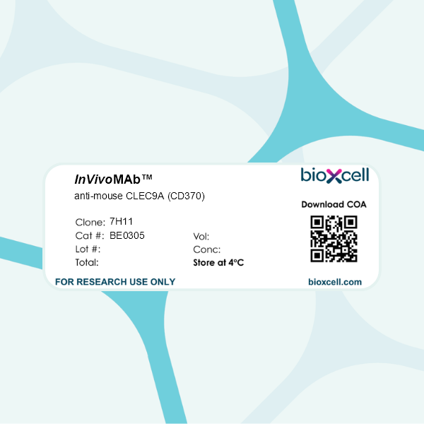
InVivoMAb anti-mouse CLEC9A (CD370)
Product Description
Specifications
| Isotype | Rat IgG1, κ |
|---|---|
| Recommended Isotype Control(s) | InVivoMAb rat IgG1 isotype control, anti-horseradish peroxidase |
| Recommended Dilution Buffer | InVivoPure pH 7.0 Dilution Buffer |
| Conjugation | This product is unconjugated. Conjugation is available via our Antibody Conjugation Services. |
| Immunogen | RBL-2H3 cells expressing mouse CLEC9A fused to an HA epitope |
| Reported Applications |
in vivo CLEC9A blockade in vivo Ag targeting to CLEC9A+ DCs Western blot ELISA Immunoprecipitation Immunofluorescence Flow cytometry |
| Formulation |
PBS, pH 7.0 Contains no stabilizers or preservatives |
| Endotoxin |
≤1EU/mg (≤0.001EU/μg) Determined by LAL assay |
| Purity |
≥95% Determined by SDS-PAGE |
| Sterility | 0.2 µm filtration |
| Production | Purified from cell culture supernatant in an animal-free facility |
| Purification | Protein G |
| RRID | AB_2721034 |
| Molecular Weight | 150 kDa |
| Storage | The antibody solution should be stored at the stock concentration at 4°C. Do not freeze. |
| Need a Custom Formulation? | See All Antibody Customization Options |
Application References
-
Snyder, A. G., et al (2019). "Intratumoral activation of the necroptotic pathway components RIPK1 and RIPK3 potentiates antitumor immunity" Sci Immunol 4(36).
PubMed
Although the signaling events that induce different forms of programmed cell death are well defined, the subsequent immune responses to dying cells in the context of cancer remain relatively unexplored. Necroptosis occurs downstream of the receptor-interacting protein kinases RIPK1 and RIPK3, whose activation leads to lytic cell death accompanied by de novo production of proinflammatory mediators. Here, we show that ectopic introduction of necroptotic cells to the tumor microenvironment promotes BATF3(+) cDC1- and CD8(+) leukocyte-dependent antitumor immunity accompanied by increased tumor antigen loading by tumor-associated antigen-presenting cells. Furthermore, we report the development of constitutively active forms of the necroptosis-inducing enzyme RIPK3 and show that delivery of a gene encoding this enzyme to tumor cells using adeno-associated viruses induces tumor cell necroptosis, which synergizes with immune checkpoint blockade to promote durable tumor clearance. These findings support a role for RIPK1/RIPK3 activation as a beneficial proximal target in the initiation of tumor immunity. Considering that successful tumor immunotherapy regimens will require the rational application of multiple treatment modalities, we propose that maximizing the immunogenicity of dying cells within the tumor microenvironment through specific activation of the necroptotic pathway represents a beneficial treatment approach that may warrant further clinical development.
-
Picco, G., et al (2014). "Targeting DNGR-1 (CLEC9A) with antibody/MUC1 peptide conjugates as a vaccine for carcinomas" Eur J Immunol 44(7): 1947-1955.
PubMed
DCs are the most potent APCs and are the focus of many immunotherapeutic approaches for the treatment of cancer, although most of these approaches require the ex vivo generation and pulsing of DCs. We have targeted a subset of DCs in vivo using an Ab to DNGR-1, a C-type lectin dedicated to the cross-presentation of Ag expressed by subsets of DCs. HLA-A2 epitopes from the tumour-associated Ag, MUC1, were coupled to the anti-DNGR-1 Ab, and their efficacy in generating a Th1-cell response and inhibiting tumour growth was evaluated in a clinically relevant double transgenic mouse model expressing human MUC1 and A2K/b. Using this strategy, we demonstrate that an effective immune response to MUC1 can be generated, which results in a significant delay in the growth of MUC1-expressing tumours in both prophylactic and therapeutic settings. In addition, we also show, using PBMCs isolated from healthy volunteer blood, that target an MUC1 HLA-A2 epitope to human DNGR-1 in vitro can induce an MUC1-specific CD8(+) -T-cell response, which confirms the relevance of our in vivo murine results in the human setting.
-
Joffre, O. P., et al (2010). "Efficient and versatile manipulation of the peripheral CD4+ T-cell compartment by antigen targeting to DNGR-1/CLEC9A" Eur J Immunol 40(5): 1255-1265.
PubMed
DC NK lectin group receptor-1 (DNGR-1, also known as CLEC9A) is a C-type lectin receptor expressed by mouse CD8alpha+ DC and by their putative equivalents in human. DNGR-1 senses necrosis and regulates CD8+ T-cell cross-priming to dead-cell-associated antigens. In addition, DNGR-1 is a target for selective in vivo delivery of antigens to DC and the induction of CD8+ T-cell and Ab responses. In this study, we evaluated whether DNGR-1 targeting can be additionally used to manipulate antigen-specific CD8+ T lymphocytes. Injection of small amounts of antigen-coupled anti-DNGR-1 mAb into mice promoted MHC class II antigen presentation selectively by CD8alpha+ DC. In the steady state, this was sufficient to induce proliferation of antigen-specific naive CD4+ T cells and to drive their differentiation into Foxp3+ regulatory lymphocytes. Co-administration of adjuvants prevented this induction of tolerance and promoted immunity. Notably, distinct adjuvants allowed qualitative modulation of CD4+ T-cell behavior: poly I:C induced a strong IL-12-independent Th1 response, whereas curdlan led to the priming of Th17 cells. Thus, antigen targeting to DNGR-1 is a versatile approach for inducing functionally distinct CD4+ T-cell responses. Given the restricted pattern of expression of DNGR-1 across species, this strategy could prove useful for developing immunotherapy protocols in humans.
-
Sancho, D., et al (2008). "Tumor therapy in mice via antigen targeting to a novel, DC-restricted C-type lectin" J Clin Invest 118(6): 2098-2110.
PubMed
The mouse CD8alpha+ DC subset excels at cross-presentation of antigen, which can elicit robust CTL responses. A receptor allowing specific antigen targeting to this subset and its equivalent in humans would therefore be useful for the induction of antitumor CTLs. Here, we have characterized a C-type lectin of the NK cell receptor group that we named DC, NK lectin group receptor-1 (DNGR-1). DNGR-1 was found to be expressed in mice at high levels by CD8+ DCs and at low levels by plasmacytoid DCs but not by other hematopoietic cells. Human DNGR-1 was also restricted in expression to a small subset of blood DCs that bear similarities to mouse CD8alpha+ DCs. The selective expression pattern and observed endocytic activity of DNGR-1 suggested that it could be used for antigen targeting to DCs. Consistent with this notion, antigen epitopes covalently coupled to an antibody specific for mouse DNGR-1 were selectively cross-presented by CD8alpha+ DCs in vivo and, when given with adjuvants, induced potent CTL responses. When the antigens corresponded to tumor-expressed peptides, treatment with the antibody conjugate and adjuvant could prevent development or mediate eradication of B16 melanoma lung pseudometastases. We conclude that DNGR-1 is a novel, highly specific marker of mouse and human DC subsets that can be exploited for CTL cross-priming and tumor therapy.
Product Citations
-
MAIT cells protect against sterile lung injury.
In Cell Rep on 25 February 2025 by Zhang, X., Li, S., et al.
PubMed
Mucosal-associated invariant T (MAIT) cells, the most abundant unconventional T cells in the lung, can exhibit a wide range of functional responses to different triggers via their T cell receptor (TCR) and/or cytokines. Their role, especially in sterile lung injury, is unknown. Using single-cell RNA sequencing (scRNA-seq), spectral analysis, and adoptive transfer in a bleomycin-induced sterile lung injury, we found that bleomycin activates murine pulmonary MAIT cells and is associated with a protective role against bleomycin-induced lung injury. MAIT cells drive the accumulation of type 1 conventional dendritic cells (cDC1s), limiting tissue damage in a DNGR-1-dependent manner. Human scRNA-seq data revealed that MAIT cells were activated, with increased cDC populations in idiopathic pulmonary fibrosis patients. Thus, MAIT cells enhance defense against sterile lung injury by fostering cDC1-driven anti-fibrotic pathways.
-
MAIT cells protect against sterile lung injury
In bioRxiv on 8 January 2024 by Zhang, X. W., Li, S., et al.
-
Intratumoral activation of the necroptotic pathway components RIPK1 and RIPK3 potentiates antitumor immunity.
In Sci Immunol on 21 June 2019 by Snyder, A. G., Hubbard, N., et al.
PubMed
Although the signaling events that induce different forms of programmed cell death are well defined, the subsequent immune responses to dying cells in the context of cancer remain relatively unexplored. Necroptosis occurs downstream of the receptor-interacting protein kinases RIPK1 and RIPK3, whose activation leads to lytic cell death accompanied by de novo production of proinflammatory mediators. Here, we show that ectopic introduction of necroptotic cells to the tumor microenvironment promotes BATF3+ cDC1- and CD8+ leukocyte-dependent antitumor immunity accompanied by increased tumor antigen loading by tumor-associated antigen-presenting cells. Furthermore, we report the development of constitutively active forms of the necroptosis-inducing enzyme RIPK3 and show that delivery of a gene encoding this enzyme to tumor cells using adeno-associated viruses induces tumor cell necroptosis, which synergizes with immune checkpoint blockade to promote durable tumor clearance. These findings support a role for RIPK1/RIPK3 activation as a beneficial proximal target in the initiation of tumor immunity. Considering that successful tumor immunotherapy regimens will require the rational application of multiple treatment modalities, we propose that maximizing the immunogenicity of dying cells within the tumor microenvironment through specific activation of the necroptotic pathway represents a beneficial treatment approach that may warrant further clinical development.