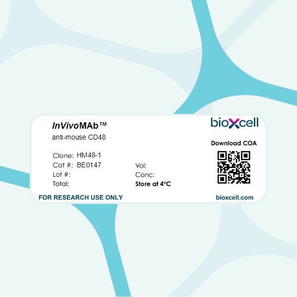
InVivoMAb anti-mouse CD48
Product Description
Specifications
| Isotype | Armenian hamster IgG |
|---|---|
| Recommended Isotype Control(s) | InVivoMAb polyclonal Armenian hamster IgG |
| Recommended Dilution Buffer | InVivoPure pH 7.0 Dilution Buffer |
| Conjugation | This product is unconjugated. Conjugation is available via our Antibody Conjugation Services. |
| Immunogen | MBL2 mouse T lymphoma cells |
| Reported Applications |
in vivo CD48 blockade in vitro CD48 blocking |
| Formulation |
PBS, pH 7.0 Contains no stabilizers or preservatives |
| Endotoxin |
≤1EU/mg (≤0.001EU/μg) Determined by LAL assay |
| Purity |
≥95% Determined by SDS-PAGE |
| Sterility | 0.2 µm filtration |
| Production | Purified from cell culture supernatant in an animal-free facility |
| Purification | Protein G |
| RRID | AB_10949470 |
| Molecular Weight | 150 kDa |
| Storage | The antibody solution should be stored at the stock concentration at 4°C. Do not freeze. |
| Need a Custom Formulation? | See All Antibody Customization Options |
Application References
in vivo CD48 blockade
Page, N., et al (2018). "Expression of the DNA-Binding Factor TOX Promotes the Encephalitogenic Potential of Microbe-Induced Autoreactive CD8(+) T Cells" Immunity 48(5): 937-950.e938.
PubMed
Infections are thought to trigger CD8(+) cytotoxic T lymphocyte (CTL) responses during autoimmunity. However, the transcriptional programs governing the tissue-destructive potential of CTLs remain poorly defined. In a model of central nervous system (CNS) inflammation, we found that infection with lymphocytic choriomeningitis virus (LCMV), but not Listeria monocytogenes (Lm), drove autoimmunity. The DNA-binding factor TOX was induced in CTLs during LCMV infection and was essential for their encephalitogenic properties, and its expression was inhibited by interleukin-12 during Lm infection. TOX repressed the activity of several transcription factors (including Id2, TCF-1, and Notch) that are known to drive CTL differentiation. TOX also reduced immune checkpoint sensitivity by restraining the expression of the inhibitory checkpoint receptor CD244 on the surface of CTLs, leading to increased CTL-mediated damage in the CNS. Our results identify TOX as a transcriptional regulator of tissue-destructive CTLs in autoimmunity, offering a potential mechanistic link to microbial triggers.
in vivo CD48 blockade
Jing, W., et al (2015). "Combined immune checkpoint protein blockade and low dose whole body irradiation as immunotherapy for myeloma" J Immunother Cancer 3(1): 2.
PubMed
80%). The increased survival rates correlated with increased frequencies of tumor-reactive CD8 and CD4 T cells. When stimulated in vitro with myeloma cells, CD8 T cells from treated mice produced elevated levels proinflammatory cytokines. Cytokines were spontaneously released from CD4 T cells isolated from mice treated with PD-L1 plus CTLA4 blocking antibodies. CONCLUSIONS: These data indicate that blocking PD-1/PD-L1 interactions in conjunction with other immune checkpoint proteins provides synergistic anti-tumor efficacy following lymphodepletive doses of whole body irradiation. This strategy is a promising combination strategy for myeloma and other hematologic malignancies.”}” data-sheets-userformat=”{“2″:14851,”3”:{“1″:0},”4”:{“1″:2,”2″:16777215},”12″:0,”14”:{“1″:2,”2″:1521491},”15″:”Roboto, sans-serif”,”16″:12}”>BACKGROUND: Multiple myeloma is characterized by the presence of transformed neoplastic plasma cells in the bone marrow and is generally considered to be an incurable disease. Successful treatments will likely require multi-faceted approaches incorporating conventional drug therapies, immunotherapy and other novel treatments. Our lab previously showed that a combination of transient lymphodepletion (sublethal whole body irradiation) and PD-1/PD-L1 blockade generated anti-myeloma T cell reactivity capable of eliminating established disease. We hypothesized that blocking a combination of checkpoint receptors in the context of low-dose, lymphodepleting whole body radiation would boost anti-tumor immunity. METHODS: To test our central hypothesis, we utilized a 5T33 murine multiple myeloma model. Myeloma-bearing mice were treated with a low dose of whole body irradiation and combinations of blocking antibodies to PD-L1, LAG-3, TIM-3, CD48 (the ligand for 2B4) and CTLA4. RESULTS: Temporal phenotypic analysis of bone marrow from myeloma-bearing mice demonstrated that elevated percentages of PD-1, 2B4, LAG-3 and TIM-3 proteins were expressed on T cells. When PD-L1 blockade was combined with blocking antibodies to LAG-3, TIM-3 or CTLA4, synergistic or additive increases in survival were observed (survival rates improved from ~30% to >80%). The increased survival rates correlated with increased frequencies of tumor-reactive CD8 and CD4 T cells. When stimulated in vitro with myeloma cells, CD8 T cells from treated mice produced elevated levels proinflammatory cytokines. Cytokines were spontaneously released from CD4 T cells isolated from mice treated with PD-L1 plus CTLA4 blocking antibodies. CONCLUSIONS: These data indicate that blocking PD-1/PD-L1 interactions in conjunction with other immune checkpoint proteins provides synergistic anti-tumor efficacy following lymphodepletive doses of whole body irradiation. This strategy is a promising combination strategy for myeloma and other hematologic malignancies.
in vitro CD48 blocking
Erickson, J. J., et al (2014). "Programmed death-1 impairs secondary effector lung CD8(+) T cells during respiratory virus reinfection" J Immunol 193(10): 5108-5117.
PubMed
Reinfections with respiratory viruses are common and cause significant clinical illness, yet precise mechanisms governing this susceptibility are ill defined. Lung Ag-specific CD8(+) T cells (T(CD8)) are impaired during acute viral lower respiratory infection by the inhibitory receptor programmed death-1 (PD-1). To determine whether PD-1 contributes to recurrent infection, we first established a model of reinfection by challenging B cell-deficient mice with human metapneumovirus (HMPV) several weeks after primary infection, and found that HMPV replicated to high titers in the lungs. A robust secondary effector lung TCD8 response was generated during reinfection, but these cells were more impaired and more highly expressed the inhibitory receptors PD-1, LAG-3, and 2B4 than primary T(CD8). In vitro blockade demonstrated that PD-1 was the dominant inhibitory receptor early after reinfection. In vivo therapeutic PD-1 blockade during HMPV reinfection restored lung T(CD8) effector functions (i.e., degranulation and cytokine production) and enhanced viral clearance. PD-1 also limited the protective efficacy of HMPV epitope-specific peptide vaccination and impaired lung T(CD8) during heterotypic influenza virus challenge infection. Our results indicate that PD-1 signaling may contribute to respiratory virus reinfection and evasion of vaccine-elicited immune responses. These results have important implications for the design of effective vaccines against respiratory viruses.
Product Citations
-
-
Immunology and Microbiology
Imaging of biphasic signalosomes constructed by checkpoint receptor 2B4 in conventional and chimeric antigen receptor-T cells.
In iScience on 17 January 2025 by Matsushima, R., Wakamatsu, E., et al.
PubMed
A co-signaling receptor, 2B4, has dual effects in immune cells, but its actual functions in T cells remain elusive. Here, using super-resolution imaging technology with an immunological synapse model, we showed that 2B4 forms "2B4 microclusters" immediately after 2B4-CD48 binding. A lipid phosphatase, SHIP-1, subsequently combined with 2B4 to form coinhibitory signalosomes, leading to the suppression of cytokine production. An activating adapter, SLAM-associated protein (SAP), attenuated the clustering of SHIP-1 and recruited a kinase, Fyn, enhancing the Vav1 signaling pathway as costimulatory signalosomes. Furthermore, we found that a chimeric antigen receptor with a 2B4 tail (2B4-CAR) retained the original signal transduction mechanism of 2B4. With endogenous levels of SAP expression, 2B4-CAR-T cells exposed sufficient antitumor efficacy in vivo without excess cytokine production. Our results may help explain the biphasic feature of 2B4 in T cell responses from the viewpoint of the signalosome and provide a new candidate for CAR development.
-
-
-
Neutralization experiments
-
Mus musculus (Mouse)
-
Genetics
-
Immunology and Microbiology
Expression of the DNA-Binding Factor TOX Promotes the Encephalitogenic Potential of Microbe-Induced Autoreactive CD8+ T Cells.
In Immunity on 15 May 2018 by Page, N., Klimek, B., et al.
PubMed
Infections are thought to trigger CD8+ cytotoxic T lymphocyte (CTL) responses during autoimmunity. However, the transcriptional programs governing the tissue-destructive potential of CTLs remain poorly defined. In a model of central nervous system (CNS) inflammation, we found that infection with lymphocytic choriomeningitis virus (LCMV), but not Listeria monocytogenes (Lm), drove autoimmunity. The DNA-binding factor TOX was induced in CTLs during LCMV infection and was essential for their encephalitogenic properties, and its expression was inhibited by interleukin-12 during Lm infection. TOX repressed the activity of several transcription factors (including Id2, TCF-1, and Notch) that are known to drive CTL differentiation. TOX also reduced immune checkpoint sensitivity by restraining the expression of the inhibitory checkpoint receptor CD244 on the surface of CTLs, leading to increased CTL-mediated damage in the CNS. Our results identify TOX as a transcriptional regulator of tissue-destructive CTLs in autoimmunity, offering a potential mechanistic link to microbial triggers.
-
-
-
Immunology and Microbiology
Anti-CD48 Monoclonal Antibody Attenuates Experimental Autoimmune Encephalomyelitis by Limiting the Number of Pathogenic CD4+ T Cells.
In J Immunol on 15 October 2016 by McArdel, S. L., Brown, D. R., et al.
PubMed
CD48 (SLAMF2) is an adhesion and costimulatory molecule constitutively expressed on hematopoietic cells. Polymorphisms in CD48 have been linked to susceptibility to multiple sclerosis (MS), and altered expression of the structurally related protein CD58 (LFA-3) is associated with disease remission in MS. We examined CD48 expression and function in experimental autoimmune encephalomyelitis (EAE), a mouse model of MS. We found that a subpopulation of CD4+ T cells highly upregulated CD48 expression during EAE and were enriched for pathogenic CD4+ T cells. These CD48++CD4+ T cells were predominantly CD44+ and Ki67+, included producers of IL-17A, GM-CSF, and IFN-γ, and were most of the CD4+ T cells in the CNS. Administration of anti-CD48 mAb during EAE attenuated clinical disease, limited accumulation of lymphocytes in the CNS, and reduced the number of pathogenic cytokine-secreting CD4+ T cells in the spleen at early time points. These therapeutic effects required CD48 expression on CD4+ T cells but not on APCs. Additionally, the effects of anti-CD48 were partially dependent on FcγRs, as anti-CD48 did not ameliorate EAE or reduce the number of cytokine-producing effector CD4+ T cells in Fcεr1γ-/- mice or in wild-type mice receiving anti-CD16/CD32 mAb. Our data suggest that anti-CD48 mAb exerts its therapeutic effects by both limiting CD4+ T cell proliferation and preferentially eliminating pathogenic CD48++CD4+ T cells during EAE. Our findings indicate that high CD48 expression is a feature of pathogenic CD4+ T cells during EAE and point to CD48 as a potential target for immunotherapy.
-

