InVivoMAb anti-mouse CD172a (SIRPα)
Product Description
Specifications
| Isotype | Rat IgG1, κ |
|---|---|
| Recommended Isotype Control(s) | InVivoMAb rat IgG1 isotype control, anti-horseradish peroxidase |
| Recommended Dilution Buffer | InVivoPure pH 7.0 Dilution Buffer |
| Conjugation | This product is unconjugated. Conjugation is available via our Antibody Conjugation Services. |
| Immunogen | Mouse brain membrane protein |
| Reported Applications |
in vivo SIRPα blocking In vitro SIRPα blocking Western blot Immunoprecipitation Flow cytometry |
| Formulation |
PBS, pH 7.0 Contains no stabilizers or preservatives |
| Endotoxin |
≤1EU/mg (≤0.001EU/μg) Determined by LAL assay |
| Purity |
≥95% Determined by SDS-PAGE |
| Sterility | 0.2 µm filtration |
| Production | Purified from cell culture supernatant in an animal-free facility |
| Purification | Protein A |
| RRID | AB_2819049 |
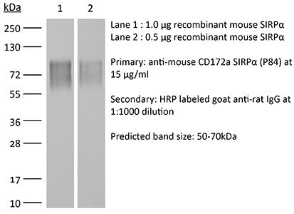
| Molecular Weight | 150 kDa |
| Storage | The antibody solution should be stored at the stock concentration at 4°C. Do not freeze. |
| Need a Custom Formulation? | See All Antibody Customization Options |
Application References
-
Yanagita, T., et al (2017). "Anti-SIRPalpha antibodies as a potential new tool for cancer immunotherapy" JCI Insight 2(1): e89140.
PubMed
Tumor cells are thought to evade immune surveillance through interaction with immune cells. Much recent attention has focused on the modification of immune responses as a basis for new cancer treatments. SIRPalpha is an Ig superfamily protein that inhibits phagocytosis in macrophages upon interaction with its ligand CD47 expressed on the surface of target cells. Here, we show that SIRPalpha is highly expressed in human renal cell carcinoma and melanoma. Furthermore, an anti-SIRPalpha Ab that blocks the interaction with CD47 markedly suppressed tumor formation by renal cell carcinoma or melanoma cells in immunocompetent syngeneic mice. This inhibitory effect of the Ab appeared to be mediated by dual mechanisms: direct induction of Ab-dependent cellular phagocytosis of tumor cells by macrophages and blockade of CD47-SIRPalpha signaling that negatively regulates such phagocytosis. The antitumor effect of the Ab was greatly attenuated by selective depletion not only of macrophages but also of NK cells or CD8(+) T cells. In addition, the anti-SIRPalpha Ab also enhances the inhibitory effects of Abs against CD20 and programmed cell death 1 (PD-1) on tumor formation in mice injected with SIRPalpha-nonexpressing tumor cells. Anti-SIRPalpha Abs thus warrant further study as a potential new therapy for a broad range of cancers.
-
Koskinen, C., et al (2013). "Lack of CD47 impairs bone cell differentiation and results in an osteopenic phenotype in vivo due to impaired signal regulatory protein alpha (SIRPalpha) signaling" J Biol Chem 288(41): 29333-29344.
PubMed
Here, we investigated whether the cell surface glycoprotein CD47 was required for normal formation of osteoblasts and osteoclasts and to maintain normal bone formation activity in vitro and in vivo. In parathyroid hormone or 1alpha,25(OH)2-vitamin D3 (D3)-stimulated bone marrow cultures (BMC) from CD47(-/-) mice, we found a strongly reduced formation of multinuclear tartrate-resistant acid phosphatase (TRAP)(+) osteoclasts, associated with reduced expression of osteoclastogenic genes (nfatc1, Oscar, Trap/Acp, ctr, catK, and dc-stamp). The production of M-CSF and RANKL (receptor activator of nuclear factor kappabeta ligand) was reduced in CD47(-/-) BMC, as compared with CD47(+/+) BMC. The stromal cell phenotype in CD47(-/-) BMC involved a blunted expression of the osteoblast-associated genes osterix, Alp/Akp1, and alpha-1-collagen, and reduced mineral deposition, as compared with that in CD47(+/+) BMC. CD47 is a ligand for SIRPalpha (signal regulatory protein alpha), which showed strongly reduced tyrosine phosphorylation in CD47(-/-) bone marrow stromal cells. In addition, stromal cells lacking the signaling SIRPalpha cytoplasmic domain also had a defect in osteogenic differentiation, and both CD47(-/-) and non-signaling SIRPalpha mutant stromal cells showed a markedly reduced ability to support osteoclastogenesis in wild-type bone marrow macrophages, demonstrating that CD47-induced SIRPalpha signaling is critical for stromal cell support of osteoclast formation. In vivo, femoral bones of 18- or 28-week-old CD47(-/-) mice showed significantly reduced osteoclast and osteoblast numbers and exhibited an osteopenic bone phenotype. In conclusion, lack of CD47 strongly impairs SIRPalpha-dependent osteoblast differentiation, deteriorate bone formation, and cause reduced formation of osteoclasts.
-
Teraoka, Y., et al (2013). "Expression of recipient CD47 on rat insulinoma cell xenografts prevents macrophage-mediated rejection through SIRPalpha inhibitory signaling in mice" PLoS One 8(3): e58359.
PubMed
We have previously proven that the interspecies incompatibility of CD47 is responsible for in vitro phagocytosis of xenogeneic cells by host macrophages. Utilizing an in vivo model in the present study, we investigated whether genetically engineered expression of mouse CD47 in rat insulinoma cells (INS-1E) could inhibit macrophage-mediated xenograft rejection. INS-1E cells transfected with the pRc/CMV-mouse CD47 vector (mCD47-INS-1E) induced SIRPalpha-tyrosine phosphorylation in mouse macrophages in vitro, whereas cells transfected with the control vector (cont-INS-1E) did not. When these cells were injected into the peritoneal cavity of streptozotocin-induced diabetic Rag2(-/-)gamma chain (-/-) mice, which lack T, B, and NK cells, the expression of mouse CD47 on the INS-1E cells markedly reduced the susceptibility of these cells to phagocytosis by macrophages. Moreover, these mice became normoglycemic after receiving mCD47-INS-1E, whereas the mice that received cont-INS-1E failed to achieve normoglycemia. Furthermore, injection of an anti-mouse SIRPalpha blocking monoclonal antibody into the mouse recipients of mCD47-INS-1E cells prevented achievement of normoglycemia. These results demonstrate that interspecies incompatibility of CD47 significantly contributes to in vivo rejection of xenogeneic cells by macrophages. Thus, genetic induction of the expression of recipient CD47 on xenogeneic donor cells could provide inhibitory signals to recipient macrophages via SIPRalpha; this constitutes a novel approach for preventing macrophage-mediated xenograft rejection.
-
Zen, K., et al (2013). "Inflammation-induced proteolytic processing of the SIRPalpha cytoplasmic ITIM in neutrophils propagates a proinflammatory state" Nat Commun 4: 2436.
PubMed
Signal regulatory protein alpha (SIRPalpha), an immunoreceptor tyrosine-based inhibitory motif (ITIM)-containing receptor, is an essential negative regulator of leukocyte inflammatory responses. Here we report that SIRPalpha cytoplasmic signalling ITIMs in neutrophils are cleaved during active inflammation and that the loss of SIRPalpha ITIMs enhances the polymorphonuclear leukocyte (PMN) inflammatory response. Using human leukocytes and two inflammatory models in mice, we show that the cleavage of SIRPalpha ITIMs in PMNs but not monocytes occurs at the post-acute stage of inflammation and correlates with increased PMN recruitment to inflammatory loci. Enhanced transmigration of PMNs and PMN-associated tissue damage are confirmed in mutant mice expressing SIRPalpha but lacking the ITIMs. Moreover, the loss of SIRPalpha ITIMs in PMNs during colitis is blocked by an anti-interleukin-17 (IL-17) antibody. These results demonstrate a SIRPalpha-based mechanism that dynamically regulates PMN inflammatory responses by generating a CD47-binding but non-signalling SIRPalpha ‘decoy’.
Product Citations
-
Myokine SIRPα exacerbates kidney disease in diabetes.
In JCI Insight on 9 February 2026 by Wu, J., Russo, E., et al.
PubMed
Mechanisms responsible for skeletal muscle kidney crosstalk have not been defined. We have determined that a circulating mediator, signal regulatory protein α (SIRPα), impairs intracellular insulin-mediated functions. To elucidate the effect of myokine SIRPα on diabetic kidney disease (DKD), flox mice and muscle-specific (m-specific) SIRPα-KO mice were subjected to an obesity-induced model of diabetes, high-fat diet (HFD; 60%) or insulin-deficient hyperglycemia model, streptozotocin (STZ), and were subsequently exposed to anti-SIRPα monoclonal antibodies. In the obesity-induced diabetic mice, serum SIRPα increased. Genetic deletion of muscle SIRPα protected against obesity and improved intracellular insulin signaling in muscle and adipose tissue, with reduced intramuscular fat deposition when compared with flox mice on HFD. Moreover, mSIRPα-KO mice displayed enhanced kidney tubular fatty acid oxidation (FAO) expression with suppressed intraorgan triglycerides deposition, and importantly, protection against DKD. Conversely, exogenous SIRPα impaired kidney proximal tubular cell FAO, ATP production, and exacerbated fibrosis. Finally, suppressing SIRPα in skeletal muscles or treatment with anti-SIRPα monoclonal antibodies in STZ-treated mice mitigated cachexia, hyperlipidemia, kidney triglyceride deposition, and renal dysfunction in spite of significant hyperglycemia. Importantly, serum SIRPα was upregulated in patients with DKD. In conclusion, SIRPα serves as a potential biomarker and therapeutic target in DKD.
-
Loss of NR2F6 Protects from Salmonella Typhimurium Infection.
In Adv Sci (Weinh) on 1 October 2025 by Woelk, J., Pfeifhofer-Obermair, C., et al.
PubMed
Nuclear receptors regulate key functions of mononuclear phagocytes and are critical components of the innate immune system, acting as regulators of organ health and disease. In healthy mice, the loss of the nuclear orphan receptor NR2F6 alters tissue-resident macrophage populations in the liver, lung, and spleen. In response to Salmonella Typhimurium infection, Nr2f6-deficient mice exhibit improved clinical outcomes, characterized by reduced weight loss, bacterial loads in the spleen and liver, and decreased plasma pro-inflammatory cytokines. Despite unchanged basal iron metabolism in the spleen and liver, iron regulatory proteins and the interleukin (IL)-6-hepcidin axis are altered in Nr2f6-deficient mice during Salmonella infection, reducing hypoferremia. Transcriptomic analysis of splenic red pulp macrophages reveals significant alterations of phagocytosis-related genes, including upregulation of signal-regulatory protein alpha (Sirpa). In vitro, phagocytosis of red blood cells, regulated by the inhibitory CD47-Sirpα axis, and Salmonella Typhimurium phagocytosis are significantly impaired in Nr2f6-deficient splenic macrophages. Blocking Sirpα in vitro restores the phagocytic activity of Nr2f6-deficient macrophages to wild-type levels. In vivo, Salmonella Typhimurium loads are partially increased post-infection in anti-Sirpα treated Nr2f6-deficient mice. These findings uncover a previously unrecognized role of NR2F6 in host-pathogen interactions, positioning it as a potential therapeutic target for infectious diseases.
-
Hematopoietic loss of Y chromosome activates immune checkpoints and contributes to impaired senescent cell clearance and renal disease.
In Sci Transl Med on 6 August 2025 by Arai, Y., Chavkin, N., et al.
PubMed
The accumulation of senescent cells contributes to morbidity and mortality; however, common mechanisms underpinning this age-associated phenomenon remain elusive. Hematopoietic loss of the Y chromosome (LOY) is the most frequently acquired somatic mutation in males, and this condition has been associated with various age-associated diseases and reduced lifespan. Therefore, we investigated the role of hematopoietic LOY in promoting cellular senescence, focusing on kidney disease because of its well-documented connection with aging and senescence. Herein, a prospective cohort study revealed that LOY in blood is associated with an increased incidence of kidney diseases. Analyses of transcriptional signatures in human kidneys found that immune cell LOY is enriched in patients with kidney disease and associated with greater amounts of cellular senescence. In male mice reconstituted with bone marrow lacking the Y chromosome, renal dysfunction was accompanied by senescent cell accumulation in models of kidney injury and advanced age. Treatment with a senolytic agent promoted senolysis and preferentially inhibited the progression of renal dysfunction in LOY mice. Hematopoietic LOY led to up-regulation of multiple immune inhibitory receptors, and treatment with the combination of antibodies targeting PD-1 (programmed cell death protein 1) and SIRPα (signal regulatory protein α) reduced senescent cell accumulation and rescued the renal pathology conferred by hematopoietic LOY in the kidney injury model. Collectively, these data indicate that hematopoietic LOY contributes to pathological conditions by impairing the clearance of senescent cells through up-regulation of immune checkpoint proteins.
-
SIRPα blockade therapy potentiates immunotherapy by inhibiting PD-L1+ myeloid cells in hepatocellular carcinoma.
In Cell Death Dis on 16 June 2025 by Huang, D., Xu, M., et al.
PubMed
Tumor-infiltrating myeloid cells (TIMs) are pivotal cell populations involved in the immunosuppressive tumor immune microenvironment (TIME). However, there has been little success in large-scale clinical trials of myeloid cell modulators. We aim to investigate potential molecular targets for TIMs and disclose the underlying mechanism. Using mass cytometry by time of flight (CyTOF), we analyzed 24 spontaneous HCC tissues from mouse. Orthotopic and subcutaneous tumor models were established with or without anti-SIRPα antibody treatment. Patient-derived tumor xenografts model (PDX) was used to identify the CD47-SIRPα axis blocked therapy. In 24 murine spontaneous HCC tissues, we observed that the proportion of myeloid-derived suppressor cells (MDSCs) plus macrophages accounts for 40-90% of TIMs and SIRPα was highly expressed in TIMs, especially in macrophages and MDSCs. Through in vivo experiments, we showed that anti-SIRPα therapy inhibited tumor growth, accompanied by increased CD8+ T cells infiltration and decreased TIMs including MDSCs and macrophages. We found that anti-SIRPα inhibited immunosuppressive function, migration and PD-L1 expression of myeloid cells. In a series of in vivo experiments, we demonstrated the anti-tumor and immune-active effect of SIRPα-blocked therapy. Mechanistically, anti-SIRPα inhibited the immunosuppressive function and PD-L1 expression of TIMs through downregulating PI3K/AKT signaling in myeloid cells. At last, anti-SIRPα enhanced the antitumor effect of anti-PD-L1 therapy in orthotopic and spontaneous murine models. Together, SIRPα blocked therapy reversed the immunosuppressive TIME, which provides a promising therapeutic rationale for increasing the efficacy of anti-PD-L1 therapy in treating HCC.