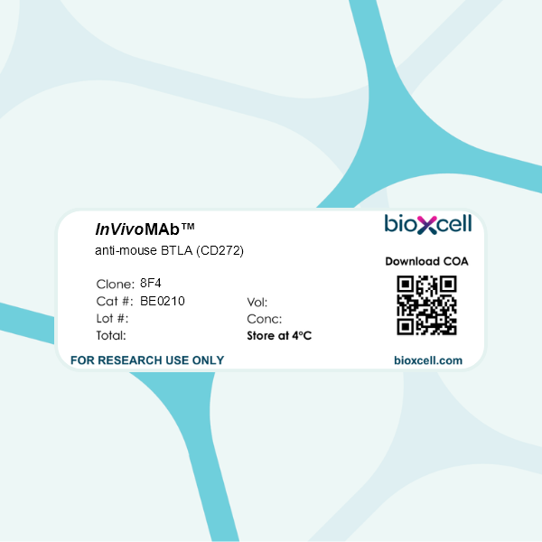
InVivoMAb anti-mouse BTLA (CD272)
Product Description
Specifications
| Isotype | Mouse IgG1, κ |
|---|---|
| Recommended Isotype Control(s) | InVivoMAb mouse IgG1 isotype control, unknown specificity |
| Recommended Dilution Buffer | InVivoPure pH 7.0 Dilution Buffer |
| Conjugation | This product is unconjugated. Conjugation is available via our Antibody Conjugation Services. |
| Immunogen | C57BL/6 mouse BTLA Ig domain |
| Reported Applications |
in vivo blockade of BTLA/HVEM signaling Flow cytometry |
| Formulation |
PBS, pH 7.0 Contains no stabilizers or preservatives |
| Endotoxin |
≤1EU/mg (≤0.001EU/μg) Determined by LAL assay |
| Purity |
≥95% Determined by SDS-PAGE |
| Sterility | 0.2 µm filtration |
| Production | Purified from cell culture supernatant in an animal-free facility |
| Purification | Protein G |
| RRID | AB_10948994 |
| Molecular Weight | 150 kDa |
| Storage | The antibody solution should be stored at the stock concentration at 4°C. Do not freeze. |
| Need a Custom Formulation? | See All Antibody Customization Options |
Application References
in vivo blockade of BTLA/HVEM signaling
Xu X, Shang B, Wu H, Jin X, Wang J, Li J, Li D, Liang B, Wang X, Su L, You W, Jiang S (2025). "FXR shapes an immunosuppressive microenvironment in PD-L1lo/- non-small cell lung cancer by upregulating HVEM" JCI Insight 10(18):e190716.
PubMed
Immune checkpoint therapy has changed cancer treatment, including non-small cell lung cancer (NSCLC). The unresponsiveness of PD-L1lo/- tumors to anti-PD-1/PD-L1 immunotherapy is attributed to alternative immune evasion mechanisms that remain elusive. We previously reported that farnesoid X receptor (FXR) was increased in PD-L1lo/- NSCLC. Herein, we found that immune checkpoint HVEM was positively correlated with FXR but inversely correlated with PD-L1 in NSCLC. HVEM was highly expressed in FXRhiPD-L1lo NSCLC. Consistently, clinically relevant FXR antagonist dose-dependently inhibited HVEM expression in NSCLC. FXR inhibited cytokine production and cytotoxicity of cocultured CD8+ T cells in vitro, and it shaped an immunosuppressive tumor microenvironment (TME) in mouse tumors in vivo through the HVEM/BTLA pathway. Clinical investigations show that the FXR/HVEM axis was associated with immunoevasive TME and inferior survival outcomes in patients with NSCLC. Mechanistically, FXR upregulated HVEM via transcriptional activation, intracellular Akt, Erk1/2 and STAT3 signals, and G1/S cycle progression in NSCLC cells. In vivo treatment experiments demonstrated that anti-BTLA immunotherapy reinvigorated antitumor immunity in TME, resulting in enhanced tumor inhibition and survival improvement in FXRhiPD-L1lo mouse Lewis lung carcinomas. In summary, our findings establish the FXR/HVEM axis as an immune evasion mechanism in PD-L1lo/- NSCLC, providing translational implications for future immunotherapy in this subgroup of patients.
Flow Cytometry
Shao, L., et al (2015). "Aberrant germinal center formation, follicular T-helper cells, and germinal center B-cells were involved in chronic graft-versus-host disease" Ann Hematol 94(9): 1493-1504.
PubMed
Chronic graft-versus-host disease (cGVHD) is an important complication after allogeneic hematopoietic stem cell transplantation (HSCT). To define the roles of T-cells and B-cells in cGVHD, a murine minor histocompatibility complex-mismatched HSCT model was used. Depletion of donor splenocyte CD4(+) T-cells and B220(+) B-cells alleviated cGVHD. Allogeneic recipients had significantly increased splenic germinal centers (GCs), with significant increases in follicular T-helper (Tfh) cells and GC B-cells. There were increased expressions in Tfh cells of inducible T-cell co-stimulator (ICOS), interleukin (IL)-4 and IL-17, and in GC B-cells of B-cell activating factor receptor and ICOS ligand. Depletion of donor splenocyte CD4(+) T-cells abrogated aberrant GC formation and suppressed Tfh cells and GC B-cells. Interestingly, depletion of donor splenocyte B200(+) B-cells also suppressed Tfh cells in addition to GC B-cells. These results suggested that in cGVHD, both Tfh and GC B-cells were involved, and their developments were mutually dependent. The mammalian target of rapamycin (mTOR) inhibitor everolimus was effective in suppressing cGVHD, Tfh cells, and GC B-cells, either as a prophylaxis or when cGVHD had established. These results implied that therapeutic targeting of both T-cells and B-cells in cGVHD might be effective. Signaling via mTOR may be another useful target in cGVHD.
Flow Cytometry
Vaeth, M., et al (2014). "Follicular regulatory T cells control humoral autoimmunity via NFAT2-regulated CXCR5 expression" J Exp Med 211(3): 545-561.
PubMed
Maturation of high-affinity B lymphocytes is precisely controlled during the germinal center reaction. This is dependent on CD4(+)CXCR5(+) follicular helper T cells (TFH) and inhibited by CD4(+)CXCR5(+)Foxp3(+) follicular regulatory T cells (TFR). Because NFAT2 was found to be highly expressed and activated in follicular T cells, we addressed its function herein. Unexpectedly, ablation of NFAT2 in T cells caused an augmented GC reaction upon immunization. Consistently, however, TFR cells were clearly reduced in the follicular T cell population due to impaired homing to B cell follicles. This was TFR-intrinsic because only in these cells NFAT2 was essential to up-regulate CXCR5. The physiological relevance for humoral (auto-)immunity was corroborated by exacerbated lupuslike disease in the presence of NFAT2-deficient TFR cells.
Product Citations
-
-
Cancer Research
FXR shapes an immunosuppressive microenvironment in PD-L1lo/- non-small cell lung cancer by upregulating HVEM.
In JCI Insight on 23 September 2025 by Xu, X., Shang, B., et al.
PubMed
Immune checkpoint therapy has changed cancer treatment, including non-small cell lung cancer (NSCLC). The unresponsiveness of PD-L1lo/- tumors to anti-PD-1/PD-L1 immunotherapy is attributed to alternative immune evasion mechanisms that remain elusive. We previously reported that farnesoid X receptor (FXR) was increased in PD-L1lo/- NSCLC. Herein, we found that immune checkpoint HVEM was positively correlated with FXR but inversely correlated with PD-L1 in NSCLC. HVEM was highly expressed in FXRhiPD-L1lo NSCLC. Consistently, clinically relevant FXR antagonist dose-dependently inhibited HVEM expression in NSCLC. FXR inhibited cytokine production and cytotoxicity of cocultured CD8+ T cells in vitro, and it shaped an immunosuppressive tumor microenvironment (TME) in mouse tumors in vivo through the HVEM/BTLA pathway. Clinical investigations show that the FXR/HVEM axis was associated with immunoevasive TME and inferior survival outcomes in patients with NSCLC. Mechanistically, FXR upregulated HVEM via transcriptional activation, intracellular Akt, Erk1/2 and STAT3 signals, and G1/S cycle progression in NSCLC cells. In vivo treatment experiments demonstrated that anti-BTLA immunotherapy reinvigorated antitumor immunity in TME, resulting in enhanced tumor inhibition and survival improvement in FXRhiPD-L1lo mouse Lewis lung carcinomas. In summary, our findings establish the FXR/HVEM axis as an immune evasion mechanism in PD-L1lo/- NSCLC, providing translational implications for future immunotherapy in this subgroup of patients.
-

