InVivoMAb anti-human EGFR
Product Description
Specifications
| Isotype | Mouse IgG2a |
|---|---|
| Recommended Isotype Control(s) | InVivoMAb mouse IgG2a isotype control, unknown specificity |
| Recommended Dilution Buffer | InVivoPure pH 7.0 Dilution Buffer |
| Conjugation | This product is unconjugated. Conjugation is available via our Antibody Conjugation Services. |
| Immunogen | Purified EGFR from A431 cells |
| Reported Applications |
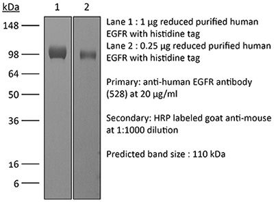
in vitro EGFR blockade in vivo EGFR blockade in xenografts Western blot Functional assays Immunoprecipitation Immunohistochemistry (paraffin) Immunofluorescence Flow cytometry |
| Formulation |
PBS, pH 7.0 Contains no stabilizers or preservatives |
| Endotoxin |
≤1EU/mg (≤0.001EU/μg) Determined by LAL assay |
| Purity |
≥95% Determined by SDS-PAGE |
| Sterility | 0.2 µm filtration |
| Production | Purified from cell culture supernatant in an animal-free facility |
| Purification | Protein A |
| RRID | AB_2687802 |
| Molecular Weight | 150 kDa |
| Storage | The antibody solution should be stored undiluted at 4°C, and protected from prolonged exposure to light. Do not freeze. |
| Need a Custom Formulation? | See All Antibody Customization Options |
Application References
-
Dong, A., et al (2015). "Epidermal growth factor receptor (EGFR) signaling requires a specific endoplasmic reticulum thioredoxin for the post-translational control of receptor presentation to the cell surface" J Biol Chem 290(13): 8016-8027.
PubMed
The epidermal growth factor receptor (EGFR) is a well characterized receptor-tyrosine kinase that functions in development and serves a vital role in many human cancers. Understanding EGFR regulatory mechanisms, and hence approaches for clinical intervention, has focused on ligand-receptor interactions and tyrosine kinase activity. Here, we show using the NCI-H460 lung and A431 epidermoid human cancer cell lines that EGFR binding to anterior gradient homolog 2 (AGR2) in the endoplasmic reticulum is required for receptor delivery to the plasma membrane and thus EGFR signaling. Reduced AGR2 protein levels or mutation of an essential cysteine in the active site result in decreased cell surface EGFR and a concomitant decrease in signaling as reflected by AREG, EGR1, and FOS expression. Similar to previously described EGFR nulls, an AGR2 null also resulted in embryonic lethality. Consistent with its role in regulating EGFR-mediated signaling, AGR2 expression is also enhanced in many human cancers and promotes the transformed phenotype. Furthermore, EGFR-mediated signaling in NCI-H460 cells, which are resistant to the tyrosine kinase inhibitor AG1478, is also disrupted with reduced AGR2 expression. The results provide insights into why cancer prognosis or response to therapy often does not correlate with EGFR protein or RNA levels because they do not reflect delivery to the cell surface where signaling is initiated. AGR2, therefore, represents a novel post-translational regulator of EGFR-mediated signaling and a promising target for treating human cancers.
-
Raimondi, F., et al (2008). "Bile acids modulate tight junction structure and barrier function of Caco-2 monolayers via EGFR activation" Am J Physiol Gastrointest Liver Physiol 294(4): G906-913.
PubMed
Intestinal and systemic illnesses have been linked to increased gut permeability. Bile acids, whose luminal profile can be altered in human disease, modulate intestinal paracellular permeability. We investigated the mechanism by which selected bile acids increase gut permeability using a validated in vitro model. Human intestinal Caco-2 cells were grown in monolayers and challenged with a panel of bile acids. Transepithelial electrical resistance and luminal-to-basolateral fluxes of 10-kDa Cascade blue-conjugated dextran were used to monitor paracellular permeability. Immunoprecipitation and immunoblot analyses were employed to investigate the intracellular pathway. Redistribution of tight junction proteins was studied by confocal laser microscopy. Micromolar concentrations of cholic acid, deoxycholic acid (DCA), and chenodeoxycholic acid (CDCA) but not ursodeoxycholic acid decreased transepithelial electrical resistance and increased dextran flux in a reversible fashion. Coincubation of 50 muM CDCA or DCA with EGF, anti-EGF monoclonal antibody, or specific src inhibitor 4-Amino-5-(4-chlorophenyl)-7-(t-butyl)pyrazolo[3,4-d]pyrimidine (PP-2) abolished the effect. A concentration of 50 muM of either CDCA or DCA also induced EGF receptor phosphorylation, occludin dephosphorylation, and occludin redistribution at the tight junction level in the same time frame and in a reversible fashion. We conclude that selected bile acids modulate intestinal permeability via EGF receptor autophosphorylation, occludin dephosphorylation, and rearrangement at the tight junction level. The effect is mediated by the src family kinases and is abolished by EGF treatment. These data also support the role of bile acids in the genesis of necrotizing enterocolitis and the protective effect of EGF treatment.
-
Kurai, J., et al (2007). "Antibody-dependent cellular cytotoxicity mediated by cetuximab against lung cancer cell lines" Clin Cancer Res 13(5): 1552-1561.
PubMed
PURPOSE: Epidermal growth factor receptor (EGFR) is commonly overexpressed in lung cancer. Cetuximab is a chimeric mouse-human antibody targeted against EGFR. Compared with its inhibitory properties, its immunologic mechanisms have not been well studied. In this study, we investigated the antibody-dependent cellular cytotoxicity (ADCC) activity of cetuximab against lung cancer cell lines. EXPERIMENTAL DESIGN: We studied the correlation between EGFR expression in lung cancer cell lines and the ADCC activity of cetuximab as well as the influence of interleukin-2 and chemotherapy on the ADCC activity. EGFR expression was measured by a quantitative flow cytometric analysis and immunohistochemistry. The ADCC activity was assessed by a 4-h (51)Cr release assay. Peripheral blood mononuclear cells, purified T cells, natural killer (NK) cells, and monocytes from healthy donors or lung cancer patients were used as effector cells. RESULTS: Fresh peripheral blood mononuclear cells exhibited cetuximab-mediated ADCC activity against lung cancer cell lines at a low concentration of cetuximab (0.25 microg/mL). A logarithmic correlation was observed between the number of EGFRs and ADCC activity. Even low EGFR expression, which was weakly detectable by immunohistochemistry, was sufficient for maximum ADCC activity, and further increases in EGFR expression on the target cells had no further effect on the ADCC activity. In addition, ADCC activity was enhanced by interleukin-2 mainly through activation of NK cells and was less susceptible to immunosuppression by chemotherapy than NK activity in lung cancer patients. CONCLUSIONS: These observations suggest the importance of ADCC activity as an immunologic mechanism of cetuximab in biological therapy for lung cancer patients.
-
Nakamura, T., et al (2005). "Rescue and propagation of fully retargeted oncolytic measles viruses" Nat Biotechnol 23(2): 209-214.
PubMed
Live attenuated measles viruses of the Edmonston lineage (MV-Edm) have potent anti-tumor activity but are not entirely tumor-specific owing to widespread distribution of their native receptors, CD46 and SLAM. We have therefore developed a pseudoreceptor system that allows rescue and propagation of fully retargeted viruses displaying single-chain antibody fragments. Viruses retargeted to tumor-selective CD38, epidermal growth factor receptor (EGFR) or EGFR mutant vIII (EGFRvIII) efficiently entered cells through their respective targeted receptors in vitro and in vivo, but not through CD46 and SLAM. When administered intratumorally or intravenously to mice bearing human CD38 or EGFR-positive human tumor xenografts, the targeted viruses demonstrated specific receptor-mediated anti-tumor activity. These data provide an in vivo demonstration of antibody-directed tumor destruction by retargeted oncolytic viruses.
Product Citations
-
Exploring the Sensitivity of Antibody-Drug Conjugate Efficacy to the Selection of Payload, Antibody, and Cell line.
In Bioconjug Chem on 17 January 2024 by Rao, M., Murali, S., et al.
PubMed
Antibody-drug conjugates (ADCs) make up a growing class of targeted therapeutics with important applications in cancer treatment. ADCs are highly modular in nature and thus can be engineered to target any cancer type, but their efficacy is strongly influenced by the specific choice of payload, antibody, and target cell. Considering the number of possible antibody-payload combinations, ADC development would benefit from an efficient method to narrow the number of ADC compositions to those with the highest and most universal potency prior to assessing pharmacokinetics and pharmacodynamics in animal models. To facilitate the identification of optimal ADC compositions, we describe the use of photoreactive antibody-binding domain-drug conjugates (known commercially as oYo-Link) to enable the site-specific labeling of off-the-shelf antibodies. This approach allows for the rapid generation of ADCs with a drug-to-antibody ratio of ∼2 with no subsequent purification required. As a demonstration of this approach, ADCs were generated with different combinations of tubulin-inhibitor drugs (DM1, DM4, VcMMAE, and VcMMAF) and anti-EGFR antibodies (cetuximab, panitumumab, anti-EGFR clone 425, and anti-EGFR clone 528) and were delivered to three EGFR-expressing cell lines (A431, A549, and MDA-MB-231). Real-time cytolysis assays indicated that the most effective antibody varied based on the choice of cell line: cetuximab was most potent against A431 cells, while 425 and 528 led to the greatest cytotoxicity against A549 and MDA-MB-231 cells. These results did not correlate with differences in measured anti-EGFR binding affinity as cetuximab had the highest affinity across all three cell lines, while 425 and 528 had the lowest affinities for all three cell lines. Panitumumab, which had the second-highest anti-EGFR affinity, exhibited the least effective cytolysis across A431, A549, and MDA-MB-231 cells. By demonstrating that ADC potency toward a given target is dependent on both the antibody and drug chosen, these findings can guide the selection of ADCs for further in vivo analysis.
-
Multiplexed live-cell profiling with Raman probes.
In Nat Commun on 7 June 2021 by Chen, C., Zhao, Z., et al.
PubMed
Single-cell multiparameter measurement has been increasingly recognized as a key technology toward systematic understandings of complex molecular and cellular functions in biological systems. Despite extensive efforts in analytical techniques, it is still generally challenging for existing methods to decipher a large number of phenotypes in a single living cell. Herein we devise a multiplexed Raman probe panel with sharp and mutually resolvable Raman peaks to simultaneously quantify cell surface proteins, endocytosis activities, and metabolic dynamics of an individual live cell. When coupling it to whole-cell spontaneous Raman micro-spectroscopy, we demonstrate the utility of this technique in 14-plexed live-cell profiling and phenotyping under various drug perturbations. In particular, single-cell multiparameter measurement enables powerful clustering, correlation, and network analysis with biological insights. This profiling platform is compatible with live-cell cytometry, of low instrument complexity and capable of highly multiplexed measurement in a robust and straightforward manner, thereby contributing a valuable tool for both basic single-cell biology and translation applications such as high-content cell sorting and drug discovery.
-
Single-step Enzymatic Glycoengineering for the Construction of Antibody-cell Conjugates
In bioRxiv on 10 March 2018 by Li, J., Chen, M., et al.