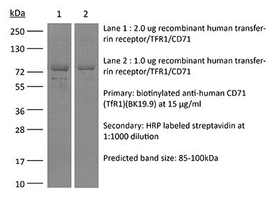
InVivoMAb anti-human CD71 (TfR1)
Product Description
Specifications
| Isotype | Mouse IgG1, κ |
|---|---|
| Recommended Isotype Control(s) | InVivoMAb mouse IgG1 isotype control, unknown specificity |
| Recommended Dilution Buffer | InVivoPure pH 7.0 Dilution Buffer |
| Conjugation | This product is unconjugated. Conjugation is available via our Antibody Conjugation Services. |
| Immunogen | Not available or unknown |
| Reported Applications | Immunohistochemistry (frozen) |
| Formulation |
PBS, pH 7.0 Contains no stabilizers or preservatives |
| Endotoxin |
≤1EU/mg (≤0.001EU/μg) Determined by LAL assay |
| Purity |
≥95% Determined by SDS-PAGE |
| Sterility | 0.2 µm filtration |
| Purification | Protein G |
| Molecular Weight | 150 kDa |
| Storage | The antibody solution should be stored at the stock concentration at 4°C. Do not freeze. |
| Need a Custom Formulation? | See All Antibody Customization Options |
Application References
Immunohistochemistry (frozen)
Sciot R, van Eyken P, Facchetti F, Callea F, van der Steen K, van Dijck H, van Parys G, Desmet VJ (1989). "Hepatocellular transferrin receptor expression in secondary siderosis" Liver 9(1):52-61.
PubMed
We investigated the hepatocellular transferrin receptor expression in 55 human liver specimens with secondary siderosis, with an indirect immunoperoxidase technique on frozen sections using 3 monoclonal anti-transferrin receptor antibodies. For comparison, specimens were also stained with the monoclonal antibody BK19.9, recognizing an antigen which is biochemically similar to the transferrin receptor, and with a monoclonal antibody against the epidermal growth factor receptor. The degree of iron overload was estimated semi-quantitatively, taking into account hepatocellular and Kupffer cell iron deposition. In 47 out of 55 specimens hepatocellular transferrin receptor expression was present. The positivity was predominantly localized on hemosiderin-free hepatocytes. With increasing hepatocellular iron deposition, the proportion of cases with absent transferrin receptor immunoreactivity increased. This supports the previously reported disappearance of hepatocellular transferrin receptor expression in primary hemochromatosis cases with severe iron deposition. However, the transferrin receptor negative cases included four specimens in which Kupffer cell iron deposition clearly exceeded hepatocyte iron load. This finding suggests that in addition to hepatocellular iron load other factors may regulate the expression of parenchymal transferrin receptors in iron overload diseases. These may include plasma levels of various iron sources and/or Kupffer cell iron load. The iron deposition did not influence the staining of the hepatocellular epidermal growth factor receptor nor the Kupffer cell staining by the BK19.9 antibody. This confirms the specificity of the findings concerning the behaviour of the transferrin receptor in secondary siderosis.
Immunohistochemistry (frozen)
Gatter KC, Brown G, Trowbridge IS, Woolston RE, Mason DY (1983). "Transferrin receptors in human tissues: their distribution and possible clinical relevance" J Clin Pathol 36(5):539-45.
PubMed
The distribution of transferrin receptors (TR) has been studied in a range of normal and malignant tissues using four monoclonal antibodies, BK19.9, B3/25, T56/14 and T58/1. In normal tissues TR was found in a limited number of sites, notably basal epidermis, the endocrine pancreas, hepatocytes, Kupffer cells, testis and pituitary. This restricted pattern of distribution may be relevant to the characteristic pattern of iron deposition in primary haemachromatosis. In contrast to this limited pattern of expression in normal tissue, the receptor was widely distributed in carcinomas, sarcomas and in samples from cases of Hodgkin's disease. This malignancy-associated expression of the receptor may play a role in the anaemia of advanced malignancy by competing with the bone marrow for serum iron.

