InVivoMAb anti-rat CD4
Product Details
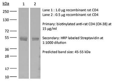
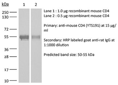
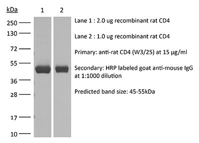
The W3/25 monoclonal antibody reacts with rat CD4. CD4 is a 55 kDa type I membrane glycoprotein from the immunoglobulin superfamily, and as a cell surface receptor protein, it acts as a co-receptor (with TCR) for class II MHC molecules displayed by antigen-presenting cells (APC). CD4 is expressed by the majority of thymocytes, helper T cells, a subset of NK-T cells, and weakly by dendritic cells and macrophages. CD4 plays an important role in the development of T cells and is required for the optimal functioning of mature T cells. The clone W3/25 is used as a “helper T-cell marker." The clone W3/25 is a nondepleting monoclonal antibody, but it has been shown to neutralize CD4 in vitro. Several in vitro studies also showed that the W3/25 monoclonal antibody inhibits CD4+ T cell activation by downregulating the CD4 molecule on the surface of lymphocytes. The W3/25 monoclonal antibody also inhibits antigen-induced T cell proliferation and IL2 production in MLR experiments. In prophylactic and experimental therapeutic in vivo investigations, this antibody has been extensively used to modulate disease-related and mechanistic parameters in experimental allergic encephalomyelitis (EAE), adjuvant arthritis, etc.Specifications
| Isotype | Mouse IgG1, κ |
|---|---|
| Recommended Isotype Control(s) | InVivoMAb rat IgG2b isotype control, anti-keyhole limpet hemocyanin |
| Recommended Dilution Buffer | InVivoPure pH 7.0 Dilution Buffer |
| Reported Applications |
in vivo down-regulation of surface CD4 in vitro neutralization of CD4 Flow cytometry Immunohistochemistry (paraffin) Immunohistochemistry (frozen) |
| Formulation |
PBS, pH 7.0 Contains no stabilizers or preservatives |
| Endotoxin |
<2EU/mg (<0.002EU/μg) Determined by LAL gel clotting assay |
| Purity |
>95% Determined by SDS-PAGE |
| Sterility | 0.2 µm filtration |
| Production | Purified from cell culture supernatant in an animal-free facility |
| Purification | Protein G |
| Molecular Weight | 150 kDa |
| Storage | The antibody solution should be stored at the stock concentration at 4°C. Do not freeze. |
Recommended Products
-
Recommended Isotype Control(s)
InVivoMAb rat IgG2b isotype control, anti-keyhole limpet hemocyanin
-

Recommended Dilution Buffer
InVivoPure pH 7.0 Dilution Buffer
in vivo down-regulation of surface CD4, Flow Cytometry
Acroute DV, Lafitte S, Dive D, Khalife J, Pierrot C. (2010). "A role for CD4+ and CD8+ cells and not for CD25+ cells in the control of Plasmodium berghei Anka blood stage parasites in rats" Parasite 10.1051/parasite/2010171053. PubMed
In previous studies of the infection of rats by P. berghei Anka, we have shown that primary blood stage infection induced the expansion of CD4+ T cells and CD8+ T cells in adult resistant rats while the number of CD4+CD25+ cells was found to be higher in young susceptible rats. In this work, the respective contribution of each cell population was determined in young and adult rats treated with monoclonal antibodies. Down-regulation of surface CD25 molecules, including those expressed by CD4+ cells did not significantly enhance the capacity of young rats to control the development of erythrocytic stages or modify the course of infection in adult infected rats. However, we observed a significant loss of protection when adult rats were treated with anti-CD4 mAb (W3/25) with higher blood parasitemia levels and approximately 50% of rats succumbed to infection. More importantly and in contrast to earlier studies performed in mice, we found a significant increase in blood parasite levels and a significant delay in parasite clearance in adult rats treated with anti-CD8 mAb OX8, known to deplete CD8+ cells. These results suggest that CD8+ cells play a critical role in the development of immune responses in rats to control the replication of blood stage parasites.
Immunohistochemistry (frozen)
Wu Z, Toh K, Nagata K, Kukita T, Iijima T. (2004). "Effect of the resection of the sciatic nerve on the Th1/Th2 balance in the synovia of the ankle joint of adjuvant arthritic rats" Histochem Cell Biol 121(2):141-7. PubMed
Inflamed synovia of the ankle joint after 2-4 weeks of adjuvant injection receives dense sensory innervation. To study the role of sensory nerves on the local inflammation, the relative expression of T helper 1 and 2 lymphocyte (Th1 and Th2) markers was investigated on both axotomized adjuvant arthritic (AA) rats, whose sciatic nerves were resected before adjuvant injection, and on sham-operated ones. Immunohistochemical expressions of CXC chemokine receptor 3 (CXCR3) and CC chemokine receptor 4 (CCR4) were examined and compared with those of Th1 cytokine (interferon-gamma, IFN-gamma), Th2 cytokine (interleukin-4, IL-4), and anti-T cell antibody (W3/25). Double-positive cells for IFN-gamma/CXCR3 and for IL-4/CCR4 were greater than 90% and greater than 95%, respectively. The reciprocal combinations, IL-4/CXCR3 and IFN-gamma/CCR4, however, yielded less than 10% and less than 5% of double-positive cells. CXCR3 and CCR4 thus appear to be available as markers for Th1/Th2 subsets in the synovia of AA rats. Using these markers, it became clear that the percentage of Th1 cells to total Th cells was higher than that of Th2 cells in axotomized AA rats at weeks 2-4, whereas in sham-operated AA rats, the percentage of Th1 cells to total Th cells was higher than that of Th2 cells at week 2 and the latter exceeded the former at week 4. Our observations strongly suggested the presence of the anti-inflammatory action of sensory nerves in rats with adjuvant arthritis.
in vivo down-regulation of surface CD4
Thullen TD, Ashbaugh AD, Daly KR, Linke MJ, Steele PE, Walzer PD. (2003). "New rat model of Pneumocystis pneumonia induced by anti-CD4(+) T-lymphocyte antibodies" Infect Immun 10.1128/IAI.71.11.6292-6297.2003. PubMed
The CD4(+) T lymphocyte plays a central role in host defense against Pneumocystis pneumonia but has received only limited attention in rats. CD4(+) T-cell-depleting (OX-38) and nondepleting (W3/25) monoclonal antibodies, which recognize an identical or adjacent epitope, were administered for up to 14 weeks to Lewis rats that had been exposed to PNEUMOCYSTIS: While OX-38 produced a greater decrease in circulating CD4(+) cells than W3/25, both antibody treatments resulted in similar effects on the health of the rats and the levels of Pneumocystis pneumonia, which were milder than those found with corticosteroids. W3/25 also did not enhance the severity of Pneumocystis pneumonia achieved with corticosteroids alone. We conclude that CD4(+) cell function is more important than CD4(+) cell number in host defense against Pneumocystis in the rat and that this new model permits study of opportunistic infections in the rat without the confounding effects of corticosteroids.
in vivo down-regulation of surface CD4, Flow Cytometry
Pohlers D, Schmidt-Weber CB, Franch A, Kuhlmann J, Bräuer R, Emmrich F, Kinne RW. (2002). "Differential clinical efficacy of anti-CD4 monoclonal antibodies in rat adjuvant arthritis is paralleled by differential influence on NF-kappaB binding activity and TNF-alpha secretion of T cells" Arthritis Res 10.1186/ar404. PubMed
The aim of this study was to analyze the differential effects of three anti-CD4 monoclonal antibodies (mAbs) (with distinct epitope specifities) in the treatment of rat adjuvant arthritis (AA) and on T-cell function and signal transduction. Rat AA was preventively treated by intraperitoneal injection of the anti-CD4 mAbs W3/25, OX35, and RIB5/2 (on days -1, 0, 3, and 6, i.e. 1 day before AA induction, on the day of induction [day 0], and thereafter). The effects on T-cell reactivity in vivo (delayed-type hypersensitivity), ex vivo (ConA-induced proliferation), and in vitro (mixed lymphocyte culture) were assessed. The in vitro effects of anti-CD4 preincubation on T-cell receptor (TCR)/CD3-induced cytokine production and signal transduction were also analyzed. While preventive treatment with OX35 and W3/25 significantly ameliorated AA from the onset, treatment with RIB5/2 even accelerated the onset of AA by approximately 2 days (day 10), and ameliorated the arthritis only in the late phase (day 27). Differential clinical effects at the onset of AA were paralleled by a differential influence of the mAbs on T-cell functions, i.e. in comparison with OX35 and W3/25, the 'accelerating' mAb RIB5/2 failed to increase the delayed-type hypersentivity (DTH) to Mycobacterium tuberculosis, increased the in vitro tumor necrosis factor (TNF)-alpha secretion, and more strongly induced NF-kappaB binding activity after anti-CD4 preincubation and subsequent TCR/CD3-stimulation. Depending on their epitope specificity, different anti-CD4 mAbs differentially influence individual proinflammatory functions of T cells. This fine regulation may explain the differential efficacy in the treatment of AA and may contribute to the understanding of such treatments in other immunopathologies.
in vivo down-regulation of surface CD4, Flow Cytometry
Pelegrí C, Castell M, Serra M, Rabanal M, Rodríguez-Palmero M, Castellote C, Franch A. (2001). "Prevention of adjuvant arthritis by the W3/25 anti-CD4 monoclonal antibody is associated with a decrease of blood CD4(+)CD45RC(high) T cells" Clin Exp Immunol 10.1046/j.1365-2249.2001.01592.x. PubMed
Imbalance between Th1 and Th2 functions is considered to play a key role in the induction and development of several autoimmune diseases, and the correction of that imbalance has led to effective therapies of some experimental pathologies. To examine whether CD4(+)CD45RC(high) (Th1-like) and CD4(+)CD45RC(low) (Th2-like) lymphocytes play a role in the pathogenesis of adjuvant arthritis (AA) and in its prevention by anti-CD4 antibody, CD45RC expression on CD4(+) T cells was determined in arthritic rats and in animals treated with an anti-CD4 MoAb (W3/25) during the latency period of AA. The phenotype of regional lymph node lymphocytes from arthritic rats in the active phase of the disease was determined by flow cytometry. Peripheral blood lymphocytes from rats treated with W3/25 MoAb were also analysed for 2 weeks after immunotherapy finished. IgG2a and IgG1 isotypes of sera antibodies against the AA-inducing mycobacteria, considered to be associated with Th1 and Th2 responses, respectively, were also determined by ELISA techniques. Fourteen days after arthritis induction, regional lymph nodes presented an increase in CD4+CD45RC(high) T cell proportion. Preventive immunotherapy with W3/25 MoAb inhibited the external signs of arthritis and produced a specific decrease in blood CD4(+)CD45RC(high) T cells and a diminution of antibodies against mycobacteria, more marked for IgG2a than for IgG1 isotype. These results indicate a possible role of CD4(+)CD45RC(high) T lymphocytes in the pathogenesis of AA, and suggest that the success of anti-CD4 treatment is due to a specific effect on CD4(+)CD45RC(high) T subset that could be associated with a decrease in Th1 activity.
in vitro neutralization of CD4
Mannie MD, Norris MS. (2001). "MHC class-II-restricted antigen presentation by myelin basic protein-specific CD4+ T cells causes prolonged desensitization and outgrowth of CD4- responders" Cell Immunol 10.1006/cimm.2001.1843. PubMed
T cells express MHC class II glycoproteins under various conditions of activation or inflammation. To assess whether T cell APC (T-APC) activity had long-term tolerogenic consequences, myelin basic protein (MBP)-specific rat T cells were induced to acquire MBP-derived I-A complexes to promote reciprocal antigen presentation. T-T antigen presentation caused extensive cell death among T-APC and MBP-specific T responders and caused long-term desensitization of surviving responders. Addition of the anti-I-A mAb OX6 to activated I-A+ responders inhibited T-APC activity, accelerated recovery from postactivation refractoriness, and prevented long-term loss of reactivity in responder T cells. Antigenic activation of responder T cells with irradiated T-APC induced profound losses in reactivity that lasted for over 1 month of propagation in IL-2 and was associated with preferential outgrowth of CD4- T cells. Antigen-activated CD4- T cells exhibited more rapid IL-2-dependent growth that eventually normalized compared to CD4+ T cells 1-2 months after antigen exposure. In conclusion, expression of T-APC activity by activated T cells represents an important negative feedback pathway that depletes antigen-reactive T cells and causes long-term desensitization of surviving T cells. Hence, T cell APC may be an important mechanism of self-tolerance.
in vitro neutralization of CD4, Flow Cytometry
Wiegers GJ, Stec IE, Klinkert WE, Reul JM. (2000). "Glucocorticoids regulate TCR-induced elevation of CD4: functional implications" J Immunol 164(12):6213-20. PubMed
CD4 serves as a coreceptor during Ag recognition by the TCR. This interaction results in a marked increase in the sensitivity of a T cell to Ag presented by MHC class II molecules. Here we report that activation of T cells either by plate-bound mAb (anti-TCR, anti-CD3) or soluble activators (staphylococcal enterotoxin A, Con A) is associated with an (up to 3-fold) increase in CD4 cell surface expression on CD25+ cells, which was maximal after 72-96 h. Incubation with the glucocorticoid hormone corticosterone (CORT) shifted the enhancement of CD4 expression to a point about 24 h earlier than that observed in control cultures. In parallel, the proliferative response of these CORT-treated cells was profoundly enhanced. An involvement of increased CD4 expression in this enhanced proliferative response was evidenced by the observation that T cell proliferation in CORT-treated cultures was much less sensitive to inhibition by an inhibitory, nondepleting anti-CD4 mAb than that in control cultures. TCR down-regulation was, however, not affected by CORT. Thus, based on this study and previous reports we propose that both TCR-mediated signals and glucocorticoids are important physiological regulators of CD4 expression. In addition, these findings may be of significance for the sensitivity of CD4+ cells to HIV infection upon T cell activation, as the efficacy of primary patient HIV entry depends on the level of surface CD4.
Flow Cytometry, Immunohistochemistry (paraffin)
Reynolds J, Tam FW, Chandraker A, Smith J, Karkar AM, Cross J, Peach R, Sayegh MH, Pusey CD. (2000). "CD28-B7 blockade prevents the development of experimental autoimmune glomerulonephritis" J Clin Invest 105(5):643-51. PubMed
Experimental autoimmune glomerulonephritis (EAG), an animal model of Goodpasture's disease, can be induced in Wistar Kyoto (WKY) rats by a single injection of rat glomerular basement membrane (GBM) in adjuvant. EAG is characterized by circulating and deposited anti-GBM antibodies, accompanied by focal necrotizing glomerulonephritis with crescent formation. The role of T cells in the pathogenesis of EAG remains unclear. T-cell costimulation is provided by ligation of CD28 with either B7.1 (CD80) or B7.2 (CD86) on antigen-presenting cells, and can be inhibited by a soluble form of CTLA4 (CTLA4-Ig) that binds to both B7.1 and B7.2. We examined the effect of CD28-B7 blockade on the development of EAG using native CTLA4-Ig or mutant CTLA4-Ig (Y100F-Ig), which selectively blocks B7.1. Native CTLA4-Ig treatment ameliorated EAG by several measures, including the levels of circulating anti-GBM antibodies, albuminuria, the deposition of IgG and fibrin in the glomeruli, the severity of glomerular abnormalities, and the numbers of infiltrating T cells and macrophages. Y100F-Ig resulted in a similar reduction in the severity of nephritis, but produced no overall reduction in circulating anti-GBM antibodies, although there was a reduction in IgG2a antibodies. We concluded that CD28-B7 blockade reduced autoantibody production and cellular infiltration of glomeruli, and prevented target organ injury. Our results suggest a key role for B7. 1 in costimulation of Th1-like autoimmune responses in the rat, and show that glomerular injury in EAG is largely dependent on cell-mediated mechanisms.
Immunohistochemistry (paraffin)
Ysebaert DK, De Greef KE, Vercauteren SR, Ghielli M, Verpooten GA, Eyskens EJ, De Broe ME. (2000). "Identification and kinetics of leukocytes after severe ischaemia/reperfusion renal injury" Nephrol Dial Transplant 15(10):1562-74. PubMed
Background: Leukocyte adhesion/infiltration in response to renal ischaemia/reperfusion (I/R) injury is a well-known but poorly understood phenomenon. The identification, kinetics, and exact role of these inflammatory cells in I/R injury and regeneration are still matters of debate. Methods: Uninephrectomized rats were submitted to 60 min renal ischaemia by clamping of renal vessels. Results: Severe acute renal failure was observed, with maximum functional impairment on day 2. By 12 h after the ischaemic event, up to 80% of proximal tubular cells in the outer stripe of outer medulla (OSOM) were already severely damaged. Proliferation (proliferating cell nuclear antigen (PCNA) staining) started after 24 h, reaching maximum activity on day 3. Regeneration of tubular morphology started on the 3rd day, and after 10 days 50% of tubules had regenerated completely. Interstitial leukocytes (OX-1 immunohistochemical staining) were already prominent at day 1, thereafter gradually increasing with time. The so-called neutrophil-specific identification methods (myeloperoxidase (MPO), chloroacetate esterase, mAb HIS-48) proved to be non-specific, since they also stained for macrophages, as demonstrated by flow cytometry and the combination of these stainings with the macrophage-specific ED-1 staining. MPO activity was already significantly increased at 1 h post-I/R (439+/-34%, P<0.005), reaching its maximum activity after 12 h of I/R (1159+/-138%, P<0.0005), declining thereafter. On the other hand, neutrophil presence investigated by H&E staining revealed only a few neutrophils in glomeruli, medullary rays, and OSOM at 24 h after the ischaemic event (4.7+/-4.2 cells/mm(2) vs controls=2.3+/-2.0 cells/mm(2) (n.s.)), and remained unchanged over the next 10 days. In contrast, significant monocyte/macrophage adhesion/infiltration (ED-1 staining) occurred at the OSOM at 24 h post-ischaemia (at 24 h, 120+/-46 cells/mm(2) vs. sham=18+/-4 cells/mm(2) (P<0.05)), became prominent at day 5 (1034+/-161 cells/mm(2) vs sham=18+/-18 cells/mm(2) (P<0.05)), and almost disappeared after 10 days. CD4(+) cells (W3/25) gradually increased from day 5, reaching a maximum at day 10. A few CD8(+) cells (OX-8) were apparent from days 3 until 10, but no B-cells (OX-33) were observed. Conclusions: After severe warm I/R renal injury, a pronounced acute tubular necrosis occurs during the first 12-24 h in the absence of a marked cellular infiltrate, but with an important renal MPO activity, reflecting the activation of the adhering inflammatory cells (polymorphonuclear cells (PMNs) and mainly monocytes/macrophages). Only later at the time and site (OSOM) of regeneration a sequential accumulation of monocytes/macrophages and T cells becomes prominent, in contrast with the low number of neutrophils found in the kidney during the 10-day post-ischaemic period. The non-specificity of the so-called neutrophil-specific identification methods (MPO activity, naphthol AS-D chloroacetate esterase, or mAb HIS-48 staining), cross-reacting with monocytes/macrophages, explains the controversy in literature concerning the number of PMNs in post-ischaemic injury.
Immunohistochemistry (paraffin)
Remuzzi G, Zoja C, Gagliardini E, Corna D, Abbate M, Benigni A. (1999). "Combining an antiproteinuric approach with mycophenolate mofetil fully suppresses progressive nephropathy of experimental animals" J Am Soc Nephrol 10(7):1542-9. PubMed
Chronic renal diseases progress to organ insufficiency, which may require replacement therapy within one to three decades even independently of the type of initial insults. In the majority of cases, the degrees of proteinuria and interstitial leukocyte infiltration and scarring are strictly correlated with the rate of disease progression. This study tests the hypothesis that excess intrarenal protein traffic may cause lymphocyte-dependent interstitial injury that, while not fully controlled by antiproteinuric therapy, can be further inhibited by concomitant immunosuppression. A primarily nonimmune model was used to reproduce progressive renal disease due to a critical loss of nephron mass. Angiotensin-converting enzyme (ACE) inhibitor limited proteinuria, interstitial inflammation, MHC class II antigen expression, and severe lesions. Combined treatment with ACE inhibitor and a specific antilymphocyte agent, mycophenolate mofetil, dramatically attenuated macrophage and T cell infiltration, MHC-class II overexpression, dendritic cells, and all manifestations of the disease. Evidence of lymphocyte-mediated renal injury in the setting of excess protein traffic provides the basis for combining ACE inhibition and immunosuppression to halt progression of proteinuric kidney disease and minimize the need for dialysis or transplantation.
in vivo down-regulation of surface CD4, Flow Cytometry
Caballero F, Pelegrí C, Castell M, Franch A, Castellote C. (1998). "Kinetics of W3/25 anti-rat CD4 monoclonal antibody. Studies on optimal doses and time-related effects" Immunopharmacology 10.1016/s0162-3109(98)00011-3. PubMed
Although anti-CD4 monoclonal antibodies (MoAb) have been proven successful in preventing or treating adjuvant arthritis, little is known about the duration of the effects of these MoAb and their pharmacokinetics. In this work, we report the effects of a mouse anti-rat CD4 MoAb, named W3/25, on peripheral blood lymphocytes from female Wistar rats. Animals received a single dose of W3/25, from 1 to 3 mg, and blood was sampled at different time points from 0 h to 15 days after MoAb administration. After erythrocyte lysis, samples were stained by indirect immunofluorescence and analyzed by flow cytometry. Pharmacokinetic data were studied by assessing plasma levels of mouse IgG1 by ELISA-sandwich. W3/25 produced the down-regulation of surface CD4 molecule as early as 20 min after its administration at doses of 2 and 3 mg. The same effect was seen 30 min after a dose of 1 mg. The recovery of lymphocytes with normal expression of CD4 also depended of the dose administered. Thus, CD4+ lymphocytes were recovered at 48, 72 and 96 h in rats treated with 1, 2 or 3 mg of W3/25, respectively. Plasma levels of free antibody were detectable from 20 min to 72 h, 60 min to 48 h and 60 min to 24 h after administration of 3, 2 and 1 mg, respectively, of W3/25. The mouse IgG1 MoAb used in this study followed a two-compartment model and its behavior was linear.
in vitro neutralization of CD4, Flow Cytometry
Mannie MD, White GA, Nardella JP, Davidian DK, Arnold PY. (1998). "Partial agonism elicits an enduring phase of T-cell-medicated antigen presentation" Cell Immunol 186(2):83-93. PubMed
Previous studies have shown that the anti-CD4 mAb W3/25 strongly enhances T cell APC (T-APC) activity. In this study, single positive CD4+ and double negative (DN) (CD4-CD8-) T-helper cells specific for the 55-69 or 72-86 sequence of guinea pig (GP) myelin basic protein (GPMBP) were used to study CD4 regulation of T-APC activity. Clones were cultured with irradiated SPL and GPMBP or rat (R) MBP for 2-3 days, were propagated in IL-2 for another 1-3 days, were irradiated, and were used as T-APC. DN T cells specific for GP55-69 effectively presented GPMBP and were superior APC compared to other CD4+ T cells for presentation of this antigen. In contrast, DN T cells specific for the dominant encephalitogenic 72-86 determinant did not effectively present the agonist GPMBP but potently presented the partial agonist RMBP. The heightened APC activity of DN T cells reflected the lack of CD4 because the anti-CD4 mAb W3/25 promoted T-APC activity of CD4+ T cells to those levels expressed by DN T cells. Overall, T cells with potent reactivity to GPMBP or RMBP were subsequently unable to present that antigen, whereas T cells exhibiting partial or low antigen reactivities were highly effective APC for presentation of that antigen. The unrelated antigen conalbumin was presented by MBP-specific clones only when added to culture with a specific partial agonist. Together, these data indicate that partially agonistic MHC ligands promote prolonged expression of T-APC activity and that DN T cells may be specialized to mediate postactivational antigen presentation.
in vivo down-regulation of surface CD4, Flow Cytometry
Pelegrí C, Paz Morante M, Castellote C, Castell M, Franch A. (1995). "Administration of a nondepleting anti-CD4 monoclonal antibody (W3/25) prevents adjuvant arthritis, even upon rechallenge: parallel administration of a depleting anti-CD8 monoclonal antibody (OX8) does not modify the effect of W3/25" Cell Immunol 10.1006/cimm.1995.1203. PubMed
The aim of this study was to determine the effects of the anti-CD4 monoclonal antibody (mAb) W3/25, found to be nondepleting, on the onset of rat adjuvant arthritis (AA), and, in addition, to ascertain whether depletion of CD8+ cells during the same period could interfere with those effects. Female Wistar rats in which AA had been induced were treated with W3/25 and/or OX8 (anti-rat CD8) mAb during the latency period of arthritis. W3/25 alone or in combination with OX8 prevented the inflammatory process of AA. When the protected groups were rechallenged with a second dose of Mycobacterium butyricum no arthritis was observed. Protected and nonprotected arthritic animals developed the same anti-mycobacteria antibody levels as the arthritic control group. This study indicates that a nondepleting anti-CD4 mAb can prevent AA, while CD8+ lymphocytes do not appear relevant for the development of AA and do not seem to have a regulatory role for CD4+ cells.
in vitro neutralization of CD4
Mannie MD, Rosser JM, White GA. (1995). "Autologous rat myelin basic protein is a partial agonist that is converted into a full antagonist upon blockade of CD4. Evidence for the integration of efficacious and nonefficacious signals during T cell antigen recognition" J Immunol 154(6):2642-54. PubMed
A central question of TCR function is based on the observation that some MHC ligands may bind TCR without stimulating biologic activity. To address the role of CD4 in this mechanism, we studied the interactions of the anti-CD4 mAb W3/25 with an encephalitogenic line of T helper cells. Proliferative responses to guinea pig (GP) myelin basic protein (GPMBP) were mediated by distinct W3/25-sensitive and W3/25-insensitive mechanisms whereas responses to autologous rat (R) MBP (RMBP) were exclusively mediated by W3/25-sensitive pathways. In assays of IL-2 production, RMBP was a partial agonist that stimulated an intermediate level of IL-2 production but antagonized high levels of GPMBP-stimulated IL-2 production to that intermediate level. In the presence of W3/25, RMBP lacked stimulatory activity but instead exhibited inhibitory activity that completely blocked GPMBP-stimulated proliferative responses. The inhibitory mechanism did not involve antigenic competition for MHC glycoproteins, a blockade of mitogenic signaling, or induction of high zone tolerance. Rather, the mechanism involved specific occupancy of TCR with antagonistic MHC ligands derived from the 72-86 region of RMBP. In proliferative assays, GPMBP was approximately 10-fold more active than RMBP. In the presence of W3/25 however, GPMBP-induced agonism and RMBP-induced antagonism exhibited overlapping dose-response curves. RMBP and W3/25 not only fully inhibited GPMBP-stimulated proliferation, this synergistic combination also elicited an extended phase of T cell energy. In conclusion, RMBP-derived MHC ligands occupy TCR to exhibit full efficacy for CD4-dependent signaling pathways while simultaneously lacking efficacy for W3/25-resistant signaling pathways. These data support an "integrative" model of T cell Ag recognition whereby MHC glycoproteins actively guide specific clustering of MHC-ligated TCR to enable quantitative comparisons of efficacious (nonself) and nonefficacious (self) signals by T cells.
Immunohistochemistry (paraffin), Immunohistochemistry (frozen)
Whiteland JL, Nicholls SM, Shimeld C, Easty DL, Williams NA, Hill TJ. (1995). "Immunohistochemical detection of T-cell subsets and other leukocytes in paraffin-embedded rat and mouse tissues with monoclonal antibodies" J Histochem Cytochem 43(3):313-20. PubMed
We describe a method for immunohistochemical localization of T-cells, CD4+ T-cells, CD8+ T-cells, B-cells, activated lymphocytes, major histocompatibility complex (MHC) class II antigens, macrophages, dendritic cells, and granulocytes in rat and mouse tissue fixed in periodate-lysine-paraformaldehyde (PLP) and embedded in paraffin. Rat and mouse spleen and eyes were fixed in PLP for 18-24 hr, rapidly dehydrated, infiltrated under vacuum with paraffin at 54 degrees C, sectioned, and stained with appropriate monoclonal antibodies (MAbs). Sections of PLP-fixed, paraffin-embedded spleen were compared with acetone-fixed frozen spleen sections with respect to morphology and staining quality. Nine of 10 MAbs to rat antigens and eight of nine MAbs to mouse antigens stained paraffin sections equally or more intensely than frozen sections. The two MAbs that showed weaker staining still gave good staining on paraffin sections. Paraffin-embedded rat and mouse eyes were easier to section serially than frozen eyes, showed superior morphology, and individually stained cells were readily identified. Therefore, a combination of PLP fixation and low-temperature paraffin embedding permits detection of the major types of immune cell in rat and mouse tissues while maintaining good morphology, particularly in diseased, damaged, or delicate tissues.