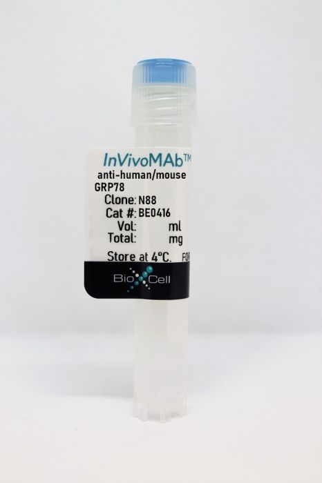
InVivoMAb anti-human/mouse GRP78
Product Details
The N88 monoclonal antibody reacts with human and mouse Glucose Regulated Protein 78 (GRP78) also known as BiP or HSPA5. GRP78 is a stress inducible, pro-survival, endoplasmic reticulum chaperone in the HSP70 family. Cell Surface GRP78 acts as a multifunctional receptor that affects both cell proliferation and viability. GRP78 is involved in many cellular processes, including translocating newly synthesized polypeptides across the ER membrane, facilitating the folding and assembly of proteins, targeting misfolded proteins for ER-associated degradation (ERAD), regulating calcium homeostasis, and serving as an ER stress sensor. Expression of cell surface GRP78 is associated with increased malignancy and resistance to chemotherapy and radiotherapy in various cancers, particularly prostate cancer. The N88 antibody binds to the N‐terminal domain of GRP78 and has been shown to accelerate tumor growth in a B16F1 melanoma tumor model.Specifications
| Isotype | Mouse IgG1, κ |
|---|---|
| Recommended Isotype Control(s) | InVivoMAb mouse IgG1 isotype control, unknown specificity |
| Recommended Dilution Buffer | InVivoPure pH 7.0 Dilution Buffer |
| Conjugation | This product is unconjugated. Conjugation is available via our Antibody Conjugation Services. |
| Immunogen | Full-length recombinant GRP78 protein |
| Reported Applications |
in vivo administration Functional assays |
| Formulation |
PBS, pH 7.0 Contains no stabilizers or preservatives |
| Endotoxin |
<2EU/mg (<0.002EU/μg) Determined by LAL gel clotting assay |
| Purity |
>95% Determined by SDS-PAGE |
| Sterility | 0.2 µm filtration |
| Production | Purified from cell culture supernatant in an animal-free facility |
| Purification | Protein G |
| Molecular Weight | 150 kDa |
| Storage | The antibody solution should be stored at the stock concentration at 4°C. Do not freeze. |
Recommended Products
-
Recommended Isotype Control(s)
InVivoMAb mouse IgG1 isotype control, unknown specificity
-

Recommended Dilution Buffer
InVivoPure pH 7.0 Dilution Buffer
in vivo administration, Functional Assays
de Ridder GG, Ray R, Pizzo SV. (2012). "A murine monoclonal antibody directed against the carboxyl-terminal domain of GRP78 suppresses melanoma growth in mice" Melanoma Res 22(3):225-35. PubMed
The HSP70 family member GRP78 is a selective tumor marker upregulated on the surface of many tumor cell types, including melanoma, where it acts as a growth factor receptor-like protein. Receptor-recognized forms of the proteinase inhibitor α2-macroglobulin (α2M*) are the best-characterized ligands for GRP78, but in melanoma and other cancer patients, autoantibodies arise against the NH2-terminal domain of GRP78 that react with tumor cell-surface GRP78. This causes the activation of signaling cascades that are proproliferative and antiapoptotic. Antibodies directed against the COOH-terminal domain of GRP78, however, upregulate p53-mediated proapoptotic signaling, leading to cell death. Here, we describe the binding characteristics, cell signaling properties, and downstream cellular effects of three novel murine monoclonal antibodies. The NH2-terminal domain-reactive antibody, N88, mimics α2M* as a ligand and drives PI 3-kinase-dependent activation of Akt and the subsequent stimulation of cellular proliferation in vitro. The COOH-terminal domain-reactive antibody, C38, acts as an antagonist of both α2M* and N88, whereas another, C107, directly induces apoptosis in vitro. In a murine B16F1 melanoma flank tumor model, we demonstrate the acceleration of tumor growth by treatment with N88, whereas C107 significantly slowed tumor growth whether administered before (P<0.005) or after (P<0.05) tumor implantation.
- Immunology and Microbiology,
- Cancer Research
Inhibition of Microsomal Prostaglandin E2 Synthase Reduces Collagen Deposition in Melanoma Tumors and May Improve Immunotherapy Efficacy by Reducing T-cell Exhaustion.
In Cancer Res Commun on 1 July 2023 by Fukuda, Y., Kim, S. H., et al.
PubMed
The arachidonic acid pathway participates in immunosuppression in various types of cancer. Our previous observation detailed that microsomal prostaglandin E2 synthase 1 (mPGES-1), an enzyme downstream of cyclooxygenase 2 (COX-2), limited antitumor immunity in melanoma; in addition, genetic depletion of mPGES-1 specifically enhanced immune checkpoint blockade therapy. The current study set out to distinguish the roles of mPGES-1 from those of COX-2 in tumor immunity and determine the potential of mPGES-1 inhibitors for reinforcing immunotherapy in melanoma. Genetic deletion of mPGES-1 showed different profiles of prostaglandin metabolites from that of COX-2 deletion. In our syngeneic mouse model, mPGES-1-deficient cells exhibited similar tumorigenicity to that of COX-2-deficient cells, despite a lower ability to suppress PGE2 synthesis by mPGES-1 depletion, indicating the presence of factors other than PGE2 that are likely to regulate tumor immunity. RNA-sequencing analysis revealed that mPGES-1 depletion reduced the expressions of collagen-related genes, which have been found to be associated with immunosuppressive signatures. In our mouse model, collagen was reduced in mPGES-1-deficient tumors, and phenotypic analysis of tumor-infiltrating lymphocytes indicated that mPGES-1-deficient tumors had fewer TIM3+ exhausted CD8+ T cells compared with COX-2-deficient tumors. CAY10678, an mPGES-1 inhibitor, was equivalent to celecoxib, a selective COX-2 inhibitor, in reinforcing anti-PD-1 treatment. Our study indicates that mPGES-1 inhibitors represent a promising adjuvant for immunotherapies in melanoma by reducing collagen deposition and T-cell exhaustion. Collagen is a predominant component of the extracellular matrix that may influence the tumor immune microenvironment for cancer progression. We present here that mPGES-1 has specific roles in regulating tumor immunity, associated with several collagen-related genes and propose that pharmacologic inhibition of mPGES-1 may hold therapeutic promise for improving immune checkpoint-based therapies. © 2023 The Authors; Published by the American Association for Cancer Research.
- Cancer Research,
- Immunology and Microbiology
Angiogenic inhibitor pre-administration improves the therapeutic effects of immunotherapy.
In Cancer Medicine on 1 April 2023 by Sato, M., Maishi, N., et al.
PubMed
In lung cancer, immune checkpoint inhibitors (ICIs) are often inadequate for tumor growth inhibition. Angiogenic inhibitors (AIs) are required to normalize tumor vasculature for improved immune cell infiltration. However, in clinical practice, ICIs and cytotoxic antineoplastic agents are simultaneously administered with an AI when tumor vessels are abnormal. Therefore, we examined the effects of pre-administering an AI for lung cancer immunotherapy in a mouse lung cancer model. Using DC101, an anti-vascular endothelial growth factor receptor 2 (VEGFR2) monoclonal antibody, a murine subcutaneous Lewis lung cancer (LLC) model was used to determine the timing of vascular normalization. Microvessel density (MVD), pericyte coverage, tissue hypoxia, and CD8-positive cell infiltration were analyzed. The effects of an ICI and paclitaxel after DC101 pre-administration were investigated. On Day 3, increased pericyte coverage and alleviated tumor hypoxia represented the highest vascular normalization. CD8+ T-cell infiltration was also highest on Day 3. When combined with an ICI, DC101 pre-administration significantly reduced PD-L1 expression. When combined with an ICI and paclitaxel, only DC101 pre-administration significantly inhibited tumor growth, but simultaneous administration did not. AI pre-administration, and not simultaneous administration, may increase the therapeutic effects of ICIs due to improved immune cell infiltration. © 2023 The Authors. Cancer Medicine published by John Wiley & Sons Ltd.
- Cancer Research
The COX2 Effector Microsomal PGE2 Synthase 1 is a Regulator of Immunosuppression in Cutaneous Melanoma.
In Clinical Cancer Research on 1 March 2019 by Kim, S. H., Roszik, J., et al.
PubMed
Microsomal prostaglandin E2 synthase 1 (mPGES1) was evaluated as an important downstream effector of the COX2 pathway responsible for tumor-mediated immunosuppression in melanoma. The analysis of a stage III melanoma tissue microarray (n = 91) was performed to assess the association between mPGES1, COX2, CD8, and patient survival. Pharmacologic inhibitors and syngeneic mouse models using PTGES-knockout (KO) mouse melanoma cell lines were used to evaluate the mPGES1-mediated immunosuppressive function. We observed correlations in expression and colocalization of COX2 and mPGES1, which are associated with increased expression of immunosuppressive markers in human melanoma. In a syngeneic melanoma mouse model, PTGES KO increased melanoma expression of PD-L1, increased infiltration of CD8a+ T cells, and CD8a+ dendritic cells into tumors and suppressed tumor growth. Durable tumor regression was observed in mice bearing PTGES KO tumors that were given anti-PD-1 therapy. Analysis of a stage III melanoma tissue microarray revealed significant associations between high mPGES1 expression and low CD8+ infiltration, which correlated with a shorter patient survival. Our results are the first to illustrate a potential role for mPGES1 inhibition in melanoma immune evasion and selective targeting in supporting the durability of response to PD-1 checkpoint immunotherapy. More research effort in this drug development space is needed to validate the use of mPGES1 inhibitors as safe treatment options. ©2018 American Association for Cancer Research.