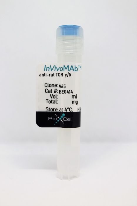
InVivoMAb anti-rat TCR γ/δ
Product Details
The V65 monoclonal antibody reacts with rat γ/δ TCR (gamma delta T cell receptor). The γ/δ TCR is expressed by a subset of T cells found in the thymus, peripheral lymphoid tissues, intestinal epithelium, epidermis, and peritoneum. The exact function, ligand, and specificity of γ/δ TCR-expressing T cells are not completely understood. Studies suggest that these cells recognize bacterial ligands and some tumor cells in the context of MHC class I-like gene products and play a role in regulating the immune response during bacterial infection.Specifications
| Isotype | Mouse IgG1, κ |
|---|---|
| Recommended Isotype Control(s) | InVivoMAb mouse IgG1 isotype control, unknown specificity |
| Recommended Dilution Buffer | InVivoPure pH 7.0 Dilution Buffer |
| Conjugation | This product is unconjugated. Conjugation is available via our Antibody Conjugation Services. |
| Immunogen | Rat T Blasts |
| Reported Applications |
in vivo γ/δ T cell depletion Flow cytometry |
| Formulation |
PBS, pH 7.0 Contains no stabilizers or preservatives |
| Endotoxin |
<2EU/mg (<0.002EU/μg) Determined by LAL gel clotting assay |
| Purity |
>95% Determined by SDS-PAGE |
| Sterility | 0.2 µm filtration |
| Production | Purified from cell culture supernatant in an animal-free facility |
| Purification | Protein G |
| Molecular Weight | 150 kDa |
| Storage | The antibody solution should be stored at the stock concentration at 4°C. Do not freeze. |
Recommended Products
-
Recommended Isotype Control(s)
InVivoMAb mouse IgG1 isotype control, unknown specificity
-

Recommended Dilution Buffer
InVivoPure pH 7.0 Dilution Buffer
Flow Cytometry
Ando T, Wu H, Watson D, Hirano T, Hirakata H, Fujishima M, Knight JF. (2001). "Infiltration of canonical Vgamma4/Vdelta1 gammadelta T cells in an adriamycin-induced progressive renal failure model" J Immunol 167(7):3740-5. PubMed
We have previously reported an infiltration of renal interstitial gammadelta T cells in Adriamycin-induced progressive glomerulosclerosis in the rat kidney. The TCR repertoire and sequences used by these gammadelta T cells have now been studied. Two injections of Adriamycin 14 days apart caused segmental glomerulosclerosis, massive interstitial infiltration of mononuclear cells, and end-stage renal failure. Flow cytometry of lymphocyte subpopulations with Abs to CD3, the gammadelta TCR, and the alphabeta TCR showed that gammadelta T cells as a proportion of CD3(+) cells were increased in Adriamycin-treated kidneys (8.5 +/- 5.4%), but not in lymph nodes (1.3 +/- 0.4%). A semiquantitative score of glomerular damage (r = 0.65; p < 0.01) and creatinine (r = 0.62; p < 0.01) correlated significantly with the presence of gammadelta T cells. TCR Vgamma repertoire analysis by RT-PCR and Southern blotting showed that Vgamma2 was the dominant subfamily in lymph nodes, whereas Vgamma4 became the predominant subfamily in advanced stages of the rat Adriamycin-treated kidney. Sequencing of the Vgamma4-Jgamma junctional region showed an invariant sequence. The amino acid sequence of the junctional region of the Vgamma4 TCR was the same as the reported mouse canonical Vgamma4 TCR sequence. Analysis of the kidney Vdelta repertoire showed dominant expression of Vdelta1, and sequencing again revealed the selective expression of a canonical Vdelta1 gene. Semiquantitative RT-PCR for cytokine gene expression showed that gammadelta T cells from the kidneys expressed TGF-beta, but not IL-4, IL-10, or IFN-gamma. These results suggest that the predominant gammadelta T cells in the Adriamycin kidney use an invariant Vgamma4/Vdelta1 receptor.
in vivo γ/δ T cell depletion
Jansson AM, Lorentzen JC, Bucht A. (2000). "CD8+ cells suppress oil-induced arthritis" Clin Exp Immunol 120(3):532-6. PubMed
Oil-induced arthritis is a genetically restricted polyarthritis that develops in the DA rat after injection of the mineral oil Freund's incomplete adjuvant. Here, we investigated the role of the potentially disease-limiting cell populations CD8+ T cells, gammadelta T cells, natural killer (NK) cells and NK T cells in inguinal lymph nodes for the development of this adjuvant-induced arthritis. Flow cytometry analysis before and at disease onset revealed a higher proportion of lymph node T cells expressing NKR-P1 in the disease-resistant LEW.1AV1 compared with the disease-susceptible DA strain, suggesting that NK T cells might be disease protective. However, prophylactic in vivo administration of an anti-NKR-P1 MoAb (clone 10/78) did not consistently affect the disease course. The proportion of CD8+ T cells and the ratio CD4+/CD8+ T cells in inguinal lymph nodes did not differ significantly between DA and LEW.1AV1 rats before or at disease onset. Nevertheless, prophylactic in vivo depletion of CD8+ cells by the OX8 MoAb in the DA strain resulted in an earlier disease onset compared with the control group, demonstrating that CD8+ cells regulate arthritis development. In vivo depletion of gammadelta T cells by the V65 MoAb did not alter the disease course, indicating that the disease-suppressive CD8+ cells are alphabeta T cells or NK cells.
Flow Cytometry
Knudsen E, Seierstad T, Vaage JT, Naper C, Benestad HB, Rolstad B, Maghazachi AA. (1997). "Cloning, functional activities and in vivo tissue distribution of rat NKR-P1+ TCR alpha beta + cells" Int Immunol 9(7):1043-51. PubMed
We have successfully cloned nine NKR-P1+ TCR alpha beta + cells from PVG rat spleens, utilizing murine macrophage inflammatory protein-1 alpha (MIP-1 alpha) and IL-2. These clones are either double negative (DN, CD4-CD8-), which included clones 3.31, 3.71, 4.19, 4.59 and 4.65, or single positive (SP, CD4+CD8-), which included clones 1.64, 3.8, 3.76 and 3.78. No CD8+ clone was recovered. All nine clones are restricted in terms of their expression of the V beta antigens, since they express V beta 8.2 but not V beta 8.5, V beta 10 or V beta 16. These clones are agranular and they fall to generate NK or LAK activity upon incubation with IL-2, IL-12 or their combination. On the basis of their production of intracellular cytokines they can be divided into three categories: (I) SP clones (1.64, 3.8, 3.76 and 3.78) do not produce IL-2 or IL-4, but produce IFN-gamma and IL-12, and they vary in their production of IL-1, RANTES or tumor necrosis factor (TNF)-alpha; (II) DN clones 4.59 and 4.65 produce IL-1 alpha and IFN-gamma only, and fall to produce other cytokines; and (III) DN clones 3.31, 3.71 and 4.19 produce IL-1 alpha, IL-1 beta, IL-2, IL-12, IFN-gamma, RANTES and TNF-alpha. From all the clones examined only DN clones 3.31 and to a lesser degree 4.19 produce IL-4. In vivo tissue localization of clones 3.8, 3.31 and 4.59 shows that these cells distribute into the liver and bone marrow 24 h post i.v. administration. Their accumulation in the liver and bone marrow along with their ability to secrete various cytokines suggest that these cells may influence the generation, differentiation or apoptosis of immune or hematopoietic cells.
Kühnlein P, Park JH, Herrmann T, Elbe A, Hünig T. (1994). "Identification and characterization of rat gamma/delta T lymphocytes in peripheral lymphoid organs, small intestine, and skin with a monoclonal antibody to a constant determinant of the gamma/delta T cell receptor" J Immunol 153(3):979-86. PubMed
A mAb called V65 was raised to a CD3+, TCR-alpha/beta- rat/mouse T cell hybrid that selectively reacts with all CD3+, TCR-alpha/beta- rat lymphocytes. Both anti-CD3 and V65 precipitate a 48- to 50-kDa heterodimeric protein from digitonin-lysed surface-iodinated cells. V65+ but not V65- T cells and T cell hybridoma cells express TCR-gamma mRNA. Together, these results show that V65 detects a constant determinant of the rat TCR-gamma/delta. In the presence of either IL-2 or IL-4, V65 stimulates proliferation in peripheral rat gamma/delta T cells. Approximately 90% of gamma/delta T cells from peripheral lymphoid organs have the same cell surface phenotype as thymus-derived MHC class I-restricted alpha/beta T cells, i.e., they are CD4- but express the CD8 alpha/beta heterodimer together with CD2 and CD5. In contrast, gamma/delta T cells from the epithelium of the small intestine lack CD2, CD4, and CD5 and express CD8 alpha only. Finally, V65 directly identifies a dense network of dendritic cells in the epidermis as gamma/delta T cells. These dendritic epidermal T cells are absent from athymic rats, indicating that like their mouse counterparts, they are thymus dependent.