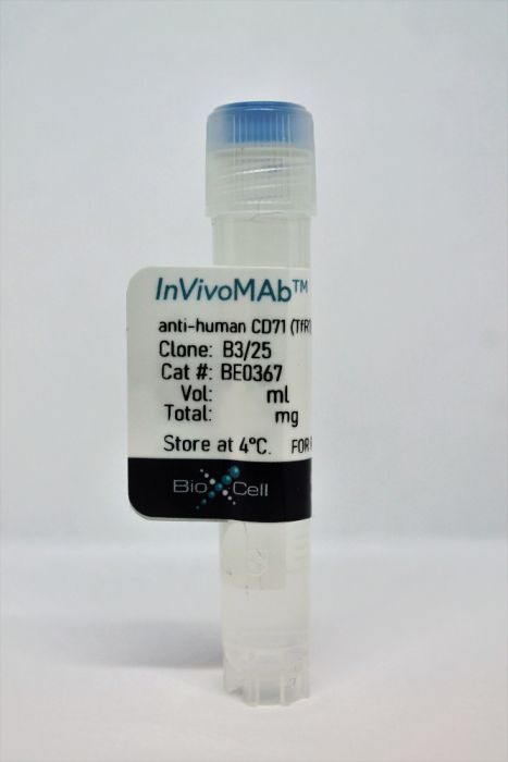
InVivoMAb anti-human CD71 (TfR1)
Product Details
The B3/25 monoclonal antibody reacts with human CD71 also known as transferrin receptor or TfR1. CD71 is a type II homodimeric transmembrane glycoprotein which is expressed on the surface of proliferating cells, reticulocytes, and erythroid precursors. CD71 plays a role in the control of cellular proliferation and is required for iron import from transferrin into cells. CD71 is expressed on malignant cells at high levels and its expression correlates with cancer progression. The B3/25 antibody is a non-neutralizing antibody that does not inhibit the binding of transferrin but does result in decreased transferrin binding sites. B3/25 has been shown to inhibit the in vitro proliferation of human myeloid leukemia cells as well as used for the delivery of toxins into cancer cells. in vivo studies carried out in nude mice showed that one intravenous injection of B3/25 antibody alone inhibits growth of human melanoma M21 xenografts.Specifications
| Isotype | Mouse IgG1, κ |
|---|---|
| Recommended Isotype Control(s) | InVivoMAb mouse IgG1 isotype control, unknown specificity |
| Recommended Dilution Buffer | InVivoPure pH 7.0 Dilution Buffer |
| Conjugation | This product is unconjugated. Conjugation is available via our Antibody Conjugation Services. |
| Immunogen | Human hematopoietic cell line K562 |
| Reported Applications |
in vitro CD71 targeting in vivo CD71 targeting Immunofluorescence |
| Formulation |
PBS, pH 7.0 Contains no stabilizers or preservatives |
| Endotoxin |
<2EU/mg (<0.002EU/μg) Determined by LAL gel clotting assay |
| Purity |
>95% Determined by SDS-PAGE |
| Sterility | 0.2 μM filtered |
| Purification | Protein G |
| RRID | AB_2927504 |
| Molecular Weight | 150 kDa |
| Storage | The antibody solution should be stored at the stock concentration at 4°C. Do not freeze. |
Recommended Products
Immunofluorescence
Das, S. and P. E. Pellett. (2011). "Spatial relationships between markers for secretory and endosomal machinery in human cytomegalovirus-infected cells versus those in uninfected cells" J Virol 85(12): 5864-5879. PubMed
Human cytomegalovirus (HCMV) induces extensive remodeling of the secretory apparatus to form the cytoplasmic virion assembly compartment (cVAC), where virion tegumentation and envelopment take place. We studied the structure of the cVAC by confocal microscopy to assess the three-dimensional distribution of proteins specifically associated with individual secretory organelles. In infected cells, early endosome antigen 1 (EEA1)-positive vesicles are concentrated at the center of the cVAC and, as previously seen, are distinct from structures visualized by markers for the endoplasmic reticulum, Golgi apparatus, and trans-Golgi network (TGN). EEA1-positive vesicles can be strongly associated with markers for recycling endosomes, to a lesser extent with markers associated with components of the endosomal sorting complex required for transport III (ESCRT III) machinery, and then with markers of late endosomes. In comparisons of uninfected and infected cells, we found significant changes in the structural associations and colocalization of organelle markers, as well as in net organelle volumes. These results provide new evidence that the HCMV-induced remodeling of the membrane transport apparatus involves much more than simple relocation and expansion of preexisting structures and are consistent with the hypothesis that the shift in identity of secretory organelles in HCMV-infected cells results in new functional profiles.
in vitro CD71 targeting
Rybak, S. M., et al. (1991). "Cytotoxic potential of ribonuclease and ribonuclease hybrid proteins" J Biol Chem 266(31): 21202-21207. PubMed
Pancreatic RNase injected into Xenopus oocytes abolishes protein synthesis at concentrations comparable to the toxin ricin yet has no effect on oocyte protein synthesis when added to the extracellular medium. Therefore RNase behaves like a potent toxin when directed into a cell. To explore the cytotoxic potential of RNase toward mammalian cells, bovine pancreatic ribonuclease A was coupled via a disulfide bond to human transferrin or antibodies to the transferrin receptor. The RNase hybrid proteins were cytotoxic to K562 human erythroleukemia cells in vitro with an IC50 around 10(-7) M whereas greater than 10(-5) M native RNase was required to inhibit protein synthesis. Cytotoxicity requires both components of the conjugate since excess transferrin or ribonuclease inhibitors added to the medium protected the cells from the transferrin-RNase toxicity. Compounds that interfere with transferrin receptor cycling and compartmentalization such as ammonium chloride decreased the cytotoxicity of transferrin-RNase. After a dose-dependent lag period inactivation of protein synthesis by transferrin-RNase followed a first-order decay constant. In a clonogenic assay that measures the extent of cell death 1 x 10(-6) M transferrin-RNase killed at least 4 logs or 99.99% of the cells whereas 70 x 10(-6) M RNase was nontoxic. These results show that RNase coupled to a ligand can be cytotoxic. Human ribonucleases coupled to antibodies also may exhibit receptor-mediated toxicities providing a new approach to selective cell killing possibly with less systemic toxicity and importantly less immunogenicity than the currently employed ligand-toxin conjugates.
in vitro CD71 targeting
Taetle, R., et al. (1983). "Effects of anti-transferrin receptor antibodies on growth of normal and malignant myeloid cells" Int J Cancer 32(3): 343-349. PubMed
The effects of three monoclonal antibodies (B3/25, 43/31, and 42/6) reactive with human transferrin (Tf) receptors on growth of normal and malignant myeloid cells were examined using in vitro culture techniques. When added directly to cultures, all three antibodies caused dose-dependent inhibition of normal granulocyte/macrophage progenitor (CFU-GM) growth. Monoclonal antibody 42/6 was by far the most potent of the three, with an ID50 of less than 5 micrograms/ml. Identical effects were seen on CFU-GM from three patients with chronic myelogenous leukemia. Growth of colonies from two myeloid leukemia cells lines (KG-I, HL60) was also inhibited by all three antibodies, and these cells were generally more sensitive than normal CFU-GM. Blast colony-forming cells from three patients with acute non-lymphocytic leukemia were relatively resistant to the antibodies, and CFU-GM from a patient with myeloid metaplasia were resistant (ID50 greater than 50 micrograms/ml) to 42/6. In liquid culture, growth of the leukemic cell lines was inhibited by saturating concentrations of the three antibodies, although in both liquid and colony culture recovery was seen even after exposure to antibody for periods of up to 72 h. Analysis of the cell-cycle status of these cells showed that the antibodies did not cause accumulation of cells in any particular phase of the cell cycle. Addition to cultures of large quantities of human Tf failed to reverse the inhibitory effects of the antibodies. Competitive binding studies on the leukemia cell lines showed that only 42/6 inhibited binding of Tf to its receptor, although all three antibodies inhibited cell growth. Addition of Fe chelate (as ferric nitriloacetic acid, FeNTA) failed to reverse the inhibitory effects of the antibodies on CFU-GM and HL60 cells, but had variable effects on KG-I cell growth. FeNTA fully reversed inhibitory effects of 42/6 on KG-I cells. We conclude that monoclonal antibodies to Tf receptors can inhibit growth of both normal and malignant myeloid cells. Overall, no selectivity for malignant vs normal cells is apparent, although malignant cells from one individual were more sensitive to colony inhibition by 43/31 monoclonal antibody than normal CFU-GM.

