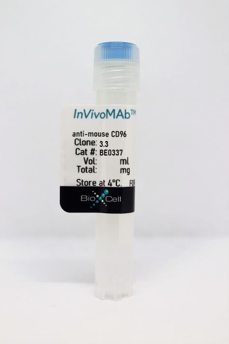
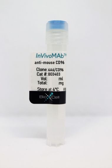
InVivoMAb anti-mouse CD96
Product Details
The 3.3 monoclonal antibody reacts with CD96, also known as TACTILE (T cell activation increased late expression). CD96 is a type I transmembrane glycoprotein and member of the Ig superfamily. CD96 is expressed at low levels on resting NK cells and T cells and at high levels on activated NK and T cells. CD96 binds to its ligand, CD155 to mediate NK cell adhesion to target cells and cytotoxicity. CD96 has recently been identified as a novel target for cancer immunotherapy and has been shown to play a role in metastasis. The 3.3 antibody has been shown to suppress primary tumor growth in a number of experimental mouse tumor models.Specifications
| Isotype | Rat IgG1, λ |
|---|---|
| Recommended Isotype Control(s) | InVivoMAb rat IgG1 isotype control, anti-horseradish peroxidase |
| Recommended Dilution Buffer | InVivoPure pH 7.0 Dilution Buffer |
| Conjugation | This product is unconjugated. Conjugation is available via our Antibody Conjugation Services. |
| Immunogen | Not available or unknown |
| Reported Applications |
in vivo CD96 blocking in vitro CD96 blocking Flow cytometry |
| Formulation |
PBS, pH 7.0 Contains no stabilizers or preservatives |
| Endotoxin |
<2EU/mg (<0.002EU/μg) Determined by LAL gel clotting assay |
| Purity |
>95% Determined by SDS-PAGE |
| Sterility | 0.2 µm filtration |
| Production | Purified from cell culture supernatant in an animal-free facility |
| Purification | Protein G |
| RRID | AB_2894757 |
| Molecular Weight | 150 kDa |
| Storage | The antibody solution should be stored at the stock concentration at 4°C. Do not freeze. |
Recommended Products
in vivo CD96 blocking, Flow Cytometry
Mittal, D., et al. (2019). "CD96 Is an Immune Checkpoint That Regulates CD8(+) T-cell Antitumor Function" Cancer Immunol Res 7(4): 559-571. PubMed
CD96 is a novel target for cancer immunotherapy shown to regulate NK cell effector function and metastasis. Here, we demonstrated that blocking CD96 suppressed primary tumor growth in a number of experimental mouse tumor models in a CD8(+) T cell-dependent manner. DNAM-1/CD226, Batf3, IL12p35, and IFNgamma were also critical, and CD96-deficient CD8(+) T cells promoted greater tumor control than CD96-sufficient CD8(+) T cells. The antitumor activity of anti-CD96 therapy was independent of Fc-mediated effector function and was more effective in dual combination with blockade of a number of immune checkpoints, including PD-1, PD-L1, TIGIT, and CTLA-4. We consistently observed coexpression of PD-1 with CD96 on CD8(+) T lymphocytes in tumor-infiltrating leukocytes both in mouse and human cancers using mRNA analysis, flow cytometry, and multiplex IHF. The combination of anti-CD96 with anti-PD-1 increased the percentage of IFNgamma-expressing CD8(+) T lymphocytes. Addition of anti-CD96 to anti-PD-1 and anti-TIGIT resulted in superior antitumor responses, regardless of the ability of the anti-TIGIT isotype to engage FcR. The optimal triple combination was also dependent upon CD8(+) T cells and IFNgamma. Overall, these data demonstrate that CD96 is an immune checkpoint on CD8(+) T cells and that blocking CD96 in combination with other immune-checkpoint inhibitors is a strategy to enhance T-cell activity and suppress tumor growth.
in vivo CD96 blocking, Flow Cytometry
Roman Aguilera, A., et al. (2018). "CD96 targeted antibodies need not block CD96-CD155 interactions to promote NK cell anti-metastatic activity" Oncoimmunology 7(5): e1424677. PubMed
3.3 > 8B10. The metastatic burden control of all of the anti-CD96 clones was highly dependent on NK cells and IFN-gamma. Consistent with its inability to block CD96-CD155 interactions, 8B10 retained anti-metastatic activity in CD155-deficient mice, whereas 3.3 and 6A6 lost potency in CD155-deficient mice. Furthermore, 8B10 retained most of its anti-metastatic activity in IL-12p35-deficient mice whereas the activity of 3.3 and 6A6 were partially lost. All three mAbs were inactive in CD226-deficient mice. Altogether, these data demonstrate anti-CD96 need not block CD96-CD155 interactions (ie. immune checkpoint blockade) to promote NK cell anti-metastatic activity.”}” data-sheets-userformat=”{“2″:14851,”3”:{“1″:0},”4”:{“1″:2,”2″:16777215},”12″:0,”14”:{“1″:2,”2″:1521491},”15″:”Roboto, sans-serif”,”16″:12}”>CD96 is a transmembrane glycoprotein Ig superfamily receptor, expressed on various T cell subsets and NK cells, that interacts with nectin and nectin-like proteins, including CD155/polio virus receptor (PVR). Here, we have compared three rat anti-mouse CD96 mAbs, including two that block CD96-CD155 (3.3 and 6A6) and one that does not block CD96-CD155 (8B10). Using flow cytometry, we demonstrated that both mAbs 3.3 and 6A6 bind to the first Ig domain of mouse CD96 and compete with CD155 binding, while mAb 8B10 binds to the second Ig domain and does not block CD155. While Fc isotype was irrelevant concerning the anti-metastatic activity of 3.3 mAb, in four different experimental metastases models and one spontaneous metastasis model, the relative order of anti-metastatic potency was 6A6 > 3.3 > 8B10. The metastatic burden control of all of the anti-CD96 clones was highly dependent on NK cells and IFN-gamma. Consistent with its inability to block CD96-CD155 interactions, 8B10 retained anti-metastatic activity in CD155-deficient mice, whereas 3.3 and 6A6 lost potency in CD155-deficient mice. Furthermore, 8B10 retained most of its anti-metastatic activity in IL-12p35-deficient mice whereas the activity of 3.3 and 6A6 were partially lost. All three mAbs were inactive in CD226-deficient mice. Altogether, these data demonstrate anti-CD96 need not block CD96-CD155 interactions (ie. immune checkpoint blockade) to promote NK cell anti-metastatic activity.
in vivo CD96 blocking
Blake, S. J., et al. (2016). "Suppression of Metastases Using a New Lymphocyte Checkpoint Target for Cancer Immunotherapy" Cancer Discov 6(4): 446-459. PubMed
CD96 has recently been shown as a negative regulator of mouse natural killer (NK)-cell activity, with Cd96(-/-)mice displaying hyperresponsive NK cells upon immune challenge. In this study, we have demonstrated that blocking CD96 with a monoclonal antibody inhibited experimental metastases in three different tumor models. The antimetastatic activity of anti-CD96 was dependent on NK cells, CD226 (DNAM-1), and IFNgamma, but independent of activating Fc receptors. Anti-CD96 was more effective in combination with anti-CTLA-4, anti-PD-1, or doxorubicin chemotherapy. Blocking CD96 in Tigit(-/-)mice significantly reduced experimental and spontaneous metastases compared with its activity in wild-type mice. Co-blockade of CD96 and PD-1 potently inhibited lung metastases, with the combination increasing local NK-cell IFNgamma production and infiltration. Overall, these data demonstrate that blocking CD96 is a new and complementary immunotherapeutic strategy to reduce tumor metastases. SIGNIFICANCE: This article illustrates the antimetastatic activity and mechanism of action of an anti-CD96 antibody that inhibits the CD96-CD155 interaction and stimulates NK-cell function. Targeting host CD96 is shown to complement surgery and conventional immune checkpoint blockade.
in vivo CD96 blocking, in vitro CD96 blocking, Flow Cytometry
Chan, C. J., et al. (2014). "The receptors CD96 and CD226 oppose each other in the regulation of natural killer cell functions" Nat Immunol 15(5): 431-438. PubMed
CD96, CD226 (DNAM-1) and TIGIT belong to an emerging family of receptors that interact with nectin and nectin-like proteins. CD226 activates natural killer (NK) cell-mediated cytotoxicity, whereas TIGIT reportedly counterbalances CD226. In contrast, the role of CD96, which shares the ligand CD155 with CD226 and TIGIT, has remained unclear. In this study we found that CD96 competed with CD226 for CD155 binding and limited NK cell function by direct inhibition. As a result, Cd96(-/-) mice displayed hyperinflammatory responses to the bacterial product lipopolysaccharide (LPS) and resistance to carcinogenesis and experimental lung metastases. Our data provide the first description, to our knowledge, of the ability of CD96 to negatively control cytokine responses by NK cells. Blocking CD96 may have applications in pathologies in which NK cells are important.
- Cancer Research,
- Immunology and Microbiology
Single-Cell Landscape Highlights Heterogenous Microenvironment, Novel Immune Reaction Patterns, Potential Biomarkers and Unique Therapeutic Strategies of Cervical Squamous Carcinoma, Human Papillomavirus-Associated (HPVA) and Non-HPVA Adenocarcinoma.
In Advanced Science (Weinheim, Baden-Wurttemberg, Germany) on 1 April 2023 by Qiu, J., Qu, X., et al.
PubMed
Cervical adenocarcinomas (ADCs), including human papillomavirus (HPV)-associated (HPVA) and non-HPVA (NHPVA), though exhibiting a more malignant phenotype and poorer prognosis, are treated identically to squamous cell carcinoma (SCC). This clinical dilemma requires a deeper investigation into their differences. Herein a transcriptomic atlas of SCC, HPVA, and NHPVA-ADC using single-cell RNA (scRNA) and T-cell receptor sequencing (TCR-seq) is presented. Regarding structural cells, the malignancy origin of epithelial cells, angiogenic tip cells and two subtypes of fibroblasts is revealed. The promalignant properties of the structural cells using organoids are further confirmed. Regarding immune cells, myeloid cells with multiple functions other than antigen presentation and exhausted T lymphocytes contribute to immunosuppression. From the perspective of HPV infection, not only is HPV-dependent and independent cervical cancer oncogenesis proposed but also three immune reaction patterns mediated by T cells (coordinated/inactive/imbalanced) are identified. Strikingly, diagnostic biomarkers to distinguish ADC from SCC are discovered and prognostic biomarkers with marker genes for malignant epithelial cells, tip cells, and SPP1/C1QC macrophages are generated. Importantly, the efficacy of anti-CD96 and anti-TIGIT, not inferior to anti-PD1, in animal experiments is confirmed and targeted therapies specifically for HPV-positive SCC, HPVA and NHPVA-ADC, providing essential clues for further clinical trials, are proposed. © 2023 The Authors. Advanced Science published by Wiley-VCH GmbH.
- Mus musculus (House mouse),
- Cancer Research,
- Immunology and Microbiology
T cell-derived interleukin-22 drives the expression of CD155 by cancer cells to suppress NK cell function and promote metastasis.
In Immunity on 10 January 2023 by Briukhovetska, D., Suarez-Gosalvez, J., et al.
PubMed
Although T cells can exert potent anti-tumor immunity, a subset of T helper (Th) cells producing interleukin-22 (IL-22) in breast and lung tumors is linked to dismal patient outcome. Here, we examined the mechanisms whereby these T cells contribute to disease. In murine models of lung and breast cancer, constitutional and T cell-specific deletion of Il22 reduced metastases without affecting primary tumor growth. Deletion of the IL-22 receptor on cancer cells decreases metastasis to a degree similar to that seen in IL-22-deficient mice. IL-22 induced high expression of CD155, which bound to the activating receptor CD226 on NK cells. Excessive activation led to decreased amounts of CD226 and functionally impaired NK cells, which elevated the metastatic burden. IL-22 signaling was also associated with CD155 expression in human datasets and with poor patient outcomes. Taken together, our findings reveal an immunosuppressive circuit activated by T cell-derived IL-22 that promotes lung metastasis.Copyright © 2022 The Author(s). Published by Elsevier Inc. All rights reserved.


