InVivoMAb anti-mouse TIGIT
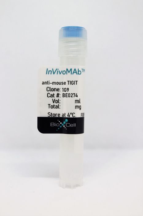
Product Details
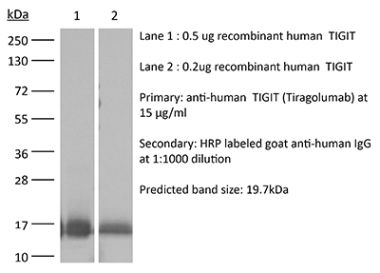
The 1G9 monoclonal antibody reacts with mouse TIGIT (T cell immunoreceptor with Ig and ITIM domains). TIGIT is a 26 kDa, type I transmembrane protein and a member of the poliovirus receptor (PVR) family. TIGIT has been found to be expressed on follicular T helper cells in mice while in humans it’s expressed by many T cell subsets including activated T cells, follicular T helper cells, memory T cells, and regulatory T cells as well as on NK cells. TIGIT can interact with certain members of the PVR and PVR-like families, including PVR, PVRL2, PVRL3, CD155, and CD112. TIGIT is thought to negatively regulate NK and T cell activation. Binding of TIGIT on T cells by dendritic cells results in their differentiation into a tolerogenic phenotype, with increased secretion of IL-10 and diminished production of IL-12. TIGIT knock-out mice are more susceptible to autoimmune disease.Specifications
| Isotype | Mouse IgG1, κ |
|---|---|
| Recommended Isotype Control(s) | InVivoMAb mouse IgG1 isotype control, unknown specificity |
| Recommended Dilution Buffer | InVivoPure pH 7.0 Dilution Buffer |
| Conjugation | This product is unconjugated. Conjugation is available via our Antibody Conjugation Services. |
| Immunogen | Mouse TIGIT |
| Reported Applications |
in vivo TIGIT stimulation Flow cytometry |
| Formulation |
PBS, pH 7.0 Contains no stabilizers or preservatives |
| Endotoxin |
<2EU/mg (<0.002EU/μg) Determined by LAL gel clotting assay |
| Purity |
>95% Determined by SDS-PAGE |
| Sterility | 0.2 µm filtration |
| Production | Purified from cell culture supernatant in an animal-free facility |
| Purification | Protein G |
| RRID | AB_2687797 |
| Molecular Weight | 150 kDa |
| Storage | The antibody solution should be stored at the stock concentration at 4°C. Do not freeze. |
Additional Formats
Recommended Products
-
Recommended Isotype Control(s)
InVivoMAb mouse IgG1 isotype control, unknown specificity
-

Recommended Dilution Buffer
InVivoPure pH 7.0 Dilution Buffer
in vivo TIGIT stimulation
Schorer, M., et al. (2020). "TIGIT limits immune pathology during viral infections" Nat Commun 11(1): 1288. PubMed
Co-inhibitory pathways have a fundamental function in regulating T cell responses and control the balance between promoting efficient effector functions and restricting immune pathology. The TIGIT pathway has been implicated in promoting T cell dysfunction in chronic viral infection. Importantly, TIGIT signaling is functionally linked to IL-10 expression, which has an effect on both virus control and maintenance of tissue homeostasis. However, whether TIGIT has a function in viral persistence or limiting tissue pathology is unclear. Here we report that TIGIT modulation effectively alters the phenotype and cytokine profile of T cells during influenza and chronic LCMV infection, but does not affect virus control in vivo. Instead, TIGIT has an important effect in limiting immune pathology in peripheral organs by inducing IL-10. Our data therefore identify a function of TIGIT in limiting immune pathology that is independent of viral clearance.
in vivo TIGIT stimulation
Dixon, K. O., et al. (2018). "Functional Anti-TIGIT Antibodies Regulate Development of Autoimmunity and Antitumor Immunity" J Immunol 200(8): 3000-3007. PubMed
Coinhibitory receptors, such as CTLA-4 and PD-1, play a critical role in maintaining immune homeostasis by dampening T cell responses. Recently, they have gained attention as therapeutic targets in chronic disease settings where their dysregulated expression contributes to suppressed immune responses. The novel coinhibitory receptor TIGIT (T cell Ig and ITIM domain) has been shown to play an important role in modulating immune responses in the context of autoimmunity and cancer. However, the molecular mechanisms by which TIGIT modulates immune responses are still insufficiently understood. We have generated a panel of monoclonal anti-mouse TIGIT Abs that show functional properties in mice in vivo and can serve as important tools to study the underlying mechanisms of TIGIT function. We have identified agonistic as well as blocking anti-TIGIT Ab clones that are capable of modulating T cell responses in vivo. Administration of either agonist or blocking anti-TIGIT Abs modulated autoimmune disease severity whereas administration of blocking anti-TIGIT Abs synergized with anti-PD-1 Abs to affect partial or even complete tumor regression. The Abs presented in this study can thus serve as important tools for detailed analysis of TIGIT function in different disease settings and the knowledge gained will provide valuable insight for the development of novel therapeutic approaches targeting TIGIT.
Flow Cytometry
Burton, B. R., et al. (2014). "Sequential transcriptional changes dictate safe and effective antigen-specific immunotherapy" Nat Commun 5: 4741. PubMed
Antigen-specific immunotherapy combats autoimmunity or allergy by reinstating immunological tolerance to target antigens without compromising immune function. Optimization of dosing strategy is critical for effective modulation of pathogenic CD4(+) T-cell activity. Here we report that dose escalation is imperative for safe, subcutaneous delivery of the high self-antigen doses required for effective tolerance induction and elicits anergic, interleukin (IL)-10-secreting regulatory CD4(+) T cells. Analysis of the CD4(+) T-cell transcriptome, at consecutive stages of escalating dose immunotherapy, reveals progressive suppression of transcripts positively regulating inflammatory effector function and repression of cell cycle pathways. We identify transcription factors, c-Maf and NFIL3, and negative co-stimulatory molecules, LAG-3, TIGIT, PD-1 and TIM-3, which characterize this regulatory CD4(+) T-cell population and whose expression correlates with the immunoregulatory cytokine IL-10. These results provide a rationale for dose escalation in T-cell-directed immunotherapy and reveal novel immunological and transcriptional signatures as surrogate markers of successful immunotherapy.
Flow Cytometry
Chan, C. J., et al. (2014). "The receptors CD96 and CD226 oppose each other in the regulation of natural killer cell functions" Nat Immunol 15(5): 431-438. PubMed
CD96, CD226 (DNAM-1) and TIGIT belong to an emerging family of receptors that interact with nectin and nectin-like proteins. CD226 activates natural killer (NK) cell-mediated cytotoxicity, whereas TIGIT reportedly counterbalances CD226. In contrast, the role of CD96, which shares the ligand CD155 with CD226 and TIGIT, has remained unclear. In this study we found that CD96 competed with CD226 for CD155 binding and limited NK cell function by direct inhibition. As a result, Cd96(-/-) mice displayed hyperinflammatory responses to the bacterial product lipopolysaccharide (LPS) and resistance to carcinogenesis and experimental lung metastases. Our data provide the first description, to our knowledge, of the ability of CD96 to negatively control cytokine responses by NK cells. Blocking CD96 may have applications in pathologies in which NK cells are important.
Flow Cytometry
Foks, A. C., et al. (2013). "Agonistic anti-TIGIT treatment inhibits T cell responses in LDLr deficient mice without affecting atherosclerotic lesion development" PLoS One 8(12): e83134. PubMed
OBJECTIVE: Co-stimulatory and co-inhibitory molecules are mainly expressed on T cells and antigen presenting cells and strongly orchestrate adaptive immune responses. Whereas co-stimulatory molecules enhance immune responses, signaling via co-inhibitory molecules dampens the immune system, thereby showing great therapeutic potential to prevent cardiovascular diseases. Signaling via co-inhibitory T cell immunoglobulin and ITIM domain (TIGIT) directly inhibits T cell activation and proliferation, and therefore represents a novel therapeutic candidate to specifically dampen pro-atherogenic T cell reactivity. In the present study, we used an agonistic anti-TIGIT antibody to determine the effect of excessive TIGIT-signaling on atherosclerosis. METHODS AND RESULTS: TIGIT was upregulated on CD4(+) T cells isolated from mice fed a Western-type diet in comparison with mice fed a chow diet. Agonistic anti-TIGIT suppressed T cell activation and proliferation both in vitro and in vivo. However, agonistic anti-TIGIT treatment of LDLr(-/-) mice fed a Western-type diet for 4 or 8 weeks did not affect atherosclerotic lesion development in comparison with PBS and Armenian Hamster IgG treatment. Furthermore, elevated percentages of dendritic cells were observed in the blood and spleen of agonistic anti-TIGIT-treated mice. Additionally, these cells showed an increased activation status but decreased IL-10 production. CONCLUSIONS: Despite the inhibition of splenic T cell responses, agonistic anti-TIGIT treatment does not affect initial atherosclerosis development, possibly due to increased activity of dendritic cells.
Flow Cytometry
Joller, N., et al. (2011). "Cutting edge: TIGIT has T cell-intrinsic inhibitory functions" J Immunol 186(3): 1338-1342. PubMed
Costimulatory molecules regulate the functional outcome of T cell activation, and disturbance of the balance between activating and inhibitory signals results in increased susceptibility to infection or the induction of autoimmunity. Similar to the well-characterized CD28/CTLA-4 costimulatory pathway, a newly emerging pathway consisting of CD226 and T cell Ig and ITIM domain (TIGIT) has been associated with susceptibility to multiple autoimmune diseases. In this study, we examined the role of the putative coinhibitory molecule TIGIT and show that loss of TIGIT in mice results in hyperproliferative T cell responses and increased susceptibility to autoimmunity. TIGIT is thought to indirectly inhibit T cell responses by the induction of tolerogenic dendritic cells. By generating an agonistic anti-TIGIT Ab, we demonstrate that TIGIT can inhibit T cell responses directly independent of APCs. Microarray analysis of T cells stimulated with agonistic anti-TIGIT Ab revealed that TIGIT can act directly on T cells by attenuating TCR-driven activation signals.
- Mus musculus (House mouse),
- Immunology and Microbiology
Combining toll-like receptor agonists with immune checkpoint blockade affects antitumor vaccine efficacy.
In Journal for Immunotherapy of Cancer on 3 May 2024 by Jeon, D., Hill, E., et al.
PubMed
T cell checkpoint receptors are expressed when T cells are activated, and modulation of the expression or signaling of these receptors can alter the function of T cells and their antitumor efficacy. We previously found that T cells activated with cognate antigen had increases in the expression of PD-1, and this was attenuated in the presence of multiple toll-like receptor (TLR) agonists, notably TLR3 plus TLR9. In the current report, we sought to investigate whether combining TLR agonists with immune checkpoint blockade can further augment vaccine-mediated T cell antitumor immunity in murine tumor models. TLR agonists (TLR3 plus TLR9) and immune checkpoint inhibitors (antibodies targeting PD-1, CTLA-4, LAG-3, TIM-3 or VISTA) were combined and delivered with vaccines or vaccine-activated CD8+T cells to E.G7-OVA or MyC-CaP tumor-bearing mice. Tumors were assessed for growth and then collected and analyzed by flow cytometry. Immunization of E.G7-OVA tumor-bearing mice with SIINFEKL peptide vaccine, coadministered with TLR agonists and αCTLA-4, demonstrated greater antitumor efficacy than immunization with TLR agonists or αCTLA-4 alone. Conversely, the antitumor efficacy was abrogated when vaccine and TLR agonists were combined with αPD-1. TLR agonists suppressed PD-1 expression on regulatory T cells (Tregs) and activated this population. Depletion of Tregs in tumor-bearing mice led to greater antitumor efficacy of this combination therapy, even in the presence of αPD-1. Combining vaccination with TLR agonists and αCTLA-4 or αLAG-3 showed greater antitumor than with combinations with αTIM-3 or αVISTA. The combination of TLR agonists and αCTLA-4 or αLAG-3 can further improve the efficacy of a cancer vaccine, an effect not observed using αPD-1 due to activation of Tregs when αPD-1 was combined with TLR3 and TLR9 agonists. These data suggest that optimal combinations of TLR agonists and immune checkpoint blockade may improve the efficacy of human anticancer vaccines. © Author(s) (or their employer(s)) 2024. Re-use permitted under CC BY-NC. No commercial re-use. See rights and permissions. Published by BMJ.
- Mus musculus (House mouse),
- Immunology and Microbiology,
- Cancer Research
Combining TIGIT blockade with IL-15 stimulation is a promising immunotherapy strategy for lung adenocarcinoma.
In Clinical and Translational Medicine on 1 January 2024 by Luo, B., Sun, Y., et al.
PubMed
T-cell immunoglobulin and immunoreceptor tyrosine-based inhibitory motif domain (TIGIT) is an immune checkpoint molecule that suppresses CD8+ T-cell function in cancer. However, the expression profile and functional significance of TIGIT in the immune microenvironment of lung adenocarcinoma (LUAD) remain elusive. Interleukin (IL)-15 has emerged as a promising candidate for enhancing CD8+ T-cell mediated tumour eradication. Exploring therapeutic strategies that combine IL-15 with TIGIT blockade in LUAD is warranted. We investigated the regulatory network involving coinhibitory TIGIT and CD96, as well as costimulatory CD226 in LUAD using clinical samples. The potential role of TIGIT in regulating the pathogenesis of LUAD was addressed through a murine model with transplanted tumours constructed in Tigit-/- mice. The therapeutic strategy that combines TIGIT blockade with IL-15 stimulation was verified using a transplanted tumour murine model and a patient-derived organoid (PDO) model. The frequency of TIGIT+ CD8+ T cells was significantly increased in LUAD. Increased TIGIT expression indicated poorer prognosis in LUAD patients. Furthermore, the effector function of TIGIT+ CD8+ tumour-infiltrating lymphocytes (TILs) was impaired in LUAD patients and TIGIT inhibited antitumour immune response of CD8+ TILs in tumour-bearing mice. Mechanistically, IL-15 enhanced the effector function of CD8+ TILs but stimulated the expression of TIGIT on CD8+ TILs concomitantly. The application of IL-15 combined with TIGIT blockade showed additive effects in enhancing the cytotoxicity of CD8+ TILs and thus further increased the antitumour immune response in LUAD. Our findings identified TIGIT as a promising therapeutic target for LUAD. LUAD could benefit more from the combined therapy of IL-15 stimulation and TIGIT blockade. © 2024 The Authors. Clinical and Translational Medicine published by John Wiley & Sons Australia, Ltd on behalf of Shanghai Institute of Clinical Bioinformatics.
- Cancer Research,
- Immunology and Microbiology
Interactions of Indoleamine 2,3-dioxygenase-expressing LAMP3+ dendritic cells with CD4+ regulatory T cells and CD8+ exhausted T cells: synergistically remodeling of the immunosuppressive microenvironment in cervical cancer and therapeutic implication
In Cancer Communications (London, England) on 1 November 2023 by Qu, X., Wang, Y., et al.
PubMed
Cervical cancer (CC) is the fourth most common cancer in women worldwide. Although immunotherapy has been applied in clinical practice, its therapeutic efficacy remains far from satisfactory, necessitating further investigation of the mechanism of CC immune remodeling and exploration of novel treatment targets. This study aimed to investigate the mechanism of CC immune remodeling and explore potential therapeutic targets. We conducted single-cell RNA sequencing on a total of 17 clinical specimens, including normal cervical tissues, high-grade squamous intraepithelial lesions, and CC tissues. To validate our findings, we conducted multicolor immunohistochemical staining of CC tissues and constructed a subcutaneous tumorigenesis model in C57BL/6 mice using murine CC cell lines (TC1) to evaluate the effectiveness of combination therapy involving indoleamine 2,3-dioxygenase 1 (IDO1) inhibition and immune checkpoint blockade (ICB). We used the unpaired two-tailed Student's t-test, Mann-Whitney test, or Kruskal-Wallis test to compare continuous data between two groups and one-way ANOVA with Tukey's post hoc test to compare data between multiple groups. Malignant cervical epithelial cells did not manifest noticeable signs of tumor escape, whereas lysosomal-associated membrane protein 3-positive (LAMP3+ ) dendritic cells (DCs) in a mature state with immunoregulatory roles were found to express IDO1 and affect tryptophan metabolism. These cells interacted with both tumor-reactive exhausted CD8+ T cells and CD4+ regulatory T cells, synergistically forming a vicious immunosuppressive cycle and mediating CC immune escape. Further validation through multicolor immunohistochemical staining showed co-localization of neoantigen-reactive T cells (CD3+ , CD4+ /CD8+ , and PD-1+ ) and LAMP3+ DCs (CD80+ and PD-L1+ ). Additionally, a combination of the IDO1 inhibitor with an ICB agent significantly reduced tumor volume in the mouse model of CC compared with an ICB agent alone. Our study suggested that a combination treatment consisting of targeting IDO1 and ICB agent could improve the therapeutic efficacy of current CC immunotherapies. Additionally, our results provided crucial insights for designing drugs and conducting future clinical trials for CC. © 2023 The Authors. Cancer Communications published by John Wiley & Sons Australia, Ltd on behalf of SUN YAT-SEN UNIVERSITY CANCER CENTER.
- IHC,
- Mus musculus (House mouse),
- Cancer Research
Peptide-based PET imaging agent of tumor TIGIT expression.
In EJNMMI Research on 2 May 2023 by Weng, D., Guo, R., et al.
PubMed
Accumulating studies have demonstrated that elevated TIGIT expression in tumor microenvironment correlates with better therapeutic response to TIGIT-based immunotherapy in pre-clinical studies. Therefore, a non-invasive method to detect tumor TIGIT expression is crucial to predict the therapeutic effect. In this study, a peptide-based PET imaging agent, 68Ga-DOTA-DTBP-3, was developed to non-invasively detect TIGIT expression by micro-PET in tumor-bearing BALB/c mice. DTBP-3, a D-peptide comprising of 12 amino acids, was radiolabeled with 68Ga through a DOTA chelator. In vitro studies were performed to evaluate the affinity of 68Ga-DOTA-DTBP-3 to TIGIT and its stability in fetal bovine serum. In vivo studies were assessed by micro-PET, biodistribution, and immunohistochemistry on tumor-bearing BALB/c mice. The in vitro studies showed the equilibrium dissociation constant of 68Ga-DOTA-DTBP-3 for TIGIT was 84.21 nM and its radiochemistry purity was 89.24 ± 1.82% in FBS at 4 h in room temperature. The results of micro-PET, biodistribution and immunohistochemistry studies indicated that 68Ga-DOTA-DTBP-3 could be specifically targeted in 4T1 tumor-bearing mice, with a highest uptake at 0.5 h. 68Ga-DOTA-DTBP-3 holds potential for non-invasively detect tumor TIGIT expression and for timely assessment of the therapeutic effect of immune checkpoint blockade. © 2023. The Author(s).
- Cancer Research,
- Immunology and Microbiology,
- Mus musculus (House mouse)
T cell-derived interleukin-22 drives the expression of CD155 by cancer cells to suppress NK cell function and promote metastasis.
In Immunity on 10 January 2023 by Briukhovetska, D., Suarez-Gosalvez, J., et al.
PubMed
Although T cells can exert potent anti-tumor immunity, a subset of T helper (Th) cells producing interleukin-22 (IL-22) in breast and lung tumors is linked to dismal patient outcome. Here, we examined the mechanisms whereby these T cells contribute to disease. In murine models of lung and breast cancer, constitutional and T cell-specific deletion of Il22 reduced metastases without affecting primary tumor growth. Deletion of the IL-22 receptor on cancer cells decreases metastasis to a degree similar to that seen in IL-22-deficient mice. IL-22 induced high expression of CD155, which bound to the activating receptor CD226 on NK cells. Excessive activation led to decreased amounts of CD226 and functionally impaired NK cells, which elevated the metastatic burden. IL-22 signaling was also associated with CD155 expression in human datasets and with poor patient outcomes. Taken together, our findings reveal an immunosuppressive circuit activated by T cell-derived IL-22 that promotes lung metastasis.Copyright © 2022 The Author(s). Published by Elsevier Inc. All rights reserved.
- In Vivo,
- Cancer Research,
- Immunology and Microbiology,
- Mass Spec,
- Mus musculus (House mouse)
Stromal Reprogramming by FAK Inhibition Overcomes Radiation Resistance to Allow for Immune Priming and Response to Checkpoint Blockade.
In Cancer Discovery on 2 December 2022 by Krisnawan, V. E., Belle, J. I., et al.
PubMed
The effects of radiotherapy (RT) on tumor immunity in pancreatic ductal adenocarcinoma (PDAC) are not well understood. To better understand if RT can prime antigen-specific T-cell responses, we analyzed human PDAC tissues and mouse models. In both settings, there was little evidence of RT-induced T-cell priming. Using in vitro systems, we found that tumor-stromal components, including fibroblasts and collagen, cooperate to blunt RT efficacy and impair RT-induced interferon signaling. Focal adhesion kinase (FAK) inhibition rescued RT efficacy in vitro and in vivo, leading to tumor regression, T-cell priming, and enhanced long-term survival in PDAC mouse models. Based on these data, we initiated a clinical trial of defactinib in combination with stereotactic body RT in patients with PDAC (NCT04331041). Analysis of PDAC tissues from these patients showed stromal reprogramming mirroring our findings in genetically engineered mouse models. Finally, the addition of checkpoint immunotherapy to RT and FAK inhibition in animal models led to complete tumor regression and long-term survival. Checkpoint immunotherapeutics have not been effective in PDAC, even when combined with RT. One possible explanation is that RT fails to prime T-cell responses in PDAC. Here, we show that FAK inhibition allows RT to prime tumor immunity and unlock responsiveness to checkpoint immunotherapy. This article is highlighted in the In This Issue feature, p. 2711. ©2022 American Association for Cancer Research.
- In Vivo,
- Mus musculus (House mouse),
- Cancer Research,
- Immunology and Microbiology
TIGIT Blockade Exerts Synergistic Effects on Microwave Ablation Against Cancer.
In Frontiers in Immunology on 25 March 2022 by Chen, Y., Huang, H., et al.
PubMed
Combination immunotherapy based on immune checkpoint inhibitors (ICIs) has shown great success in the treatment of many types of cancers and has become the mainstream in the comprehensive treatment of cancers. Ablation in combination with immunotherapy has achieved tremendous efficacy in some preclinical and clinical studies. To date, our team proved that ablation in combination with ICIs was a promising antitumor therapeutic strategy for the liver metastasis of colorectal cancer (CRC). Moreover, we found that the expression of T cell immunoglobulin and immunoreceptor tyrosine-based inhibitory motif domain (TIGIT) expression was up-regulated after microwave ablation (MWA), indicating that TIGIT was involved in immunosuppression, and the combination of MWA and TIGIT blockade represented a potential clinical treatment strategy. In the present study, we examined the expression of TIGIT using a preclinical mouse model treated with MWA. Moreover, we evaluated the antitumor functions of MWA alone or in combination with TIGIT blockade by monitoring tumor growth and survival of the mice. Besides, we also detected the numbers of tumor-infiltrating lymphocytes (TILs), and effector molecules of CD8+ T cells using flow cytometry. Finally, we analyzed the single-cell RNA sequencing (scRNA-seq) data from the MWA and MWA plus anti-TIGIT groups. The expression of TIGIT in various immune cells was up-regulated after MWA, and the addition of TIGIT blockade to MWA prolonged survival and delayed tumor growth in the MC38 tumor model. Taken together, our findings showed that TIGIT blockade in combination with MWA significantly promoted the expansion and functions of CD8+ TILs and reshaped myeloid cells in the tumor microenvironment (TME) using flow cytometry and scRNA-seq analysis. TIGIT blockade in combination with MWA was a novel treatment strategy for the liver metastasis of CRC, and this combination therapy could reprogram the TME toward an antitumor environment. Copyright © 2022 Chen, Huang, Li, Xiao, Liu, Chen, Zhu, Zheng, Wu and Chen.
- In Vivo,
- Mus musculus (House mouse)
Extracellular matrix proteins regulate NK cell function in peripheral tissues.
In Science Advances on 18 March 2022 by Bunting, M. D., Vyas, M., et al.
PubMed
Natural killer (NK) cells reject major histocompatibility complex class I (MHC-I)-deficient bone marrow through direct cytotoxicity but not solid organ transplants devoid of MHC-I. Here, we demonstrate an immediate switch in NK cell function upon exit from the circulation, characterized by a shift from direct cytotoxicity to chemokine/cytokine production. In the skin transplant paradigm, combining an NK cell-specific activating ligand, m157, with missing self MHC-I resulted in complete graft rejection, which was dependent on NK cells as potential helpers and T cells as effectors. Extracellular matrix proteins, collagen I, collagen III, and elastin, blocked NK cell cytotoxicity and promoted their chemokine/cytokine production. NK cell cytotoxicity against MHC-I-deficient melanoma in the skin was markedly increased by blocking tumor collagen deposition. MHC-I down-regulation occurred in solid human cancers but not leukemias, which could be directly targeted by circulating cytotoxic NK cells. Our findings uncover a fundamental mechanism that restricts direct NK cell cytotoxicity in peripheral tissues.
- In Vivo,
- Mus musculus (House mouse),
- Cancer Research,
- Immunology and Microbiology
Tegaserod Maleate Inhibits Breast Cancer Progression and Enhances the Sensitivity of Immunotherapy.
In Journal of Oncology on 15 February 2022 by Li, X., Wu, L., et al.
PubMed
Breast cancer (BC) is the most commonly diagnosed cancer in women worldwide. The challenge in managing this heterogeneous malignancy is that BC is highly aggressive and is always associated with chemical resistance, radiation resistance, hormone therapy resistance, and targeted therapy resistance. Therefore, there is an urgent need to find effective drugs to treat BC. Based on the Selleck drug library approved by FDA, we screened 800 drugs for anti-BC cells and found that tegaserod maleate (TM), a 5-hydroxytryptamine 4-receptor (HTR4) partial agonist had the best anti-BC effect, which was further verified. The effects of different concentrations of TM on cell proliferation, invasion, and migration were evaluated in vitro using CCK8, plate cloning, transwell, and scratch assays. The UALCAN database, Kaplan-Meier Plotter database, Human Protein Atlas, and GEPIA2 were used to explore the correlation between HTR4 expression and BC patients' clinicopathological data as well as immune response. In vivo experiments demonstrated the effect of the TM and immunotherapy drug (anti-PD1/anti-TIGIT) combination on BC tumor growth in mice. TM significantly inhibited the proliferation, invasion, and migration of BC cells, and the higher the concentration, the better the inhibition effect. HTR4 was significantly downregulated in BC tissues compared to paracancerous tissues. The downregulation of HTR4 was correlated with clinicopathological data and positively correlated with BC prognosis. Interestingly, the GEPIA2 database suggested that there was a strong positive correlation between the expression of HTR4 and effector T cells, effector memory T cells, and exhausted T cells. In vitro experiments showed that TM, anti-PD1, and anti-TIGIT could all inhibit the growth and weight of BC tumors as compared with the control group. However, when anti-PD1 or anti-TIGIT was used simultaneously with TM, the inhibition of tumors significantly exceeded that in the control group. Moreover, the combination of anti-TIGIT and TM has the best inhibitory effect. TM inhibited the progression of breast cancer, and its combination with anti-TIGIT could effectively inhibit tumor growth and improve the sensitivity of immunotherapy in breast cancer. Copyright © 2022 Xiao Li et al.
- In Vivo,
- Mus musculus (House mouse),
- Cancer Research,
- Immunology and Microbiology
CD155/TIGIT signaling regulates the effector function of tumor-infiltrating CD8+ T cell by NF-κB pathway in colorectal cancer.
In Journal of Gastroenterology and Hepatology on 1 January 2022 by Li, S., Ding, J., et al.
PubMed
CD155/T-cell immunoglobulin and ITIM domain (TIGIT) suppressed anti-cancer immunity in several cancers, but its roles in colorectal cancer (CRC) were not clear. Here, we investigated its roles in CRC. The percentages of CD8+ T cells expressing TIGIT and secreting cytokines (IL-2, TNF-α, and IFNγ) were evaluated by flow cytometry. The expression level of CD155 was determined by western blot and immunohistochemistry. The levels of cytokines were determined by enzyme-linked immunosorbent assay. The activation of the nuclear factor-kappa B (NF-κB) pathway was examined by western blot and immunofluorescent assay. T-cell immunoglobulin and ITIM domain was overexpressed on CD8+ T cells of CRC patients and mice. CD155 was overexpressed in mice CRC tissues and cells. The addition of CD155 recombinant protein could decrease the percentages of CD8+ T cells secreting cytokines. Blocking TIGIT could increase the percentages of cytokine-secreting CD8+ T cells. Coculturing with CD155-knockdown CRC cells could upregulate the percentages of CD8+ T cells secreting cytokines. Blocking TIGIT partially counteracted the effect of the knockdown of CD155. Besides, coculturing with CD155-knockdown CRC cells could promote the secretion of cytokines, activate the NF-κB pathway, and enhance the nuclear translocation of p65. And these effects were counteracted by the application of an NF-κB inhibitor. Finally, blocking TIGIT played anti-cancer roles such as suppression of tumor growth, increasing the percentages of cytokine-secreting CD8+ T cells and activation of the NF-κB signaling pathway. Suppressing CD155/TIGIT exerted anti-cancer effects against CRC, and our findings provided a potential therapeutic approach to treat CRC. © 2021 Journal of Gastroenterology and Hepatology Foundation and John Wiley Sons Australia, Ltd.
- Mus musculus (House mouse)
TIGIT as a therapeutic target of HPV-positive head and neck squamous cell carcinomas
Preprint on MedRxiv : the Preprint Server for Health Sciences on 5 December 2021 by Le, X., Dang, M., et al.
PubMed
The tumor immune microenvironment (TIME) of treatment-naïve, human papillomavirus-positive head and neck squamous cell carcinoma (HPV-positive HNSCC) was interrogated at single-cell level to identify influential immune checkpoints as therapeutic targets. Single-cell transcriptome profiling revealed enrichment of numerous cell-cell interactions mediated by TIGIT-PVR/NECTIN2 in the TIME of HPV-positive HNSCC versus normal tonsil. TIGIT was the most differentially upregulated immune checkpoint on clonally expanded CD8 + T cells and was abundant on antigen-experienced, tissue-resident memory CD8 + T cell and T-regulatory subsets. TIGIT ligands PVR / NECTIN1/2 were abundant on mature regulatory dendritic cells, immunosuppressive plasmacytoid DCs, and macrophages. TIGIT and PD-1 co-blockade in the mEER murine model of HPV-positive HNSCC significantly reduced tumor growth, improved survival, restored effector function of HPV16 E7-specific CD8 + T cells, natural killer cells, and DCs, and conferred tumor re-challenge protection. This immunogenetic analysis at single-cell resolution focusing on HPV-positive HNSCC identified TIGIT as a rational therapeutic target.
- In Vivo,
- Mus musculus (House mouse),
- Cancer Research
Elimination of acquired resistance to PD-1 blockade via the concurrent depletion of tumour cells and immunosuppressive cells.
In Nature Biomedical Engineering on 1 November 2021 by Xue, G., Wang, Z., et al.
PubMed
Antigen release resulting from the death of tumour cells induced by chemotherapies and targeted therapies can augment the antitumour responses induced by immune checkpoint blockade (ICB). However, tumours responding to ICB therapies often become resistant to them. Here we show that the specific targeting of tumour cells promotes the growth of tumour-cell variants that are resistant to ICB, and that the acquired resistance can be overcome via the concurrent depletion of tumour cells and of major types of immunosuppressive cell via a monoclonal antibody binding the enzyme CD73, which we identified as highly expressed on tumour cells and on regulatory T cells, myeloid-derived suppressor cells and tumour-associated macrophages, but not on cytolytic T lymphocytes, natural killer cells and dendritic cells. In mice with murine tumours, the systemic administration of anti-PD1 antibodies and anti-CD73 antibodies conjugated to a near-infrared dye prevented near-infrared-irradiated tumours from acquiring resistance to ICB and resulted in the eradication of advanced tumours. The elimination of immunosuppressive cells may overcome acquired resistance to ICB across a range of tumour types and combination therapies. © 2021. The Author(s), under exclusive licence to Springer Nature Limited.
- In Vivo,
- Mus musculus (House mouse),
- Cancer Research,
- Immunology and Microbiology
The CD155/TIGIT axis promotes and maintains immune evasion in neoantigen-expressing pancreatic cancer.
In Cancer Cell on 11 October 2021 by Freed-Pastor, W. A., Lambert, L. J., et al.
PubMed
The CD155/TIGIT axis can be co-opted during immune evasion in chronic viral infections and cancer. Pancreatic adenocarcinoma (PDAC) is a highly lethal malignancy, and immune-based strategies to combat this disease have been largely unsuccessful to date. We corroborate prior reports that a substantial portion of PDAC harbors predicted high-affinity MHC class I-restricted neoepitopes and extend these findings to advanced/metastatic disease. Using multiple preclinical models of neoantigen-expressing PDAC, we demonstrate that intratumoral neoantigen-specific CD8+ T cells adopt multiple states of dysfunction, resembling those in tumor-infiltrating lymphocytes of PDAC patients. Mechanistically, genetic and/or pharmacologic modulation of the CD155/TIGIT axis was sufficient to promote immune evasion in autochthonous neoantigen-expressing PDAC. Finally, we demonstrate that the CD155/TIGIT axis is critical in maintaining immune evasion in PDAC and uncover a combination immunotherapy (TIGIT/PD-1 co-blockade plus CD40 agonism) that elicits profound anti-tumor responses in preclinical models, now poised for clinical evaluation. Copyright © 2021 Elsevier Inc. All rights reserved.
- In Vivo,
- Mus musculus (House mouse),
- Immunology and Microbiology
TIGIT modulates sepsis-induced immune dysregulation in mice with preexisting malignancy.
In JCI Insight on 8 June 2021 by Zhang, W., Anyalebechi, J. C., et al.
PubMed
TIGIT is a recently identified coinhibitory receptor that is upregulated in the setting of cancer and functionally contributes to the impairment of antitumor immunity. However, its role during sepsis is unknown. Because patients with cancer are 10 times more likely to die of sepsis than previously healthy (PH) patients with sepsis, we interrogated the role of TIGIT during sepsis in the context of preexistent malignancy. PH mice or cancer (CA) mice inoculated with lung carcinoma cells were made septic by cecal ligation and puncture (CLP). We found that sepsis induced TIGIT upregulation predominantly on Tregs and NK cells in both PH and CA mice. Anti-TIGIT Ab improved the 7-d survival of CA septic mice but not PH mice after CLP. Treatment of CA septic animals but not PH septic animals with anti-TIGIT mAb significantly reversed sepsis-induced loss of CD4+ T cells, CD8+ T cells, Foxp3+ Treg, and CD19+ B cells in the spleen, which was the result of decreased caspase-3+ apoptotic cells. In sum, we found that anti-TIGIT Ab reversed sepsis-induced T cell apoptosis in CA septic mice and led to a significant survival benefit, suggesting its use as a potential immunotherapy to improve outcomes in septic patients with cancer.
- FC/FACS,
- Mus musculus (House mouse),
- Cancer Research
PET Imaging of TIGIT Expression on Tumor-Infiltrating Lymphocytes.
In Clinical Cancer Research on 1 April 2021 by Shaffer, T. M., Natarajan, A., et al.
PubMed
Therapeutic checkpoint inhibitors on tumor-infiltrating lymphocytes (TIL) are being increasingly utilized in the clinic. The T-cell immunoreceptor with Ig and ITIM domains (TIGIT) is an inhibitory receptor expressed on T and natural killer cells. The TIGIT signaling pathway is an alternative target for checkpoint blockade to current PD-1/CTLA-4 strategies. Elevated TIGIT expression in the tumor microenvironment correlates with better therapeutic responses to anti-TIGIT therapies in preclinical models. Therefore, quantifying TIGIT expression in tumors is necessary for determining whether a patient may respond to anti-TIGIT therapy. PET imaging of TIGIT expression on TILs can therefore aid diagnosis and in monitoring therapeutic responses.Antibody-based TIGIT imaging radiotracers were developed with the PET radionuclides copper-64 (64Cu) and zirconium-89 (89Zr). In vitro characterization of the imaging probes was followed by in vivo evaluation in both xenografts and syngeneic tumor models in mouse.Two anti-TIGIT probes were developed and exhibited immunoreactivity of >72%, serum stability of >95%, and specificity for TIGIT with both mouse TIGIT-expressing HeLa cells and ex vivo-activated primary splenocytes. In vivo, the 89Zr-labeled probe demonstrated superior contrast than the 64Cu probe due to 89Zr's longer half-life matching the TIGIT antibody's pharmacokinetics. The 89Zr probe was used to quantify TIGIT expression on TILs in B16 melanoma in immunocompetent mice and confirmed by ex vivo flow cytometry.This study develops and validates novel TIGIT-specific 64Cu and 89Zr PET probes for quantifying TIGIT expression on TILs for diagnosis of patient selection for anti-TIGIT therapies.©2021 American Association for Cancer Research.
- In Vivo,
- Mus musculus (House mouse),
- Cancer Research,
- Immunology and Microbiology
Reprogramming immunosuppressive myeloid cells by activated T cells promotes the response to anti-PD-1 therapy in colorectal cancer.
In Signal Transduction and Targeted Therapy on 8 January 2021 by Chen, J., Sun, H. W., et al.
PubMed
Overcoming local immunosuppression is critical for immunotherapy to produce robust anti-tumor responses. Myeloid-derived suppressor cells (MDSCs) are key regulators of immunosuppressive networks and promote tumor progression. However, it remains unclear whether and how tumor-infiltrating MDSCs are shaped in response to anti-PD-1 treatment and what their impact on therapeutic efficacy is in colorectal cancer (CRC). In this study, the levels of infiltrating MDSCs were significantly higher in the non-responding organoids and were selectively reduced in the responding group, with MDSCs showing increased apoptosis and attenuated functional activity after anti-PD-1 treatment. A negative correlation between T-cell activation and MDSC function was also observed in fresh human CRC tissues. Mechanistic studies revealed that autocrine IFN-α/β upregulated TRAIL expression on activated T cells to elicit MDSC apoptosis via the TRAIL-DR5 interaction and acted synergistically with TNF-α to inhibit MDSC function of suppressing the T-cell response through the JNK-NMDAR-ARG-1 pathway. Moreover, blockade of IFN-α/β and TNF-α abolished the therapeutic efficacy of anti-PD-1 treatment by preserving the frequency and suppressive activity of infiltrating MDSCs in a CRC mouse model. This result suggested that reprogramming MDSCs by IFN-α/β and TNF-α from activated T cells was necessary for successful anti-PD-1 treatment and might serve as a novel strategy to improve the response and efficacy of anticancer therapy.
- Cell Culture,
- Mus musculus (House mouse),
- Cancer Research
Oxysophocarpine suppresses hepatocellular carcinoma growth and sensitizes the therapeutic blockade of anti-Lag-3 via reducing FGL1 expression.
In Cancer Medicine on 1 October 2020 by Wang, J., Wei, W., et al.
PubMed
Hepatocellular carcinoma (HCC) is an aggressive malignancy with limited effective treatments and ranks as the second most lethal tumor. Immunotherapy has brought great hope for HCC treatment. Oxysophocarpine is a bioactive alkaloid which poses various pharmacological functions including neuroprotective, anti-virus, anti-convulsant, and anti-nociception. However, there is little systematic study of Oxysophocarpine against HCC and its underlying potential and mechanism combined with immunotherapy in HCC treatment remain poorly unknown. This study was aimed to investigate whether Oxysophocarpine can distinctly suppress HCC cells and sensitize the immunotherapy of CD8+ T cells against HCC. We used HepG2, Hepa1-6, and primary CD8+ T cells to perform in vitro assays and Hepa1-6 subcutaneous tumor to conduct in vivo assay. Oxysophocarpine inhibited the proliferation and increased the apoptosis of HepG2 and Hepa1-6 cells, meanwhile suppressed the migration of HepG2 and Hepa1-6 cells. Oxysophocarpine sensitized the Lag-3 immunotherapy effect of CD8+ T cells against HCC in vivo and in vitro by decreasing Fibrinogen-like protein 1 (FGL1) expression through downregulating IL-6-mediated JAK2/STAT3 signaling, whereas Oxysophocarpine treatment had a little effect of CD8+ T cells cytotoxicity function against HCC with PD-1, Tim-3, or TIGIT blockade. Our studies provided preclinical basis for clinical application of Oxysophocarpine. © 2020 The Authors. Cancer Medicine published by John Wiley Sons Ltd.
- Cell Culture,
- Mus musculus (House mouse),
- Cancer Research,
- Cell Biology
Cancer cells educate natural killer cells to a metastasis-promoting cell state.
In The Journal of Cell Biology on 7 September 2020 by Chan, I. S., Knutsdottir, H., et al.
PubMed
Natural killer (NK) cells have potent antitumor and antimetastatic activity. It is incompletely understood how cancer cells escape NK cell surveillance. Using ex vivo and in vivo models of metastasis, we establish that keratin-14+ breast cancer cells are vulnerable to NK cells. We then discovered that exposure to cancer cells causes NK cells to lose their cytotoxic ability and promote metastatic outgrowth. Gene expression comparisons revealed that healthy NK cells have an active NK cell molecular phenotype, whereas tumor-exposed (teNK) cells resemble resting NK cells. Receptor-ligand analysis between teNK cells and tumor cells revealed multiple potential targets. We next showed that treatment with antibodies targeting TIGIT, antibodies targeting KLRG1, or small-molecule inhibitors of DNA methyltransferases (DMNT) each reduced colony formation. Combinations of DNMT inhibitors with anti-TIGIT or anti-KLRG1 antibodies further reduced metastatic potential. We propose that NK-directed therapies targeting these pathways would be effective in the adjuvant setting to prevent metastatic recurrence. © 2020 Chan et al.
- In Vivo,
- Mus musculus (House mouse),
- Cancer Research,
- Immunology and Microbiology
Hepatocellular Carcinoma Cells Up-regulate PVRL1, Stabilizing PVR and Inhibiting the Cytotoxic T-Cell Response via TIGIT to Mediate Tumor Resistance to PD1 Inhibitors in Mice.
In Gastroenterology on 1 August 2020 by Chiu, D. K., Yuen, V. W., et al.
PubMed
Immune checkpoint inhibitors are effective in the treatment of some hepatocellular carcinomas (HCCs), but these tumors do not always respond to inhibitors of programmed cell death 1 (PDCD1, also called PD1). We investigated mechanisms of resistance of liver tumors in mice to infiltrating T cells.Mice were given hydrodynamic tail vein injections of clustered regularly interspaced short palindromic repeats-Cas9 (CRISPR-Cas9) and transposon vectors to disrupt Trp53 and overexpress C-Myc (Trp53KO/C-MycOE mice). Pvrl1 and Pvrl3 were knocked down in Hepa1-6 cells by using short hairpin RNAs. Hepa1-6 cells were injected into livers of C57BL/6 mice; some mice were given intraperitoneal injections of antibodies against PD1, T-cell immunoreceptor with Ig and ITIM domains (TIGIT), or CD8 before the cancer cells were injected. Liver tissues were collected from mice and analyzed by histology, immunohistochemistry, and quantitative real-time polymerase chain reaction; tumors were analyzed by mass cytometry using markers to detect T cells and other lymphocytes. We obtained HCC and nontumorous liver tissues and clinical data from patients who underwent surgery in Hong Kong and analyzed the tissues by immunohistochemistry.Trp53KO/C-MycOE mice developed liver tumors in 3-5 weeks; injections of anti-PD1 did not slow tumor development. Tumors from mice given anti-PD1 had larger numbers of memory CD8+ T cells (CD44+CD62L-KLRG1int) and T cells that expressed PD1, lymphocyte activating 3 (LAG3), and TIGIT compared with mice not given the antibody. HCC tissues from patients had higher levels of PVRL1 messenger RNA and protein than nontumorous tissues. Increased PVRL1 was associated with shorter times of disease-free survival. Knockdown of Pvrl1 in Hepa1-6 cells caused them to form smaller tumors in mice, infiltrated by higher numbers of CD8+ T cells that expressed the inhibitory protein TIGIT; these effects were not observed in mice with depletion of CD8+ T cells. In Hepa1-6 cells, PVRL1 stabilized cell surface PVR, which interacted with TIGIT on CD8+ T cells; knockdown of Pvrl1 reduced cell-surface levels of PVR but not levels of Pvr messenger RNA. In Trp53KO/C-MycOE mice and mice with tumors grown from Hepa1-6 cells, injection of the combination of anti-PD1 and anti-TIGIT significantly reduced tumor growth, increased the ratio of cytotoxic to regulatory T cells in tumors, and prolonged survival.PVRL1, which is up-regulated by HCC cells, stabilizes cell surface PVR, which interacts with TIGIT, an inhibitory molecule on CD8+ effector memory T cells. This suppresses the ant-tumor immune response. Inhibitors of PVRL1/TIGIT, along with anti-PD1 might be developed for treatment of HCC.Copyright © 2020 AGA Institute. Published by Elsevier Inc. All rights reserved.
- In Vivo,
- Mus musculus (House mouse),
- Immunology and Microbiology
Intratumoral Delivery of a PD-1-Blocking scFv Encoded in Oncolytic HSV-1 Promotes Antitumor Immunity and Synergizes with TIGIT Blockade.
In Cancer Immunology Research on 1 May 2020 by Lin, C., Ren, W., et al.
PubMed
Oncolytic virotherapy can lead to systemic antitumor immunity, but the therapeutic potential of oncolytic viruses in humans is limited due to their insufficient ability to overcome the immunosuppressive tumor microenvironment (TME). Here, we showed that locoregional oncolytic virotherapy upregulated the expression of PD-L1 in the TME, which was mediated by virus-induced type I and type II IFNs. To explore PD-1/PD-L1 signaling as a direct target in tumor tissue, we developed a novel immunotherapeutic herpes simplex virus (HSV), OVH-aMPD-1, that expressed a single-chain variable fragment (scFv) against PD-1 (aMPD-1 scFv). The virus was designed to locally deliver aMPD-1 scFv in the TME to achieve enhanced antitumor effects. This virus effectively modified the TME by releasing damage-associated molecular patterns, promoting antigen cross-presentation by dendritic cells, and enhancing the infiltration of activated T cells; these alterations resulted in antitumor T-cell activity that led to reduced tumor burdens in a liver cancer model. Compared with OVH, OVH-aMPD-1 promoted the infiltration of myeloid-derived suppressor cells (MDSC), resulting in significantly higher percentages of CD155+ granulocytic-MDSCs (G-MDSC) and monocytic-MDSCs (M-MDSC) in tumors. In combination with TIGIT blockade, this virus enhanced tumor-specific immune responses in mice with implanted subcutaneous tumors or invasive tumors. These findings highlighted that intratumoral immunomodulation with an OV expressing aMPD-1 scFv could be an effective stand-alone strategy to treat cancers or drive maximal efficacy of a combination therapy with other immune checkpoint inhibitors. ©2020 American Association for Cancer Research.