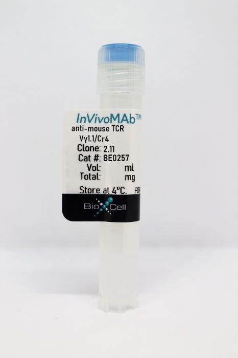
InVivoMAb anti-mouse TCR Vγ1.1/Cr4
Product Details
The 2.11 monoclonal antibody reacts with an epitope in the Cr4 domain of TCR Vγ1.1 (T cell receptor V gamma 1.1). The TCR is expressed on the surface of T lymphocytes and is responsible for recognizing fragments of antigen as peptides bound to MHC molecules. When the TCR engages with antigenic peptide and MHC the T lymphocyte is activated through signal transduction. The Vγ1Jγ4Cγ4 chain is expressed by a major population of γδ T cells in the thymus and peripheral lymphoid organs of adult mice. However, during postnatal and early life stages only a minor population of γδ T cells express Vγ1Jγ4Cγ4 during fetal and early postnatal life.Specifications
| Isotype | Armenian hamster IgG |
|---|---|
| Recommended Isotype Control(s) | InVivoMAb polyclonal Armenian hamster IgG |
| Recommended Dilution Buffer | InVivoPure pH 7.0 Dilution Buffer |
| Conjugation | This product is unconjugated. Conjugation is available via our Antibody Conjugation Services. |
| Immunogen | 3.13.1 T cell hybridoma |
| Reported Applications |
in vivo Vγ1 TCR+ cell depletion Flow cytometry |
| Formulation |
PBS, pH 7.0 Contains no stabilizers or preservatives |
| Endotoxin |
<2EU/mg (<0.002EU/μg) Determined by LAL gel clotting assay |
| Purity |
>95% Determined by SDS-PAGE |
| Sterility | 0.2 µm filtration |
| Production | Purified from cell culture supernatant in an animal-free facility |
| Purification | Protein G |
| RRID | AB_2687736 |
| Molecular Weight | 150 kDa |
| Storage | The antibody solution should be stored at the stock concentration at 4°C. Do not freeze. |
Recommended Products
-
Recommended Isotype Control(s)
InVivoMAb polyclonal Armenian hamster IgG
-

Recommended Dilution Buffer
InVivoPure pH 7.0 Dilution Buffer
in vivo Vγ1 TCR+ cell depletion, Flow Cytometry
Zheng, L., et al. (2017). "Recruitment of Neutrophils Mediated by Vγ2 γδ T Cells Deteriorates Liver Fibrosis Induced by Schistosoma japonicum Infection in C57BL/6 Mice" Infect Immun 85(8). PubMed
Conventional adaptive T cell responses contribute to the pathogenesis of Schistosoma japonicum infection, leading to liver fibrosis. However, the role of gamma-delta (γδ) T cells in this disease is less clear. γδ T cells are known to secrete interleukin-17 (IL-17) in response to infection, exerting either protective or pathogenic functions. In the present study, mice infected with S. japonicum are used to characterize the role of γδ T cells. Combined with the infection of S. japonicum, an extremely significant increase in the percentage of neutrophils in the CD45(+) cells was detected (from approximately 2.45% to 46.10% in blood and from 0.18% to 7.34% in spleen). Further analysis identified two different γδ T cell subsets that have different functions in the formation of granulomas in S. japonicum-infected mice. The Vγ1 T cells secrete gamma interferon (IFN-γ) only, while the Vγ2 T cells secrete both IL-17A and IFN-γ. Both subtypes lose the ability to secrete cytokine during the late stage of infection (12 weeks postinfection). When we depleted the Vγ2 T cells in infected mice, the percentage of neutrophils in blood and spleen decreased significantly, the liver fibrosis in the granulomas was reduced, and the level of IL-17A in the serum decreased (P < 0.05). These results suggest that during S. japonicum infection, Vγ2 T cells can recruit neutrophils and aggravate liver fibrosis by secreting IL-17A. This is the first report that a subset of γδ T cells plays a partial role in the pathological process of schistosome infection.
Flow Cytometry
Narayan, K., et al. (2012). "Intrathymic programming of effector fates in three molecularly distinct gammadelta T cell subtypes" Nat Immunol 13(5): 511-518. PubMed
Innate gammadelta T cells function in the early phase of immune responses. Although innate gammadelta T cells have often been studied as one homogenous population, they can be functionally classified into effector subsets on the basis of the production of signature cytokines, analogous to adaptive helper T cell subsets. However, unlike the function of adaptive T cells, gammadelta effector T cell function correlates with genomically encoded T cell antigen receptor (TCR) chains, which suggests that clonal TCR selection is not the main determinant of the differentiation of gammadelta effector cells. A high-resolution transcriptome analysis of all emergent gammadelta thymocyte subsets segregated on the basis of use of the TCR gamma-chain or delta-chain indicated the existence of three separate subtypes of gammadelta effector cells in the thymus. The immature gammadelta subsets were distinguished by unique transcription-factor modules that program effector function.
- Mus musculus (House mouse),
- Immunology and Microbiology
Mammary γδ T cells promote IL-17A-mediated immunity against Staphylococcus aureus-induced mastitis in a microbiota-dependent manner.
In IScience on 15 December 2023 by Pan, N., Xiu, L., et al.
PubMed
Mastitis, a common disease for female during lactation period that could cause a health risk for human or huge economic losses for animals, is mainly caused by S. aureus invasion. Here, we found that neutrophil recruitment via IL-17A-mediated signaling was required for host defense against S. aureus-induced mastitis in a mouse model. The rapid accumulation and activation of Vγ4+ γδ T cells in the early stage of infection triggered the IL-17A-mediated immune response. Interestingly, the accumulation and influence of γδT17 cells in host defense against S. aureus-induced mastitis in a commensal microbiota-dependent manner. Overall, this study, focusing on γδT17 cells, clarified innate immune response mechanisms against S. aureus-induced mastitis, and provided a specific response to target for future immunotherapies. Meanwhile, a link between commensal microbiota community and host defense to S. aureus mammary gland infection may unveil potential therapeutic strategies to combat these intractable infections. © 2023 The Author(s).
- WB,
- Mus musculus (House mouse),
- Immunology and Microbiology
Vγ1 and Vγ4 gamma-delta T cells play opposing roles in the immunopathology of traumatic brain injury in males.
In Nature Communications on 18 July 2023 by Abou-El-Hassan, H., Rezende, R. M., et al.
PubMed
Traumatic brain injury (TBI) is a leading cause of morbidity and mortality. The innate and adaptive immune responses play an important role in the pathogenesis of TBI. Gamma-delta (γδ) T cells have been shown to affect brain immunopathology in multiple different conditions, however, their role in acute and chronic TBI is largely unknown. Here, we show that γδ T cells affect the pathophysiology of TBI as early as one day and up to one year following injury in a mouse model. TCRδ-/- mice are characterized by reduced inflammation in acute TBI and improved neurocognitive functions in chronic TBI. We find that the Vγ1 and Vγ4 γδ T cell subsets play opposing roles in TBI. Vγ4 γδ T cells infiltrate the brain and secrete IFN-γ and IL-17 that activate microglia and induce neuroinflammation. Vγ1 γδ T cells, however, secrete TGF-β that maintains microglial homeostasis and dampens TBI upon infiltrating the brain. These findings provide new insights on the role of different γδ T cell subsets after brain injury and lay down the principles for the development of targeted γδ T-cell-based therapy for TBI. © 2023. The Author(s).
- Immu-depl,
- Neutralization,
- Homo sapiens (Human),
- Cancer Research,
- Immunology and Microbiology
Cancer cell-expressed BTNL2 facilitates tumour immune escape via engagement with IL-17A-producing γδ T cells.
In Nature Communications on 11 January 2022 by Du, Y., Peng, Q., et al.
PubMed
Therapeutic blockade of the immune checkpoint proteins programmed cell death protein 1 (PD-1) and cytotoxic T lymphocyte antigen 4 (CTLA4) has transformed cancer treatment. However, the overall response rate to these treatments is low, suggesting that immune checkpoint activation is not the only mechanism leading to dysfunctional anti-tumour immunity. Here we show that butyrophilin-like protein 2 (BTNL2) is a potent suppressor of the anti-tumour immune response. Antibody-mediated blockade of BTNL2 attenuates tumour progression in multiple in vivo murine tumour models, resulting in prolonged survival of tumour-bearing mice. Mechanistically, BTNL2 interacts with local γδ T cell populations to promote IL-17A production in the tumour microenvironment. Inhibition of BTNL2 reduces the number of tumour-infiltrating IL-17A-producing γδ T cells and myeloid-derived suppressor cells, while facilitating cytotoxic CD8+ T cell accumulation. Furthermore, we find high BTNL2 expression in several human tumour samples from highly prevalent cancer types, which negatively correlates with overall patient survival. Thus, our results suggest that BTNL2 is a negative regulator of anti-tumour immunity and a potential target for cancer immunotherapy. © 2022. The Author(s).
- Cancer Research,
- Immunology and Microbiology,
- Neuroscience
Ly6C defines a subset of memory-like CD27sup>+/sup> γδ T cells with inducible cancer-killing function
Preprint on BioRxiv : the Preprint Server for Biology on 8 September 2020 by Wiesheu, R., Edwards, S. C., et al.
PubMed
h4>ABSTRACT/h4> In mice, IFNγ-producing γδ T cells that express the co-stimulatory molecule, CD27, play a critical role in host defence and anti-tumour immunity. However, their phenotypic diversity, composition in peripheral and secondary lymphoid organs, similarity to αβ T cells as well as homology with human γδ T cells is poorly understood. Here, using single cell RNA sequencing, we show that CD27 + γδ T cells consist of two major clusters, which are distinguished by expression of Ly6C. We demonstrate that CD27 + Ly6C — γδ T cells exhibit a naïve T cell-like phenotype, whereas CD27 + Ly6C + γδ T cells display a memory-like phenotype, produce several NK cell-related and cytotoxic molecules and are highly similar to both mouse CD8 + T cells and mature human γδ T cells. In a breast cancer mouse model, depletion of CD27 + γδ T cells failed to affect tumour growth, but these cells could be coerced into killing cancer cells after expansion ex vivo . These results identify novel subsets of γδ T cells in mice that are comparable to human γδ T cells, opening new opportunities for γδ T cell-based cancer immunotherapy research.
- Immu-depl,
- Mus musculus (House mouse),
- Immunology and Microbiology
Recruitment of Neutrophils Mediated by Vγ2 γδ T Cells Deteriorates Liver Fibrosis Induced by Schistosoma japonicum Infection in C57BL/6 Mice.
In Infection and Immunity on 1 August 2017 by Zheng, L., Hu, Y., et al.
PubMed
Conventional adaptive T cell responses contribute to the pathogenesis of Schistosoma japonicum infection, leading to liver fibrosis. However, the role of gamma-delta (γδ) T cells in this disease is less clear. γδ T cells are known to secrete interleukin-17 (IL-17) in response to infection, exerting either protective or pathogenic functions. In the present study, mice infected with S. japonicum are used to characterize the role of γδ T cells. Combined with the infection of S. japonicum, an extremely significant increase in the percentage of neutrophils in the CD45+ cells was detected (from approximately 2.45% to 46.10% in blood and from 0.18% to 7.34% in spleen). Further analysis identified two different γδ T cell subsets that have different functions in the formation of granulomas in S. japonicum-infected mice. The Vγ1 T cells secrete gamma interferon (IFN-γ) only, while the Vγ2 T cells secrete both IL-17A and IFN-γ. Both subtypes lose the ability to secrete cytokine during the late stage of infection (12 weeks postinfection). When we depleted the Vγ2 T cells in infected mice, the percentage of neutrophils in blood and spleen decreased significantly, the liver fibrosis in the granulomas was reduced, and the level of IL-17A in the serum decreased (P < 0.05). These results suggest that during S. japonicum infection, Vγ2 T cells can recruit neutrophils and aggravate liver fibrosis by secreting IL-17A. This is the first report that a subset of γδ T cells plays a partial role in the pathological process of schistosome infection.Copyright © 2017 Zheng et al.