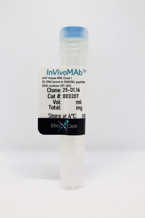
InVivoMAb anti-mouse MHC Class I (H-2Kb) bound to SIINFEKL peptide (OVA residues 257-264)
Product Details
The 25-D1.16 monoclonal antibody reacts with mouse MHC class I H-2Kb bound to the ovalbumin-derived peptide with sequence SIINFEKL. This antibody does not react with unbound MHC class I H-2Kb or MHC class I H-2Kb bound to an irrelevant peptide. The 25-D1.16 antibody is often used to track the quantity and localization of antigen-presenting cells bearing these specific molecules in vivo.Specifications
| Isotype | Mouse IgG1, κ |
|---|---|
| Recommended Isotype Control(s) | InVivoMAb mouse IgG1 isotype control, unknown specificity |
| Recommended Dilution Buffer | InVivoPure pH 7.0 Dilution Buffer |
| Conjugation | This product is unconjugated. Conjugation is available via our Antibody Conjugation Services. |
| Immunogen | SIINFEKL pulsed RMA-S cells |
| Reported Applications |
in vivo blocking of Kb -SIINFEKL Functional assays Flow cytometry |
| Formulation |
PBS, pH 7.0 Contains no stabilizers or preservatives |
| Endotoxin |
<2EU/mg (<0.002EU/μg) Determined by LAL gel clotting assay |
| Purity |
>95% Determined by SDS-PAGE |
| Sterility | 0.2 µm filtration |
| Production | Purified from cell culture supernatant in an animal-free facility |
| Purification | Protein G |
| RRID | AB_10950697 |
| Molecular Weight | 150 kDa |
| Storage | The antibody solution should be stored at the stock concentration at 4°C. Do not freeze. |
Recommended Products
in vivo blocking of Kb -SIINFEKL
Thompson, E. A., et al. (2019). "Interstitial Migration of CD8αβ T Cells in the Small Intestine Is Dynamic and Is Dictated by Environmental Cues" Cell Rep 26(11): 2859-2867.e2854. PubMed
The migratory capacity of adaptive CD8αβ T cells dictates their ability to locate target cells and exert cytotoxicity, which is the basis of immune surveillance for the containment of microbes and disease. The small intestine (SI) is the largest mucosal surface and is a primary site of pathogen entrance. Using two-photon laser scanning microscopy, we found that motility of antigen (Ag)-specific CD8αβ T cells in the SI is dynamic and varies with the environmental milieu. Pathogen-specific CD8αβ T cell movement differed throughout infection, becoming locally confined at memory. Motility was not dependent on CD103 but was influenced by micro-anatomical locations within the SI and by inflammation. CD8 T cells responding to self-protein were initially affected by the presence of self-Ag, but this was altered after complete tolerance induction. These studies identify multiple factors that affect CD8αβ T cell movement in the intestinal mucosa and show the adaptability of CD8αβ T cell motility.
Flow Cytometry
Saini, S. K., et al. (2015). "Dipeptides catalyze rapid peptide exchange on MHC class I molecules" Proc Natl Acad Sci U S A 112(1): 202-207. PubMed
Peptide ligand selection by MHC class I molecules, which occurs by iterative optimization, is the centerpiece of immunodominance in antiviral and antitumor immune responses. For its understanding, the molecular mechanisms of peptide binding and dissociation by class I molecules must be elucidated. To this end, we have investigated dipeptides that bind to the F pocket of class I molecules. We find that they accelerate the dissociation of prebound peptides of both low and high affinity, suggesting a mechanism of action for the peptide-exchange chaperone tapasin. Peptide exchange on class I molecules also has practical uses in epitope discovery and T-cell monitoring.
Flow Cytometry
Sei, J. J., et al. (2015). "Peptide-MHC-I from Endogenous Antigen Outnumber Those from Exogenous Antigen, Irrespective of APC Phenotype or Activation" PLoS Pathog 11(6): e1004941. PubMed
Naive anti-viral CD8+ T cells (TCD8+) are activated by the presence of peptide-MHC Class I complexes (pMHC-I) on the surface of professional antigen presenting cells (pAPC). Increasing the number of pMHC-I in vivo can increase the number of responding TCD8+. Antigen can be presented directly or indirectly (cross presentation) from virus-infected and uninfected cells, respectively. Here we determined the relative importance of these two antigen presenting pathways in mousepox, a natural disease of the mouse caused by the poxvirus, ectromelia (ECTV). We demonstrated that ECTV infected several pAPC types (macrophages, B cells, and dendritic cells (DC), including DC subsets), which directly presented pMHC-I to naive TCD8+ with similar efficiencies in vitro. We also provided evidence that these same cell-types presented antigen in vivo, as they form contacts with antigen-specific TCD8+. Importantly, the number of pMHC-I on infected pAPC (direct presentation) vastly outnumbered those on uninfected cells (cross presentation), where presentation only occurred in a specialized subset of DC. In addition, prior maturation of DC failed to enhance antigen presentation, but markedly inhibited ECTV infection of DC. These results suggest that direct antigen presentation is the dominant pathway in mice during mousepox. In a broader context, these findings indicate that if a virus infects a pAPC then the presentation by that cell is likely to dominate over cross presentation as the most effective mode of generating large quantities of pMHC-I is on the surface of pAPC that endogenously express antigens. Recent trends in vaccine design have focused upon the introduction of exogenous antigens into the MHC Class I processing pathway (cross presentation) in specific pAPC populations. However, use of a pantropic viral vector that targets pAPC to express antigen endogenously likely represents a more effective vaccine strategy than the targeting of exogenous antigen to a limiting pAPC subpopulation.
Flow Cytometry
Zehner, M., et al. (2015). "The translocon protein Sec61 mediates antigen transport from endosomes in the cytosol for cross-presentation to CD8(+) T cells" Immunity 42(5): 850-863. PubMed
The molecular mechanisms regulating antigen translocation into the cytosol for cross-presentation are under controversial debate, mainly because direct data is lacking. Here, we have provided direct evidence that the activity of the endoplasmic reticulum (ER) translocon protein Sec61 is essential for endosome-to-cytosol translocation. We generated a Sec61-specific intrabody, a crucial tool that trapped Sec61 in the ER and prevented its recruitment into endosomes without influencing Sec61 activity and antigen presentation in the ER. Expression of this ER intrabody inhibited antigen translocation and cross-presentation, demonstrating that endosomal Sec61 indeed mediates antigen transport across endosomal membranes. Moreover, we showed that the recruitment of Sec61 toward endosomes, and hence antigen translocation and cross-presentation, is dependent on dendritic cell activation by Toll-like receptor (TLR) ligands. These data shed light on a long-lasting question regarding antigen cross-presentation and point out a role of the ER-associated degradation machinery in compartments distinct from the ER.
Flow Cytometry
Sasaki, K., et al. (2015). "Thymoproteasomes produce unique peptide motifs for positive selection of CD8(+) T cells" Nat Commun 6: 7484. PubMed
Positive selection in the thymus provides low-affinity T-cell receptor (TCR) engagement to support the development of potentially useful self-major histocompatibility complex class I (MHC-I)-restricted T cells. Optimal positive selection of CD8(+) T cells requires cortical thymic epithelial cells that express beta5t-containing thymoproteasomes (tCPs). However, how tCPs govern positive selection is unclear. Here we show that the tCPs produce unique cleavage motifs in digested peptides and in MHC-I-associated peptides. Interestingly, MHC-I-associated peptides carrying these tCP-dependent motifs are enriched with low-affinity TCR ligands that efficiently induce the positive selection of functionally competent CD8(+) T cells in antigen-specific TCR-transgenic models. These results suggest that tCPs contribute to the positive selection of CD8(+) T cells by preferentially producing low-affinity TCR ligand peptides.
Flow Cytometry
Park, H. M., et al. (2014). "CD4 T-cells transduced with CD80 and 4-1BBL mRNA induce long-term CD8 T-cell responses resulting in potent antitumor effects" Vaccine 32(51): 6919-6926. PubMed
Therapeutic cancer vaccines are an attractive alternative to conventional therapies to treat malignant tumors, and more importantly, to prevent recurrence after primary therapy. However, the availability of professional antigen-presenting cells (APCs) has been restricted by difficulties encountered in obtaining sufficient professional APCs for clinical use. We have prepared an alternative cellular vaccine with CD4 T-cells that can be expanded easily to yield a pure and homogeneous population in vitro. To enhance their potency as a therapeutic vaccine, in vitro expanded CD4 T-cells were transfected with RNAs encoding the costimulatory ligands CD80, 4-1BBL, or both (CD80-T, 4-1BBL-T, and CD80/4-1BBL-T-cells, respectively). We observed augmented cell vitality in CD80/4-1BBL-T-cells in vitro and in vivo. Significant CD8 T-cell responses eliciting in vivo proliferation and cytotoxicity were obtained with CD80/4-1BBL-T-cell vaccination compared to CD80-T and 4-1BBL-T-cell vaccinations. In contrast, beta2m-deficient CD80/4-1BBL-T-cells were not as effective as wile-type CD80/4-1BBL-T-cells in priming CD8 T-cells. Furthermore, CD80/4-1BBL-T-cell immunization resulted in curing established EG7 tumors, resulting in the generation of memory CD8 T-cell responses, and elicited therapeutic antitumor responses against B16 melanoma. These results suggest that CD4 T-cells endowed with costimulatory ligands allow the design of effective vaccination strategies against cancer.
Functional Assays
Imai, T., et al. (2013). "CD8(+) T cell activation by murine erythroblasts infected with malaria parasites" Sci Rep 3: 1572. PubMed
Recent studies show that some human malaria parasite species Plasmodium falciparum and P. vivax parasitize erythroblasts; however, the biological and clinical significance of this is unclear. To investigate further, we generated a rodent malaria parasite (P. yoelii 17XNL) expressing GFP-ovalbumin (OVA). Its infectivity to erythroblasts was confirmed, and parasitized erythroblasts were capable of initiating malaria infections. Experiments showed that MHC class I molecules were highly expressed on parasitized erythroblasts. As CD8(+) T cells recognize MHC class I and peptide complexes on target cells, and are involved in protection or pathology against malaria, we examined whether erythroblasts are targeted by CD8(+) T cells. Purified non-parasitized erythroblasts pulsed with OVA peptides were recognized by OVA-specific CD8(+) T cells. Crucially, parasitized erythroblasts isolated from GFP-OVA-, but not GFP- infected-mice, activated OT-I CD8(+) T cells, indicating that CD8(+) T cells recognize parasitized erythroblasts in an antigen-specific manner.
- FC/FACS,
- Mus musculus (House mouse),
- Immunology and Microbiology
An input-controlled model system for identification of MHC bound peptides enabling laboratory comparisons of immunopeptidome experiments.
In Journal of Proteomics on 30 September 2020 by Klatt, M. G., Aretz, Z. E. H., et al.
PubMed
Characterization of MHC-bound peptides by mass spectrometry (MS) is an essential technique for immunologic studies. Many efforts have been made to quantify the number of MHC-presented ligands by MS and to define the limits of detection of a specific MHC ligand. However, these experiments are often complex and comparisons across different laboratories are challenging. Therefore, we compared and orthogonally validated quantitation of peptide:MHC complexes by radioimmunoassay and flow cytometry using TCR mimic antibodies in three model systems to establish a method to control the experimental input of peptide MHC:complexes for MS analysis. Following isolation of MHC-bound peptides we identified and quantified an MHC ligand of interest with high correlation to the initial input. We found that the diversity of the presented ligandome, as well as the peptide sequence itself affected the detection of the target peptide. Furthermore, results were applicable from these model systems to unmodified cell lines with a tight correlation between HLA-A*02 complex input and the number of identified HLA-A*02 ligands. Overall, this framework provides an easily accessible experimental setup that offers the opportunity to control the peptide:MHC input and in this way compare immunopeptidome experiments not only within but also between laboratories, independent of their experimental approach. SIGNIFICANCE: Although immunopeptidomics is an essential tool for the characterization of MHCbound peptides on the cell surface, there are no easily applicable established protocols available that allow comparison of immunopeptidome experiments across laboratories. Here, we demonstrate that controlling the peptide:MHC input for immunopurification and LC-MS/MS experiments by flow cytometry in pre-defined model systems allows the generation of qualitative and quantitative data that can easily be compared between investigators, independently of their methods for MHC ligand isolation for MS. Copyright © 2020 Elsevier B.V. All rights reserved.
- Mus musculus (House mouse),
- Immunology and Microbiology
T cell and chemokine receptors differentially control CD8 T cell motility behavior in the infected airways immediately before and after virus clearance in a primary infection.
In PLoS ONE on 21 August 2020 by Emo, K., Reilly, E. C., et al.
PubMed
In mice, experimental influenza virus infection stimulates CD8 T cell infiltration of the airways. Virus is cleared by day 9, and between days 8 and 9 there is an abrupt change in CD8 T cell motility behavior transitioning from low velocity and high confinement on day 8, to high velocity with continued high confinement on day 9. We hypothesized that loss of virus and/or antigen signals in the context of high chemokine levels drives the T cells into a rapid surveillance mode. Virus infection induces chemokine production, which may change when the virus is cleared. We therefore sought to examine this period of rapid changes to the T cell environment in the tissue and seek evidence on the roles of peptide-MHC and chemokine receptor interactions. Experiments were performed to block G protein coupled receptor (GPCR) signaling with Pertussis toxin (Ptx). Ptx treatment generally reduced cell velocities and mildly increased confinement suggesting chemokine mediated arrest (velocity 2 μm/min) (Friedman RS, 2005), except on day 8 when velocity increased and confinement was relieved. Blocking specific peptide-MHC with monoclonal antibody unexpectedly decreased velocities on days 7 through 9, suggesting TCR/peptide-MHC interactions promote cell mobility in the tissue. Together, these results suggest the T cells are engaged with antigen bearing and chemokine producing cells that affect motility in ways that vary with the day after infection. The increase in velocities on day 9 were reversed by addition of specific peptide, consistent with the idea that antigen signals become limiting on day 9 compared to earlier time points. Thus, antigen and chemokine signals act to alternately promote and restrict CD8 T cell motility until the point of virus clearance, suggesting the switch in motility behavior on day 9 may be due to a combination of limiting antigen in the presence of high chemokine signals as the virus is cleared.
- In Vitro,
- Mus musculus (House mouse),
- Cancer Research,
- Cell Biology,
- Immunology and Microbiology
Autophagy promotes immune evasion of pancreatic cancer by degrading MHC-I.
In Nature on 1 May 2020 by Yamamoto, K., Venida, A., et al.
PubMed
Immune evasion is a major obstacle for cancer treatment. Common mechanisms of evasion include impaired antigen presentation caused by mutations or loss of heterozygosity of the major histocompatibility complex class I (MHC-I), which has been implicated in resistance to immune checkpoint blockade (ICB) therapy1-3. However, in pancreatic ductal adenocarcinoma (PDAC), which is resistant to most therapies including ICB4, mutations that cause loss of MHC-I are rarely found5 despite the frequent downregulation of MHC-I expression6-8. Here we show that, in PDAC, MHC-I molecules are selectively targeted for lysosomal degradation by an autophagy-dependent mechanism that involves the autophagy cargo receptor NBR1. PDAC cells display reduced expression of MHC-I at the cell surface and instead demonstrate predominant localization within autophagosomes and lysosomes. Notably, inhibition of autophagy restores surface levels of MHC-I and leads to improved antigen presentation, enhanced anti-tumour T cell responses and reduced tumour growth in syngeneic host mice. Accordingly, the anti-tumour effects of autophagy inhibition are reversed by depleting CD8+ T cells or reducing surface expression of MHC-I. Inhibition of autophagy, either genetically or pharmacologically with chloroquine, synergizes with dual ICB therapy (anti-PD1 and anti-CTLA4 antibodies), and leads to an enhanced anti-tumour immune response. Our findings demonstrate a role for enhanced autophagy or lysosome function in immune evasion by selective targeting of MHC-I molecules for degradation, and provide a rationale for the combination of autophagy inhibition and dual ICB therapy as a therapeutic strategy against PDAC.
- Mus musculus (House mouse),
- Immunology and Microbiology
Antigen and G-Protein Coupled Receptor signaling differentially control CD8 T cell motility immediately before and after virus clearance in a primary infection
Preprint on BioRxiv : the Preprint Server for Biology on 16 December 2019 by Lambert-Emo, K., Reilly, E. C., et al.
PubMed
In mice, experimental influenza virus infection stimulates CD8 T cell infiltration of the airways. Virus is cleared by day 9, and between days 8 and 9 there is an abrupt change in CD8 T cell motility behavior transitioning from low velocity and high confinement on day 8, to high velocity with continued confinement on day 9. We hypothesized that it is loss of virus and/or antigen signals in the context of high chemokine levels that drives the T cells into a rapid surveillance mode. Virus infection induces chemokine production, which may change when the virus is cleared. We therefore sought to examine this period of rapid changes to the T cell environment in the tissue and seek evidence on the roles of peptide-MHC and chemokine receptor interactions. Experiments were performed to block G protein coupled receptor (GPCR) signaling with Pertussis toxin (Ptx). Ptx treatment generally reduced cell velocities and mildly increased confinement, except on day 8 when velocity increased and confinement was relieved, suggesting chemokine mediated arrest. Blocking specific peptide-MHC with monoclonal antibody unexpectedly decreased velocities on days 7 through 9, suggesting TCR/peptide-MHC interactions promote cell mobility in the tissue. Together, these results suggest the T cells are engaged with antigen bearing and chemokine producing cells that affect motility in ways that vary with the day after infection. The increase in velocities on day 9 were reversed by addition of specific peptide, consistent with the idea that antigen signals become limiting on day 9 compared to earlier time points. Thus, antigen and chemokine signals act to alternately promote and restrict CD8 T cell motility until the point of virus clearance, suggesting the switch in motility behavior on day 9 may be due to a combination of limiting antigen in the presence of high chemokine signals as the virus is cleared.
- Block,
- In Vivo,
- Mus musculus (House mouse),
- Immunology and Microbiology
Interstitial Migration of CD8αβ T Cells in the Small Intestine Is Dynamic and Is Dictated by Environmental Cues.
In Cell Reports on 12 March 2019 by Thompson, E. A., Mitchell, J. S., et al.
PubMed
The migratory capacity of adaptive CD8αβ T cells dictates their ability to locate target cells and exert cytotoxicity, which is the basis of immune surveillance for the containment of microbes and disease. The small intestine (SI) is the largest mucosal surface and is a primary site of pathogen entrance. Using two-photon laser scanning microscopy, we found that motility of antigen (Ag)-specific CD8αβ T cells in the SI is dynamic and varies with the environmental milieu. Pathogen-specific CD8αβ T cell movement differed throughout infection, becoming locally confined at memory. Motility was not dependent on CD103 but was influenced by micro-anatomical locations within the SI and by inflammation. CD8 T cells responding to self-protein were initially affected by the presence of self-Ag, but this was altered after complete tolerance induction. These studies identify multiple factors that affect CD8αβ T cell movement in the intestinal mucosa and show the adaptability of CD8αβ T cell motility.Copyright © 2019 The Author(s). Published by Elsevier Inc. All rights reserved.

