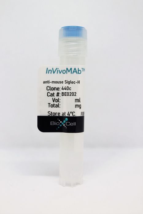
InVivoMAb anti-mouse Siglec-H
Product Details
The 440c monoclonal antibody recognizes mouse Siglec-H, a member of the CD33-related Siglec family. Siglec-H has been shown to be exclusively expressed on plasmacytoid dendritic cells (pDCs) and type I IFN-producing cells (IPC) in naïve mice. IPCs play a major role in the immune response to viruses. The 440c antibody has been shown to inhibit pDC function.Specifications
| Isotype | Rat IgG2b |
|---|---|
| Recommended Isotype Control(s) | InVivoMAb rat IgG2b isotype control, anti-keyhole limpet hemocyanin |
| Recommended Dilution Buffer | InVivoPure pH 7.0 Dilution Buffer |
| Conjugation | This product is unconjugated. Conjugation is available via our Antibody Conjugation Services. |
| Immunogen | Purified bone marrow-derived IPCs |
| Reported Applications |
in vivo administration Flow cytometry |
| Formulation |
PBS, pH 7.0 Contains no stabilizers or preservatives |
| Endotoxin |
<2EU/mg (<0.002EU/μg) Determined by LAL gel clotting assay |
| Purity |
>95% Determined by SDS-PAGE |
| Sterility | 0.2 µm filtration |
| Production | Purified from cell culture supernatant in an animal-free facility |
| Purification | Protein G |
| RRID | AB_10949014 |
| Molecular Weight | 150 kDa |
| Storage | The antibody solution should be stored at the stock concentration at 4°C. Do not freeze. |
Recommended Products
in vivo administration
Wu, V., et al. (2015). "Plasmacytoid dendritic cell-derived IFNalpha modulates Th17 differentiation during early Bordetella pertussis infection in mice" Mucosal Immunol. doi : 10.1038/mi.2015.101. PubMed
Whooping cough is a highly contagious respiratory disease caused by Bordetella pertussis (B. pertussis). T helper 17 (Th17) cells have a central role in the resolution of the infection. Emerging studies document that type I interferons (IFNs) suppress Th17 differentiation and interleukin (IL)-17 responses in models of infection and chronic inflammation. As plasmacytoid dendritic cells (pDCs) are a major source of type I IFNs, we hypothesize that during B. pertussis infection in mice, pDC-derived IFNalpha inhibits a rapid increase in Th17 cells. We found that IFNalpha-secreting pDCs appear in the lungs during the early stages of infection, while a robust rise of Th17 cells in the lungs is detected at 15 days post-infection or later. The presence of IFNalpha led to reduced Th17 differentiation and proliferation in vitro. Furthermore, in vivo blocking of IFNalpha produced by pDCs during infection with B. pertussis infection resulted in early increase of Th17 frequency, inflammation, and reduced bacterial loads in the airways of infected mice. Taken together, the experiments reported here describe an inhibitory role for pDCs and pDC-derived IFNalpha in modulating Th17 responses during the early stages of B. pertussis infection, which may explain the prolonged nature of whooping cough.Mucosal Immunology advance online publication 14 October 2015. doi:10.1038/mi.2015.101.
Flow Cytometry
Puttur, F., et al. (2013). "Absence of Siglec-H in MCMV infection elevates interferon alpha production but does not enhance viral clearance" PLoS Pathog 9(9): e1003648. PubMed
Plasmacytoid dendritic cells (pDCs) express the I-type lectin receptor Siglec-H and produce interferon alpha (IFNalpha), a critical anti-viral cytokine during the acute phase of murine cytomegalovirus (MCMV) infection. The ligands and biological functions of Siglec-H still remain incompletely defined in vivo. Thus, we generated a novel bacterial artificial chromosome (BAC)-transgenic “pDCre” mouse which expresses Cre recombinase under the control of the Siglec-H promoter. By crossing these mice with a Rosa26 reporter strain, a representative fraction of Siglec-H(+) pDCs is terminally labeled with red fluorescent protein (RFP). Interestingly, systemic MCMV infection of these mice causes the downregulation of Siglec-H surface expression. This decline occurs in a TLR9- and MyD88-dependent manner. To elucidate the functional role of Siglec-H during MCMV infection, we utilized a novel Siglec-H deficient mouse strain. In the absence of Siglec-H, the low infection rate of pDCs with MCMV remained unchanged, and pDC activation was still intact. Strikingly, Siglec-H deficiency induced a significant increase in serum IFNalpha levels following systemic MCMV infection. Although Siglec-H modulates anti-viral IFNalpha production, the control of viral replication was unchanged in vivo. The novel mouse models will be valuable to shed further light on pDC biology in future studies.
Flow Cytometry
Loschko, J., et al. (2011). "Antigen targeting to plasmacytoid dendritic cells via Siglec-H inhibits Th cell-dependent autoimmunity" J Immunol 187(12): 6346-6356. PubMed
Plasmacytoid dendritic cells (PDCs) have been shown to present Ags and to contribute to peripheral immune tolerance and to Ag-specific adaptive immunity. However, modulation of adaptive immune responses by selective Ag targeting to PDCs with the aim of preventing autoimmunity has not been investigated. In the current study, we demonstrate that in vivo Ag delivery to murine PDCs via the specifically expressed surface molecule sialic acid binding Ig-like lectin H (Siglec-H) inhibits Th cell and Ab responses in the presence of strong immune stimulation in an Ag-specific manner. Correlating with sustained low-level MHC class II-restricted Ag presentation on PDCs, Siglec-H-mediated Ag delivery induced a hyporesponsive state in CD4(+) T cells leading to reduced expansion and Th1/Th17 cell polarization without conversion to Foxp3(+) regulatory T cells or deviation to Th2 or Tr1 cells. Siglec-H-mediated delivery of a T cell epitope derived from the autoantigen myelin oligodendrocyte glycoprotein to PDCs effectively delayed onset and reduced disease severity in myelin oligodendrocyte glycoprotein-induced experimental autoimmune encephalomyelitis by interfering with the priming phase without promoting the generation or expansion of myelin oligodendrocyte glycoprotein-specific Foxp3(+) regulatory T cells. We conclude that Ag delivery to PDCs can be harnessed to inhibit Ag-specific immune responses and prevent Th cell-dependent autoimmunity.
- Cancer Research,
- Immunology and Microbiology
Redefining innate natural antibodies as important contributors to anti-tumor immunity.
In eLife on 5 October 2021 by Rawat, K., Tewari, A., et al.
PubMed
Myeloid, T, and NK cells are key players in the elimination phase of cancer immunoediting, also referred to as cancer immunosurveillance. However, the role of B cells and NAbs, which are present prior to the encounter with cognate antigens, has been overlooked. One reason is due to the popular use of a single B cell-deficient mouse model, muMT mice. Cancer models using muMT mice display a similar tumor burden as their wild-type (WT) counterparts. Empirically, we observe what others have previously reported with muMT mice. However, using two other B cell-deficient mouse models (IgHELMD4 and CD19creDTA), we show a three- to fivefold increase in tumor burden relative to WT mice. In addition, using an unconventional, non-cancer-related, immune neoantigen model where hypoxic conditions and cell clustering are absent, we provide evidence that B cells and their innate, natural antibodies (NAbs) are critical for the detection and elimination of neoantigen-expressing cells. Finally, we find that muMT mice display anti-tumor immunity because of an unexpected compensatory mechanism consisting of significantly enhanced type 1 interferon (IFN)-producing plasmacytoid dendritic cells (pDCs), which recruit a substantial number of NK cells to the tumor microenvironment compared to WT mice. Diminishing this compensatory pDC-IFN-NK cell mechanism revealed that muMT mice develop a three- to fivefold increase in tumor burden compared to WT mice. In summary, our findings suggest that NAbs are part of an early defense against not only microorganisms and dying cells, but precancerous cells as well. © 2021, Rawat et al.
- In Vivo,
- Mus musculus (House mouse),
- Immunology and Microbiology
Follicular Dendritic Cell Activation by TLR Ligands Promotes Autoreactive B Cell Responses.
In Immunity on 17 January 2017 by Das, A., Heesters, B. A., et al.
PubMed
A hallmark of autoimmunity in murine models of lupus is the formation of germinal centers (GCs) in lymphoid tissues where self-reactive B cells expand and differentiate. In the host response to foreign antigens, follicular dendritic cells (FDCs) maintain GCs through the uptake and cycling of complement-opsonized immune complexes. Here, we examined whether FDCs retain self-antigens and the impact of this process in autoantibody secretion in lupus. We found that FDCs took up and retained self-immune complexes composed of ribonucleotide proteins, autoantibody, and complement. This uptake, mediated through CD21, triggered endosomal TLR7 and led to the secretion of interferon (IFN) α via an IRF5-dependent pathway. Blocking of FDC secretion of IFN-α restored B cell tolerance and reduced the amount of GCs and pathogenic autoantibody. Thus, FDCs are a critical source of the IFN-α driving autoimmunity in this lupus model. This pathway is conserved in humans, suggesting that it may be a viable therapeutic target in systemic lupus erythematosus.Copyright © 2017 Elsevier Inc. All rights reserved.


