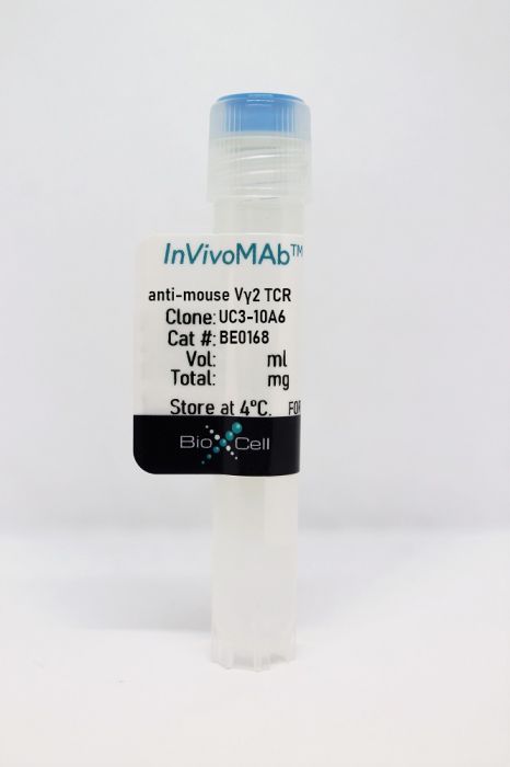
InVivoMAb anti-mouse Vγ2 TCR
Product Details
The UC3-10A6 monoclonal antibody reacts with an epitope on the delta chain of the mouse Vγ2 TCR (V gamma 2 T cell receptor). Vγ2 TCR expressing T lymphocytes make up a large proportion of the γδ T cells in late fetal and adult thymus, peripheral lymphoid tissues, lung, intestinal epithelium, and epidermis. The exact function, ligand, and specificity of γδ TCR-expressing T cells are not completely understood. Studies suggest that these cells recognize bacterial ligands and some tumor cells in the context of MHC class I-like gene products and play a role in regulating the immune response during bacterial infection. The UC3-10A6 antibody has been shown to deplete γδ T cells when administered in vivo.Specifications
| Isotype | Armenian Hamster IgG, κ |
|---|---|
| Recommended Isotype Control(s) | InVivoMAb polyclonal Armenian hamster IgG |
| Recommended Dilution Buffer | InVivoPure pH 7.0 Dilution Buffer |
| Conjugation | This product is unconjugated. Conjugation is available via our Antibody Conjugation Services. |
| Immunogen | G8 mouse T cells |
| Reported Applications |
in vivo γδ T cell depletion Flow cytometry |
| Formulation |
PBS, pH 7.0 Contains no stabilizers or preservatives |
| Endotoxin |
<2EU/mg (<0.002EU/μg) Determined by LAL gel clotting assay |
| Purity |
>95% Determined by SDS-PAGE |
| Sterility | 0.2 µm filtration |
| Production | Purified from cell culture supernatant in an animal-free facility |
| Purification | Protein G |
| RRID | AB_10950109 |
| Molecular Weight | 150 kDa |
| Storage | The antibody solution should be stored at the stock concentration at 4°C. Do not freeze. |
Recommended Products
in vivo γ/δ T cell depletion
Rezende, R. M., et al. (2018). "γδ T cells control humoral immune response by inducing T follicular helper cell differentiation" Nat Commun 9(1): 3151. PubMed
γδ T cells have many known functions, including the regulation of antibody responses. However, how γδ T cells control humoral immunity remains elusive. Here we show that complete Freund’s adjuvant (CFA), but not alum, immunization induces a subpopulation of CXCR5-expressing γδ T cells in the draining lymph nodes. TCRγδ(+)CXCR5(+) cells present antigens to, and induce CXCR5 on, CD4 T cells by releasing Wnt ligands to initiate the T follicular helper (Tfh) cell program. Accordingly, TCRδ(-/-) mice have impaired germinal center formation, inefficient Tfh cell differentiation, and reduced serum levels of chicken ovalbumin (OVA)-specific antibodies after CFA/OVA immunization. In a mouse model of lupus, TCRδ(-/-) mice develop milder glomerulonephritis, consistent with decreased serum levels of lupus-related autoantibodies, when compared with wild type mice. Thus, modulation of the γδ T cell-dependent humoral immune response may provide a novel therapy approach for the treatment of antibody-mediated autoimmunity.
in vivo γ/δ T cell depletion, Flow Cytometry
Hartwig, T., et al. (2015). "Dermal IL-17-producing gammadelta T cells establish long-lived memory in the skin" Eur J Immunol . PubMed
Conventional alphabeta T cells have the ability to form a long-lasting resident memory T-cell (TRM ) population in nonlymphoid tissues after encountering foreign antigen. Conversely, the concept of ‘innate memory’, where the ability of nonadaptive branches of the immune system to deliver a rapid, strengthened immune response upon reinfection or rechallenge, is just emerging. Using the alphabeta T-cell-independent Aldara psoriasis mouse model in combination with genetic fate-mapping and reporter systems, we identified a subset of gammadelta T cells in mice that is capable of establishing a long-lived memory population in the skin. IL-17A/F-producing Vgamma4+ Vdelta4+ T cells populate and persist in the dermis for long periods of time after initial stimulation with Aldara. Experienced Vgamma4+ Vdelta4+ cells show enhanced effector functions and mediate an exacerbated secondary inflammatory response. In addition to identifying a unique feature of gammadelta T cells during inflammation, our results have direct relevance to the human disease as this quasi-innate memory provides a mechanistic insight into relapses and chronification of psoriasis.
in vivo γ/δ T cell depletion
Suryawanshi, A., et al. (2011). "Role of IL-17 and Th17 cells in herpes simplex virus-induced corneal immunopathology" J Immunol 187(4): 1919-1930. PubMed
HSV-1 infection of the cornea leads to a blinding immunoinflammatory lesion of the eye termed stromal keratitis (SK). Recently, IL-17-producing CD4(+) T cells (Th17 cells) were shown to play a prominent role in many autoimmune conditions, but the role of IL-17 and/or of Th17 cells in virus immunopathology is unclear. In this study, we show that, after HSV infection of the cornea, IL-17 is upregulated in a biphasic manner with an initial peak production around day 2 postinfection and a second wave starting from day 7 postinfection with a steady increase until day 21 postinfection, a time point when clinical lesions are fully evident. Further studies demonstrated that innate cells, particularly gammadelta T cells, were major producers of IL-17 early after HSV infection. However, during the clinical phase of SK, the predominant source of IL-17 was Th17 cells that infiltrated the cornea only after the entry of Th1 cells. By ex vivo stimulation, the half fraction of IFN-gamma-producing CD4(+) T cells (Th1 cells) were HSV specific, whereas very few Th17 cells responded to HSV stimulation. The delayed influx of Th17 cells in the cornea was attributed to the local chemokine and cytokine milieu. Finally, HSV infection of IL-17R knockout mice as well as IL-17 neutralization in wild-type mice showed diminished SK severity. In conclusion, our results show that IL-17 and Th17 cells contribute to the pathogenesis of SK, the most common cause of infectious blindness in the Western world.
Flow Cytometry
Martin, B., et al. (2009). "Interleukin-17-producing gammadelta T cells selectively expand in response to pathogen products and environmental signals" Immunity 31(2): 321-330. PubMed
Gammadelta T cells are an innate source of interleukin-17 (IL-17), preceding the development of the adaptive T helper 17 (Th17) cell response. Here we show that IL-17-producing T cell receptor gammadelta (TCRgammadelta) T cells share characteristic features with Th17 cells, such as expression of chemokine receptor 6 (CCR6), retinoid orphan receptor (RORgammat), aryl hydrocarbon receptor (AhR), and IL-23 receptor. AhR expression in gammadelta T cells was essential for the production of IL-22 but not for optimal IL-17 production. In contrast to Th17 cells, CCR6(+)IL-17-producing gammadelta T cells, but not other gammadelta T cells, express Toll-like receptors TLR1 and TLR2, as well as dectin-1, but not TLR4 and could directly interact with certain pathogens. This process was amplified by IL-23 and resulted in expansion, increased IL-17 production, and recruitment of neutrophils. Thus, innate receptor expression linked with IL-17 production characterizes TCRgammadelta T cells as an efficient first line of defense that can orchestrate an inflammatory response to pathogen-derived as well as environmental signals long before Th17 cells have sensed bacterial invasion.
Flow Cytometry
Roark, C. L., et al. (2007). "Exacerbation of collagen-induced arthritis by oligoclonal, IL-17-producing gamma delta T cells" J Immunol 179(8): 5576-5583. PubMed
Murine gammadelta T cell subsets, defined by their Vgamma chain usage, have been shown in various disease models to have distinct functional roles. In this study, we examined the responses of the two main peripheral gammadelta T cell subsets, Vgamma1(+) and Vgamma4(+) cells, during collagen-induced arthritis (CIA), a mouse model that shares many hallmarks with human rheumatoid arthritis. We found that whereas both subsets increased in number, only the Vgamma4(+) cells became activated. Surprisingly, these Vgamma4(+) cells appeared to be Ag selected, based on preferential Vgamma4/Vdelta4 pairing and very limited TCR junctions. Furthermore, in both the draining lymph node and the joints, the vast majority of the Vgamma4/Vdelta4(+) cells produced IL-17, a cytokine that appears to be key in the development of CIA. In fact, the number of IL-17-producing Vgamma4(+) gammadelta T cells in the draining lymph nodes was found to be equivalent to the number of CD4(+)alphabeta(+) Th-17 cells. When mice were depleted of Vgamma4(+) cells, clinical disease scores were significantly reduced and the incidence of disease was lowered. A decrease in total IgG and IgG2a anti-collagen Abs was also seen. These results suggest that Vgamma4/Vdelta4(+) gammadelta T cells exacerbate CIA through their production of IL-17.
- Mus musculus (House mouse),
- Immunology and Microbiology
Mammary γδ T cells promote IL-17A-mediated immunity against Staphylococcus aureus-induced mastitis in a microbiota-dependent manner.
In IScience on 15 December 2023 by Pan, N., Xiu, L., et al.
PubMed
Mastitis, a common disease for female during lactation period that could cause a health risk for human or huge economic losses for animals, is mainly caused by S. aureus invasion. Here, we found that neutrophil recruitment via IL-17A-mediated signaling was required for host defense against S. aureus-induced mastitis in a mouse model. The rapid accumulation and activation of Vγ4+ γδ T cells in the early stage of infection triggered the IL-17A-mediated immune response. Interestingly, the accumulation and influence of γδT17 cells in host defense against S. aureus-induced mastitis in a commensal microbiota-dependent manner. Overall, this study, focusing on γδT17 cells, clarified innate immune response mechanisms against S. aureus-induced mastitis, and provided a specific response to target for future immunotherapies. Meanwhile, a link between commensal microbiota community and host defense to S. aureus mammary gland infection may unveil potential therapeutic strategies to combat these intractable infections. © 2023 The Author(s).
- WB,
- Mus musculus (House mouse),
- Immunology and Microbiology
Vγ1 and Vγ4 gamma-delta T cells play opposing roles in the immunopathology of traumatic brain injury in males.
In Nature Communications on 18 July 2023 by Abou-El-Hassan, H., Rezende, R. M., et al.
PubMed
Traumatic brain injury (TBI) is a leading cause of morbidity and mortality. The innate and adaptive immune responses play an important role in the pathogenesis of TBI. Gamma-delta (γδ) T cells have been shown to affect brain immunopathology in multiple different conditions, however, their role in acute and chronic TBI is largely unknown. Here, we show that γδ T cells affect the pathophysiology of TBI as early as one day and up to one year following injury in a mouse model. TCRδ-/- mice are characterized by reduced inflammation in acute TBI and improved neurocognitive functions in chronic TBI. We find that the Vγ1 and Vγ4 γδ T cell subsets play opposing roles in TBI. Vγ4 γδ T cells infiltrate the brain and secrete IFN-γ and IL-17 that activate microglia and induce neuroinflammation. Vγ1 γδ T cells, however, secrete TGF-β that maintains microglial homeostasis and dampens TBI upon infiltrating the brain. These findings provide new insights on the role of different γδ T cell subsets after brain injury and lay down the principles for the development of targeted γδ T-cell-based therapy for TBI. © 2023. The Author(s).
- In Vivo,
- Mus musculus (House mouse),
- Immunology and Microbiology
Interleukin-17A Serves a Priming Role in Autoimmunity by Recruiting IL-1β-Producing Myeloid Cells that Promote Pathogenic T Cells.
In Immunity on 18 February 2020 by McGinley, A. M., Sutton, C. E., et al.
PubMed
Interleukin-17A (IL-17A) is a major mediator of tissue inflammation in many autoimmune diseases. Anti-IL-17A is an effective treatment for psoriasis and is showing promise in clinical trials in multiple sclerosis. In this study, we find that IL-17A-defective mice or mice treated with anti-IL-17A at induction of experimental autoimmune encephalomyelitis (EAE) are resistant to disease and have defective priming of IL-17-secreting γδ T (γδT17) cells and Th17 cells. However, T cells from Il17a-/- mice induce EAE in wild-type mice following in vitro culture with autoantigen, IL-1β, and IL-23. Furthermore, treatment with IL-1β or IL-17A at induction of EAE restores disease in Il17a-/- mice. Importantly, mobilization of IL-1β-producing neutrophils and inflammatory monocytes and activation of γδT17 cells is reduced in Il17a-/- mice. Our findings demonstrate that a key function of IL-17A in central nervous system (CNS) autoimmunity is to recruit IL-1β-secreting myeloid cells that prime pathogenic γδT17 and Th17 cells.Copyright © 2020 Elsevier Inc. All rights reserved.
- In Vivo,
- Mus musculus (House mouse),
- Immunology and Microbiology
γδ T cells control humoral immune response by inducing T follicular helper cell differentiation.
In Nature Communications on 8 August 2018 by Rezende, R. M., Lanser, A. J., et al.
PubMed
γδ T cells have many known functions, including the regulation of antibody responses. However, how γδ T cells control humoral immunity remains elusive. Here we show that complete Freund's adjuvant (CFA), but not alum, immunization induces a subpopulation of CXCR5-expressing γδ T cells in the draining lymph nodes. TCRγδ+CXCR5+ cells present antigens to, and induce CXCR5 on, CD4 T cells by releasing Wnt ligands to initiate the T follicular helper (Tfh) cell program. Accordingly, TCRδ-/- mice have impaired germinal center formation, inefficient Tfh cell differentiation, and reduced serum levels of chicken ovalbumin (OVA)-specific antibodies after CFA/OVA immunization. In a mouse model of lupus, TCRδ-/- mice develop milder glomerulonephritis, consistent with decreased serum levels of lupus-related autoantibodies, when compared with wild type mice. Thus, modulation of the γδ T cell-dependent humoral immune response may provide a novel therapy approach for the treatment of antibody-mediated autoimmunity.
- Immunology and Microbiology
Contextual control of skin immunity and inflammation by Corynebacterium.
In The Journal of Experimental Medicine on 5 March 2018 by Ridaura, V. K., Bouladoux, N., et al.
PubMed
How defined microbes influence the skin immune system remains poorly understood. Here we demonstrate that Corynebacteria, dominant members of the skin microbiota, promote a dramatic increase in the number and activation of a defined subset of γδ T cells. This effect is long-lasting, occurs independently of other microbes, and is, in part, mediated by interleukin (IL)-23. Under steady-state conditions, the impact of Corynebacterium is discrete and noninflammatory. However, when applied to the skin of a host fed a high-fat diet, Corynebacterium accolens alone promotes inflammation in an IL-23-dependent manner. Such effect is highly conserved among species of Corynebacterium and dependent on the expression of a dominant component of the cell envelope, mycolic acid. Our data uncover a mode of communication between the immune system and a dominant genus of the skin microbiota and reveal that the functional impact of canonical skin microbial determinants is contextually controlled by the inflammatory and metabolic state of the host. © 2018 Ridaura et al.
- In Vivo,
- Mus musculus (House mouse),
- Immunology and Microbiology
Vγ4 T Cells Inhibit the Pro-healing Functions of Dendritic Epidermal T Cells to Delay Skin Wound Closure Through IL-17A.
In Frontiers in Immunology on 28 February 2018 by Li, Y., Wang, Y., et al.
PubMed
Dendritic epidermal T cells (DETCs) and dermal Vγ4 T cells engage in wound re-epithelialization and skin inflammation. However, it remains unknown whether a functional link between Vγ4 T cell pro-inflammation and DETC pro-healing exists to affect the outcome of skin wound closure. Here, we revealed that Vγ4 T cell-derived IL-17A inhibited IGF-1 production by DETCs to delay skin wound healing. Epidermal IL-1β and IL-23 were required for Vγ4 T cells to suppress IGF-1 production by DETCs after skin injury. Moreover, we clarified that IL-1β rather than IL-23 played a more important role in inhibiting IGF-1 production by DETCs in an NF-κB-dependent manner. Together, these findings suggested a mechanistic link between Vγ4 T cell-derived IL-17A, epidermal IL-1β/IL-23, DETC-derived IGF-1, and wound-healing responses in the skin.
- In Vivo,
- Mus musculus (House mouse),
- Immunology and Microbiology
γδ T Cells Support Pancreatic Oncogenesis by Restraining αβ T Cell Activation.
In Cell on 8 September 2016 by Daley, D., Zambirinis, C. P., et al.
PubMed
Inflammation is paramount in pancreatic oncogenesis. We identified a uniquely activated γδT cell population, which constituted ∼40% of tumor-infiltrating T cells in human pancreatic ductal adenocarcinoma (PDA). Recruitment and activation of γδT cells was contingent on diverse chemokine signals. Deletion, depletion, or blockade of γδT cell recruitment was protective against PDA and resulted in increased infiltration, activation, and Th1 polarization of αβT cells. Although αβT cells were dispensable to outcome in PDA, they became indispensable mediators of tumor protection upon γδT cell ablation. PDA-infiltrating γδT cells expressed high levels of exhaustion ligands and thereby negated adaptive anti-tumor immunity. Blockade of PD-L1 in γδT cells enhanced CD4(+) and CD8(+) T cell infiltration and immunogenicity and induced tumor protection suggesting that γδT cells are critical sources of immune-suppressive checkpoint ligands in PDA. We describe γδT cells as central regulators of effector T cell activation in cancer via novel cross-talk.Copyright © 2016 Elsevier Inc. All rights reserved.


