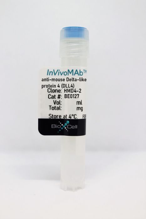
InVivoMAb anti-mouse Delta-like protein 4 (DLL4)
Product Details
The HMD4-2 monoclonal antibody reacts with mouse Delta-like protein 4 (DLL4) one of many Notch ligands. DLL4 is expressed by vascular endothelium, and plays a vital role in embryonic vascular development. The Notch pathway is an important intercellular signaling pathway that plays a major role in controlling cell fate. The HMD4-2 antibody has been shown to neutralize DLL4 in vivo.Specifications
| Isotype | Armenian Hamster IgG, κ |
|---|---|
| Recommended Isotype Control(s) | InVivoMAb polyclonal Armenian hamster IgG |
| Recommended Dilution Buffer | InVivoPure pH 7.0 Dilution Buffer |
| Conjugation | This product is unconjugated. Conjugation is available via our Antibody Conjugation Services. |
| Immunogen | Recombinant mouse DLL4 |
| Reported Applications |
in vivo DLL4 neutralization in vitro DLL4 neutralization |
| Formulation |
PBS, pH 7.0 Contains no stabilizers or preservatives |
| Endotoxin |
<2EU/mg (<0.002EU/μg) Determined by LAL gel clotting assay |
| Purity |
>95% Determined by SDS-PAGE |
| Sterility | 0.2 µm filtration |
| Production | Purified from cell culture supernatant in an animal-free facility |
| Purification | Protein G |
| RRID | AB_10950366 |
| Molecular Weight | 150 kDa |
| Storage | The antibody solution should be stored at the stock concentration at 4°C. Do not freeze. |
Recommended Products
-
Recommended Isotype Control(s)
InVivoMAb polyclonal Armenian hamster IgG
-

Recommended Dilution Buffer
InVivoPure pH 7.0 Dilution Buffer
in vitro DLL4 neutralization
Martinez-Lozada, Z. and M. B. Robinson. (2020). "Reciprocal communication between astrocytes and endothelial cells is required for astrocytic glutamate transporter 1 (GLT-1) expression" Neurochem Int 139: 104787. PubMed
Astrocytes have diverse functions that are supported by their anatomic localization between neurons and blood vessels. One of these functions is the clearance of extracellular glutamate. Astrocytes clear glutamate using two Na(+)-dependent glutamate transporters, GLT-1 (also called EAAT2) and GLAST (also called EAAT1). GLT-1 expression increases during synaptogenesis and is a marker of astrocyte maturation. Over 20 years ago, several groups demonstrated that astrocytes in culture express little or no GLT-1 and that neurons induce expression. We recently demonstrated that co-culturing endothelia with mouse astrocytes also induced expression of GLT-1 and GLAST. These increases were blocked by an inhibitor of γ-secretase. This and other observations are consistent with the hypothesis that Notch signaling is required, but the ligands involved were not identified. In the present study, we used rat astrocyte cultures to further define the mechanisms by which endothelia induce expression of GLT-1 and GLAST. We found that co-cultures of astrocytes and endothelia express higher levels of GLT-1 and GLAST protein and mRNA. That endothelia activate Hes5, a transcription factor target of Notch, in astrocytes. Using recombinant Notch ligands, anti-Notch ligand neutralizing antibodies, and shRNAs, we provide evidence that both Dll1 and Dll4 contribute to endothelia-dependent regulation of GLT-1. We also provide evidence that astrocytes secrete a factor(s) that induces expression of Dll4 in endothelia and that this effect is required for Notch-dependent induction of GLT-1. Together these studies indicate that reciprocal communication between astrocytes and endothelia is required for appropriate astrocyte maturation and that endothelia likely deploy additional non-Notch signals to induce GLT-1.
in vivo DLL4 neutralization
Sakai, M., et al. (2019). "Liver-Derived Signals Sequentially Reprogram Myeloid Enhancers to Initiate and Maintain Kupffer Cell Identity" Immunity 51(4): 655-670.e658. PubMed
Tissue environment plays a powerful role in establishing and maintaining the distinct phenotypes of resident macrophages, but the underlying molecular mechanisms remain poorly understood. Here, we characterized transcriptomic and epigenetic changes in repopulating liver macrophages following acute Kupffer cell depletion as a means to infer signaling pathways and transcription factors that promote Kupffer cell differentiation. We obtained evidence that combinatorial interactions of the Notch ligand DLL4 and transforming growth factor-b (TGF-β) family ligands produced by sinusoidal endothelial cells and endogenous LXR ligands were required for the induction and maintenance of Kupffer cell identity. DLL4 regulation of the Notch transcriptional effector RBPJ activated poised enhancers to rapidly induce LXRα and other Kupffer cell lineage-determining factors. These factors in turn reprogrammed the repopulating liver macrophage enhancer landscape to converge on that of the original resident Kupffer cells. Collectively, these findings provide a framework for understanding how macrophage progenitor cells acquire tissue-specific phenotypes.
in vivo DLL4 neutralization
Fukuda, D., et al. (2012). "Notch ligand delta-like 4 blockade attenuates atherosclerosis and metabolic disorders" Proc Natl Acad Sci U S A 109(27): E1868-1877. PubMed
Atherosclerosis and insulin resistance are major components of the cardiometabolic syndrome, a global health threat associated with a systemic inflammatory state. Notch signaling regulates tissue development and participates in innate and adaptive immunity in adults. The role of Notch signaling in cardiometabolic inflammation, however, remains obscure. We noted that a high-fat, high-cholesterol diet increased expression of the Notch ligand Delta-like 4 (Dll4) in atheromata and fat tissue in LDL-receptor-deficient mice. Blockade of Dll4-Notch signaling using neutralizing anti-Dll4 antibody attenuated the development of atherosclerosis, diminished plaque calcification, improved insulin resistance, and decreased fat accumulation. These changes were accompanied by decreased macrophage accumulation, diminished expression of monocyte chemoattractant protein-1 (MCP-1), and lower levels of nuclear factor-kappaB (NF-kappaB) activation. In vitro cell culture experiments revealed that Dll4-mediated Notch signaling increases MCP-1 expression via NF-kappaB, providing a possible mechanism for in vivo effects. Furthermore, Dll4 skewed macrophages toward a proinflammatory phenotype (“M1”). These results suggest that Dll4-Notch signaling plays a central role in the shared mechanism for the pathogenesis of cardiometabolic disorders.
in vivo DLL4 neutralization
Riella, L. V., et al. (2011). "Blockade of Notch ligand delta1 promotes allograft survival by inhibiting alloreactive Th1 cells and cytotoxic T cell generation" J Immunol 187(9): 4629-4638. PubMed
The Notch signaling pathway has been recently shown to contribute to T cell differentiation in vitro. However, the in vivo function of Notch signaling in transplantation remains unknown. In this study, we investigated the importance of Delta1 in regulating the alloimmune response in vivo. Delta1 expression was upregulated on dendritic cells and monocytes/macrophages upon transplantation in a BALB/c into B6 vascularized cardiac transplant model. Whereas administration of anti-Delta1 mAb only slightly delayed survival of cardiac allografts in this fully MHC-mismatched model, it significantly prolonged graft survival in combination with single-dose CTLA4-Ig or in CD28 knockout recipients. The prolongation of allograft survival was associated with Th2 polarization and a decrease in Th1 and granzyme B-producing cytotoxic T cells. The survival benefit of Delta1 blockade was abrogated after IL-4 neutralization and in STAT6KO recipients, but was maintained in STAT4KO recipients, reinforcing the key role of Th2 cell development in its graft-prolonging effects. To our knowledge, these data demonstrate for the first time an important role of Delta1 in alloimmunity, identifying Delta1 ligand as a potential novel target for immunomodulation in transplantation.
- Mus musculus (House mouse),
DLL4/Notch blockade disrupts mandibular advancement-induced condylar osteogenesis by inhibiting H-type angiogenesis.
In Journal of Oral Rehabilitation on 1 April 2024 by Hu, Y. & Li, H.
PubMed
Blocking Delta-like 4 (DLL4)/Notch has emerged as a promising therapeutic target for the treatment of tumours by deregulating angiogenesis. However, DLL4/Notch serves as a negative regulator of angiogenesis in multiple organs while acting as a positive regulator of H-type angiogenesis in postnatal long bones. Therefore, the effect of DLL4/Notch signalling blockade on mandibular condylar osteogenesis attracted our attention. To explore the effect of blocking DLL4/Notch on mandibular advancement (MA)-induced condylar osteogenesis. Six-week-old young male C57BL/6J mice (n = 40) were randomly divided into four groups: control group, MA group, MA + Anti-DLL4 group and MA + IgG group. Of note, IgG served as the isotype control for the anti-DLL4. The femurs, tibias and mandibular condyles were collected after sacrificing mice on Day 31 for morphology, micro-computed tomography, immunofluorescence, histology and immunohistochemistry evaluation. First, DLL4/Notch blockade shortened femoral length and reduced bone mass by inhibiting H-type angiogenesis. Second, DLL4/Notch blockade disrupted MA-induced condylar head volume and quality by inhibiting H-type angiogenesis. Mechanistically, blocking DLL4/Notch reduced the number of runt-related transcription factor 2+ (RUNX2+ ) early osteoprogenitors and the expression of Noggin protein in the condylar subchondral bone by inhibiting H-type angiogenesis. In addition, blockade of DLL4/Notch also destroyed the condylar cartilage layer. DLL4/Notch blockade results in shortened femurs and osteopenia, as well as impaired MA-induced condylar osteogenic volume and quality in growing mice by inhibiting H-type angiogenesis. Therefore, when blocking DLL4/Notch is used as a treatment target for diseases, attention should be paid to its impact on the bone mass of mandibular condyle. © 2023 John Wiley & Sons Ltd.
- Cell Culture,
- Mus musculus (House mouse),
- Immunology and Microbiology
Mycobacterium tuberculosis impedes CD40-dependent notch signaling to restrict Th17 polarization during infection.
In IScience on 20 May 2022 by Enriquez, A. B., Sia, J. K., et al.
PubMed
Early Th17 responses are necessary to provide protection against Mycobacterium tuberculosis (Mtb). Mtb impedes Th17 polarization by restricting CD40 co-stimulatory pathway on dendritic cells (DCs). We previously demonstrated that engaging CD40 on DCs increased Th17 responses. However, the molecular mechanisms that contributed to Th17 polarization were unknown. Here, we identify the Notch ligand DLL4 as necessary for Th17 polarization and demonstrate that Mtb limits DLL4 on DCs to prevent optimal Th17 responses. Although Mtb infection induced only low levels of DLL4, engaging CD40 on DCs increased DLL4 expression. Antibody blockade of DLL4 on DCs reduced Th17 polarization in vitro and in vivo. In addition, we show that the Mtb Hip1 protease attenuates DLL4 expression on lung DCs by impeding CD40 signaling. Overall, our results demonstrate that Mtb impedes CD40-dependent DLL4 expression to restrict Th17 responses and identify the CD40-DLL4 pathways as targets for developing new Th17-inducing vaccines and adjuvants for tuberculosis.© 2022 The Author(s).
- In Vivo,
- Mus musculus (House mouse),
- Stem Cells and Developmental Biology
A spatial vascular transcriptomic, proteomic, and phosphoproteomic atlas unveils an angiocrine Tie-Wnt signaling axis in the liver.
In Developmental Cell on 7 June 2021 by Inverso, D., Shi, J., et al.
PubMed
Single-cell transcriptomics (scRNA-seq) has revolutionized the understanding of the spatial architecture of tissue structure and function. Advancing the "transcript-centric" view of scRNA-seq analyses is presently restricted by the limited resolution of proteomics and genome-wide techniques to analyze post-translational modifications. Here, by combining spatial cell sorting with transcriptomics and quantitative proteomics/phosphoproteomics, we established the spatially resolved proteome landscape of the liver endothelium, yielding deep mechanistic insight into zonated vascular signaling mechanisms. Phosphorylation of receptor tyrosine kinases was detected preferentially in the central vein area, resulting in an atypical enrichment of tyrosine phosphorylation. Prototypic biological validation identified Tie receptor signaling as a selective and specific regulator of vascular Wnt activity orchestrating angiocrine signaling, thereby controlling hepatocyte function during liver regeneration. Taken together, the study has yielded fundamental insight into the spatial organization of liver endothelial cell signaling. Spatial sorting may be employed as a universally adaptable strategy for multiomic analyses of scRNA-seq-defined cellular (sub)-populations.Copyright © 2021 The Authors. Published by Elsevier Inc. All rights reserved.
- In Vitro,
- Neutralization,
- Mus musculus (House mouse),
- Neuroscience
Reciprocal communication between astrocytes and endothelial cells is required for astrocytic glutamate transporter 1 (GLT-1) expression.
In Neurochemistry International on 1 October 2020 by Martínez-Lozada, Z. & Robinson, M. B.
PubMed
Astrocytes have diverse functions that are supported by their anatomic localization between neurons and blood vessels. One of these functions is the clearance of extracellular glutamate. Astrocytes clear glutamate using two Na+-dependent glutamate transporters, GLT-1 (also called EAAT2) and GLAST (also called EAAT1). GLT-1 expression increases during synaptogenesis and is a marker of astrocyte maturation. Over 20 years ago, several groups demonstrated that astrocytes in culture express little or no GLT-1 and that neurons induce expression. We recently demonstrated that co-culturing endothelia with mouse astrocytes also induced expression of GLT-1 and GLAST. These increases were blocked by an inhibitor of γ-secretase. This and other observations are consistent with the hypothesis that Notch signaling is required, but the ligands involved were not identified. In the present study, we used rat astrocyte cultures to further define the mechanisms by which endothelia induce expression of GLT-1 and GLAST. We found that co-cultures of astrocytes and endothelia express higher levels of GLT-1 and GLAST protein and mRNA. That endothelia activate Hes5, a transcription factor target of Notch, in astrocytes. Using recombinant Notch ligands, anti-Notch ligand neutralizing antibodies, and shRNAs, we provide evidence that both Dll1 and Dll4 contribute to endothelia-dependent regulation of GLT-1. We also provide evidence that astrocytes secrete a factor(s) that induces expression of Dll4 in endothelia and that this effect is required for Notch-dependent induction of GLT-1. Together these studies indicate that reciprocal communication between astrocytes and endothelia is required for appropriate astrocyte maturation and that endothelia likely deploy additional non-Notch signals to induce GLT-1. Copyright © 2020 Elsevier Ltd. All rights reserved.
- Block,
- Mus musculus (House mouse),
- Immunology and Microbiology
Stellate Cells, Hepatocytes, and Endothelial Cells Imprint the Kupffer Cell Identity on Monocytes Colonizing the Liver Macrophage Niche.
In Immunity on 15 October 2019 by Bonnardel, J., T'Jonck, W., et al.
PubMed
Macrophages are strongly adapted to their tissue of residence. Yet, little is known about the cell-cell interactions that imprint the tissue-specific identities of macrophages in their respective niches. Using conditional depletion of liver Kupffer cells, we traced the developmental stages of monocytes differentiating into Kupffer cells and mapped the cellular interactions imprinting the Kupffer cell identity. Kupffer cell loss induced tumor necrosis factor (TNF)- and interleukin-1 (IL-1) receptor-dependent activation of stellate cells and endothelial cells, resulting in the transient production of chemokines and adhesion molecules orchestrating monocyte engraftment. Engrafted circulating monocytes transmigrated into the perisinusoidal space and acquired the liver-associated transcription factors inhibitor of DNA 3 (ID3) and liver X receptor-α (LXR-α). Coordinated interactions with hepatocytes induced ID3 expression, whereas endothelial cells and stellate cells induced LXR-α via a synergistic NOTCH-BMP pathway. This study shows that the Kupffer cell niche is composed of stellate cells, hepatocytes, and endothelial cells that together imprint the liver-specific macrophage identity.Copyright © 2019 The Authors. Published by Elsevier Inc. All rights reserved.
- In Vivo,
- Neutralization,
- Mus musculus (House mouse),
- Immunology and Microbiology
Liver-Derived Signals Sequentially Reprogram Myeloid Enhancers to Initiate and Maintain Kupffer Cell Identity.
In Immunity on 15 October 2019 by Sakai, M., Troutman, T. D., et al.
PubMed
Tissue environment plays a powerful role in establishing and maintaining the distinct phenotypes of resident macrophages, but the underlying molecular mechanisms remain poorly understood. Here, we characterized transcriptomic and epigenetic changes in repopulating liver macrophages following acute Kupffer cell depletion as a means to infer signaling pathways and transcription factors that promote Kupffer cell differentiation. We obtained evidence that combinatorial interactions of the Notch ligand DLL4 and transforming growth factor-b (TGF-β) family ligands produced by sinusoidal endothelial cells and endogenous LXR ligands were required for the induction and maintenance of Kupffer cell identity. DLL4 regulation of the Notch transcriptional effector RBPJ activated poised enhancers to rapidly induce LXRα and other Kupffer cell lineage-determining factors. These factors in turn reprogrammed the repopulating liver macrophage enhancer landscape to converge on that of the original resident Kupffer cells. Collectively, these findings provide a framework for understanding how macrophage progenitor cells acquire tissue-specific phenotypes. Copyright © 2019 Elsevier Inc. All rights reserved.