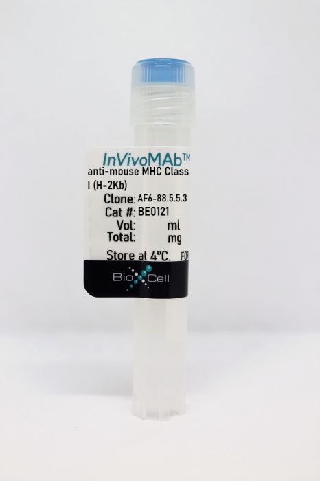
InVivoMAb anti-mouse MHC Class I (H-2Kb)
Product Details
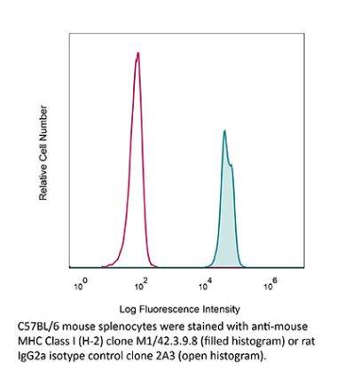
The AF6-88.5.5.3 monoclonal antibody reacts with the mouse H-2Kb MHC class I alloantigen. MHC class I antigens are heterodimers consisting of one alpha chain (44 kDa) associated with β2 microglobulin (11.5 kDa). The antigen is expressed by all nucleated cells at varying levels. MHC Class I molecules present endogenously synthesized antigenic peptides to CD8 T cells.Specifications
| Isotype | Mouse IgG2a, κ |
|---|---|
| Recommended Isotype Control(s) | InVivoMAb mouse IgG2a isotype control, unknown specificity |
| Recommended Dilution Buffer | InVivoPure pH 7.0 Dilution Buffer |
| Conjugation | This product is unconjugated. Conjugation is available via our Antibody Conjugation Services. |
| Immunogen | C57BL/6 mouse splenocytes |
| Reported Applications |
in vivo administration Flow cytometry |
| Formulation |
PBS, pH 7.0 Contains no stabilizers or preservatives |
| Endotoxin |
<2EU/mg (<0.002EU/μg) Determined by LAL gel clotting assay |
| Purity |
>95% Determined by SDS-PAGE |
| Sterility | 0.2 µm filtration |
| Production | Purified from cell culture supernatant in an animal-free facility |
| Purification | Protein G |
| RRID | AB_10950183 |
| Molecular Weight | 150 kDa |
| Storage | The antibody solution should be stored at the stock concentration at 4°C. Do not freeze. |
Recommended Products
-
Recommended Isotype Control(s)
InVivoMAb mouse IgG2a isotype control, unknown specificity
-

Recommended Dilution Buffer
InVivoPure pH 7.0 Dilution Buffer
Flow Cytometry
Paschall, A. V., et al. (2015). "IFN regulatory factor 8 represses GM-CSF expression in T cells to affect myeloid cell lineage differentiation" J Immunol 194(5): 2369-2379. PubMed
During hematopoiesis, hematopoietic stem cells constantly differentiate into granulocytes and macrophages via a distinct differentiation program that is tightly controlled by myeloid lineage-specific transcription factors. Mice with a null mutation of IFN regulatory factor 8 (IRF8) accumulate CD11b(+)Gr1(+) myeloid cells that phenotypically and functionally resemble tumor-induced myeloid-derived suppressor cells (MDSCs), indicating an essential role of IRF8 in myeloid cell lineage differentiation. However, IRF8 is expressed in various types of immune cells, and whether IRF8 functions intrinsically or extrinsically in regulation of myeloid cell lineage differentiation is not fully understood. In this study, we report an intriguing finding that, although IRF8-deficient mice exhibit deregulated myeloid cell differentiation and resultant accumulation of CD11b(+)Gr1(+) MDSCs, surprisingly, mice with IRF8 deficiency only in myeloid cells exhibit no abnormal myeloid cell lineage differentiation. Instead, mice with IRF8 deficiency only in T cells exhibited deregulated myeloid cell differentiation and MDSC accumulation. We further demonstrated that IRF8-deficient T cells exhibit elevated GM-CSF expression and secretion. Treatment of mice with GM-CSF increased MDSC accumulation, and adoptive transfer of IRF8-deficient T cells, but not GM-CSF-deficient T cells, increased MDSC accumulation in the recipient chimeric mice. Moreover, overexpression of IRF8 decreased GM-CSF expression in T cells. Our data determine that, in addition to its intrinsic function as an apoptosis regulator in myeloid cells, IRF8 also acts extrinsically to repress GM-CSF expression in T cells to control myeloid cell lineage differentiation, revealing a novel mechanism that the adaptive immune component of the immune system regulates the innate immune cell myelopoiesis in vivo.
Flow Cytometry
Penaloza-MacMaster, P., et al. (2014). "Interplay between regulatory T cells and PD-1 in modulating T cell exhaustion and viral control during chronic LCMV infection" J Exp Med 211(9): 1905-1918. PubMed
Regulatory T (T reg) cells are critical for preventing autoimmunity mediated by self-reactive T cells, but their role in modulating immune responses during chronic viral infection is not well defined. To address this question and to investigate a role for T reg cells in exhaustion of virus-specific CD8 T cells, we depleted T reg cells in mice chronically infected with lymphocytic choriomeningitis virus (LCMV). T reg cell ablation resulted in 10-100-fold expansion of functional LCMV-specific CD8 T cells. Rescue of exhausted CD8 T cells was dependent on cognate antigen, B7 costimulation, and conventional CD4 T cells. Despite the striking recovery of LCMV-specific CD8 T cell responses, T reg cell depletion failed to diminish viral load. Interestingly, T reg cell ablation triggered up-regulation of the molecule programmed cell death ligand-1 (PD-L1), which upon binding PD-1 on T cells delivers inhibitory signals. Increased PD-L1 expression was observed especially on LCMV-infected cells, and combining T reg cell depletion with PD-L1 blockade resulted in a significant reduction in viral titers, which was more pronounced than that upon PD-L1 blockade alone. These results suggest that T reg cells effectively maintain CD8 T cell exhaustion, but blockade of the PD-1 inhibitory pathway is critical for elimination of infected cells.
Flow Cytometry
Kearl, T. J., et al. (2013). "Programmed death receptor-1/programmed death receptor ligand-1 blockade after transient lymphodepletion to treat myeloma" J Immunol 190(11): 5620-5628. PubMed
Early phase clinical trials targeting the programmed death receptor-1/ligand-1 (PD-1/PD-L1) pathway to overcome tumor-mediated immunosuppression have reported promising results for a variety of cancers. This pathway appears to play an important role in the failure of immune reactivity to malignant plasma cells in multiple myeloma patients, as the tumor cells express relatively high levels of PD-L1, and T cells show increased PD-1 expression. In the current study, we demonstrate that PD-1/PD-L1 blockade with a PD-L1-specific Ab elicits rejection of a murine myeloma when combined with lymphodepleting irradiation. This particular combined approach by itself has not previously been shown to be efficacious in other tumor models. The antitumor effect of lymphodepletion/anti-PD-L1 therapy was most robust when tumor Ag-experienced T cells were present either through cell transfer or survival after nonmyeloablative irradiation. In vivo depletion of CD4 or CD8 T cells completely eliminated antitumor efficacy of the lymphodepletion/anti-PD-L1 therapy, indicating that both T cell subsets are necessary for tumor rejection. Elimination of myeloma by T cells occurs relatively quickly as tumor cells in the bone marrow were nearly nondetectable by 5 d after the first anti-PD-L1 treatment, suggesting that antimyeloma reactivity is primarily mediated by preactivated T cells, rather than newly generated myeloma-reactive T cells. Anti-PD-L1 plus lymphodepletion failed to improve survival in two solid tumor models, but demonstrated significant efficacy in two hematologic malignancy models. In summary, our results support the clinical testing of lymphodepletion and PD-1/PD-L1 blockade as a novel approach for improving the survival of patients with multiple myeloma.
in vivo administration
Takenaka, M., et al. (2012). "Complement activation is not required for obliterative airway disease induced by antibodies to major histocompatibility complex class I: Implications for chronic lung rejection" J Heart Lung Transplant 31(11): 1214-1222. PubMed
BACKGROUND: The role of non-complement activating antibodies (ncAbs) to mismatched donor human leukocyte antigen (HLA) in the pathogenesis of chronic lung rejection is not known. We used a murine model of obliterative airway disease (OAD) induced by Abs to major histocompatibility major histocompatibility complex (MHC) class I and serum from donor-specific Abs developed in human lung transplant (LTx) recipients to test the role of ncAbs in the development of OAD and bronchiolitis obliterans syndrome (BOS). METHODS: Anti-MHC ncAbs were administered intrabronchially in B.10 mice or in C3 knockout (C3KO) mice. Lungs were analyzed by histopathology. Lymphocytes secreting interleukin (IL)-17, interferon-gamma, or IL-10 to collagen V and K-alpha1 tubulin (Kalpha1T) were enumerated by enzyme-linked immunospot assay. Serum antibodies to collagen V and Kalpha1T were determined by enzyme-linked immunosorbent assay. Cytokine and growth factor expression in lungs was determined by real-time polymerase chain reaction. Donor-specific Abs from patients with BOS and control BOS-negative LTx recipients were analyzed by C1q assay. RESULTS: Administration of ncAbs in B.10 mice or C3KO resulted in OAD lesions. There were significant increases in IL-17- and interferon-gamma-secreting cells to collagen V and Kalpha1T, along with serum Abs to these antigens. There was also augmented expression of monocyte chemotactic protein-1, IL-6, IL-1beta, vascular endothelial growth factor, transforming growth factor-beta, and fibroblastic growth factor in mice administered ncAbs by Day 3. Among 5 LTx recipients with BOS, only 1 had C1q binding donor-specific Abs. CONCLUSION: Complement activation by Abs to MHC class I is not required for development of OAD and human BOS. Therefore, anti-MHC binding to epithelial and endothelial cells can directly activate pro-fibrotic and pro-inflammatory cascades leading to immune response to self-antigens and chronic rejection.
Flow Cytometry
Shaikh, S. R., et al. (2009). "Docosahexaenoic acid modifies the clustering and size of lipid rafts and the lateral organization and surface expression of MHC class I of EL4 cells" J Nutr 139(9): 1632-1639. PubMed
An emerging molecular mechanism by which docosahexaenoic acid (DHA) exerts its effects is modification of lipid raft organization. The biophysical model, based on studies with liposomes, shows that DHA avoids lipid rafts because of steric incompatibility between DHA and cholesterol. The model predicts that DHA does not directly modify rafts; rather, it incorporates into nonrafts to modify the lateral organization and/or conformation of membrane proteins, such as the major histocompatibility complex (MHC) class I. Here, we tested predictions of the model at a cellular level by incorporating oleic acid, eicosapentaenoic acid (EPA), and DHA, compared with a bovine serum albumin (BSA) control, into the membranes of EL4 cells. Quantitative microscopy showed that DHA, but not EPA, treatment, relative to the BSA control diminished lipid raft clustering and increased their size. Approximately 30% of DHA was incorporated directly into rafts without changing the distribution of cholesterol between rafts and nonrafts. Quantification of fluorescence colocalization images showed that DHA selectively altered MHC class I lateral organization by increasing the fraction of the nonraft protein into rafts compared with BSA. Both DHA and EPA treatments increased antibody binding to MHC class I compared with BSA. Antibody titration showed that DHA and EPA did not change MHC I conformation but increased total surface levels relative to BSA. Taken together, our findings are not in agreement with the biophysical model. Therefore, we propose a model that reconciles contradictory viewpoints from biophysical and cellular studies to explain how DHA modifies lipid rafts on several length scales. Our study supports the notion that rafts are an important target of DHA’s mode of action.
- Mus musculus (House mouse),
Protection of cell therapeutics from antibody-mediated killing by CD64 overexpression.
In Nature Biotechnology on 1 May 2023 by Gravina, A., Tediashvili, G., et al.
PubMed
Allogeneic cell therapeutics for cancer therapy or regenerative medicine are susceptible to antibody-mediated killing, which diminishes their efficacy. Here we report a strategy to protect cells from antibody-mediated killing that relies on engineered overexpression of the IgG receptor CD64. We show that human and mouse iPSC-derived endothelial cells (iECs) overexpressing CD64 escape antibody-dependent cellular cytotoxicity (ADCC) and complement-dependent cytotoxicity from IgG antibodies in vitro and in ADCC-enabled mice. When CD64 expression was combined with hypoimmune genetic modifications known to protect against cellular immunity, B2M-/-CIITA-/- CD47/CD64-transgenic iECs were resistant to both IgG antibody-mediated and cellular immune killing in vitro and in humanized mice. Mechanistic studies demonstrated that CD64 or its intracellularly truncated analog CD64t effectively capture monomeric IgG and occupy their Fc, and the IgG bind and occupy their target antigens. In three applications of the approach, human CD64t-engineered thyroid epithelial cells, pancreatic beta cells and CAR T cells withstood clinically relevant levels of graft-directed antibodies and fully evaded antibody-mediated killing. © 2023. The Author(s).
- Mus musculus (House mouse),
- Immunology and Microbiology
Blockade of PD-1/PD-L1 Pathway Enhances the Antigen-Presenting Capacity of Fibrocytes.
In The Journal of Immunology on 15 March 2021 by Afroj, T., Mitsuhashi, A., et al.
PubMed
Fibrocytes, a distinct population of collagen-producing, monocyte-derived cells, are involved in wound healing as well as fibrotic diseases. Recently, fibrocytes have been revealed to play a role in the tumor microenvironment, particularly under antiangiogenic therapy. In addition, combination cancer immunotherapy with immune checkpoint inhibitor and antiangiogenic agents have been developed for various cancers in the clinical setting, although the immunological background is not clear. In the current study, we aimed to determine the function of fibrocytes in tumor immunity induced by immune checkpoint inhibitor therapy. Human and murine fibrocytes were generated from PBMCs and lungs, respectively. The expression of costimulatory and inhibitory molecules on fibrocytes was examined by flow cytometry. The stimulation of CD8+ T cells by fibrocytes was examined in MLRs with a 3H-thymidine incorporation assay. Fibrocytes expressed CD80low and CD86high as a costimulatory molecule, and expressed PD-L1high, but not PD-L2, as a coinhibitory molecule. Without any stimulation, fibrocytes strongly enhanced the proliferation of CD8+ T cells in mice and humans. Treatment with anti-CD86 and -CD54 Abs inhibited the growth of CD8+ T cells induced by fibrocytes. Anti-PD-L1 Ab further enhanced the proliferation of CD8+ T cells, even in the OVA-specific MLR with OT-1Rag-/- mice. Importantly, fibrocytes derived from PBMCs of patients with lung adenocarcinoma or murine MC38 tumors augmented the proliferation of CD8+ T cells with PD-L1 blockade. These results suggest that fibrocytes infiltrating tumor sites may play a role in the antitumor immunity mediated by CD8+ T cells when the activity is further enhanced by PD-L1/PD-1 blockade. Copyright © 2021 by The American Association of Immunologists, Inc.
- Immunology and Microbiology
Activation of 4-1BBL+ B cells with CD40 agonism and IFNγ elicits potent immunity against glioblastoma.
In The Journal of Experimental Medicine on 4 January 2021 by Lee-Chang, C., Miska, J., et al.
PubMed
Immunotherapy has revolutionized the treatment of many tumors. However, most glioblastoma (GBM) patients have not, so far, benefited from such successes. With the goal of exploring ways to boost anti-GBM immunity, we developed a B cell-based vaccine (BVax) that consists of 4-1BBL+ B cells activated with CD40 agonism and IFNγ stimulation. BVax migrates to key secondary lymphoid organs and is proficient at antigen cross-presentation, which promotes both the survival and the functionality of CD8+ T cells. A combination of radiation, BVax, and PD-L1 blockade conferred tumor eradication in 80% of treated tumor-bearing animals. This treatment elicited immunological memory that prevented the growth of new tumors upon subsequent reinjection in cured mice. GBM patient-derived BVax was successful in activating autologous CD8+ T cells; these T cells showed a strong ability to kill autologous glioma cells. Our study provides an efficient alternative to current immunotherapeutic approaches that can be readily translated to the clinic. © 2020 Lee-Chang et al.
Biological and structural characterization of murine TRALI antibody reveals increased Fc-mediated complement activation.
In Blood Advances on 25 August 2020 by Zeeuw van der Laan, E. A. N., van der Velden, S., et al.
PubMed
Transfusion-related acute lung injury (TRALI) remains a leading cause of transfusion-related deaths. In most cases, anti-leukocyte antibodies in the transfusion product trigger TRALI, but not all anti-leukocyte antibodies cause TRALI. It has been shown that the anti-major histocompatibility complex (MHC) class I antibody 34-1-2S (anti-H-2Kd) causes TRALI in BALB/c mice (MHC class I haplotype H-2Kd), whereas SF1.1.10 (anti-H-2Kd) does not. In C57BL/6 mice (MHC class I haplotype H-2Kb), TRALI only occurs when anti-MHC class I antibody AF6-88.5.5.3 (anti-H-2Kb) is administered together with a high dose of 34-1-2S. It remains unknown which specific antibody characteristics are responsible for eliciting TRALI. We therefore investigated several biological and structural features of 34-1-2S compared with other anti-MHC class I antibodies, which on their own do not cause TRALI: SF1.1.10 and AF6-88.5.5.3. No substantial differences were observed between the TRALI-causing 34-1-2S and the TRALI-resistant SF1.1.10 regarding binding affinity to H-2Kd. Regarding binding affinity to H-2Kb, only AF6-88.5.5.3 potently bound to H-2Kb, whereas 34-1-2S exhibited weak but significant cross-reactivity. Furthermore, the binding affinity to FcγRs as well as the Fc glycan composition seemed to be similar for all antibodies. Similar Fc glycosylation profiles were also observed for human TRALI-causing donor anti-HLA antibodies compared with human anti-HLA antibodies from control donors. 34-1-2S, however, displayed superior complement activation capacity, which was fully Fc dependent and not significantly dependent on Fc glycosylation. We conclude that TRALI induction is not correlated with Fab- and Fc-binding affinities for antigen and FcγRs, respectively, nor with the composition of Fc glycans; but increased Fc-mediated complement activation is correlated with TRALI induction. © 2020 by The American Society of Hematology.
- In Vivo,
- Mus musculus (House mouse),
- Cancer Research,
- Immunology and Microbiology
ICOS Costimulation at the Tumor Site in Combination with CTLA-4 Blockade Therapy Elicits Strong Tumor Immunity.
In Molecular Therapy on 6 November 2019 by Soldevilla, M. M., Villanueva, H., et al.
PubMed
Cytotoxic T lymphocyte-associated protein 4 (CTLA-4) blockade therapy is able to induce long-lasting antitumor responses in a fraction of cancer patients. Nonetheless, there is still room for improvement in the quest for new therapeutic combinations. ICOS costimulation has been underscored as a possible target to include with CTLA-4 blocking treatment. Herein, we describe an ICOS agonistic aptamer that potentiates T cell activation and induces stronger antitumor responses when locally injected at the tumor site in combination with anti-CTLA-4 antibody in different tumor models. Furthermore, ICOS agonistic aptamer was engineered as a bi-specific tumor-targeting aptamer to reach any disseminated tumor lesions after systemic injection. Treatment with the bi-specific aptamer in combination with CTLA-4 blockade showed strong antitumor immunity, even in a melanoma tumor model where CTLA-4 treatment alone did not display any significant therapeutic benefit. Thus, this work provides strong support for the development of combinatorial therapies involving anti-CTLA-4 blockade and ICOS agonist tumor-targeting agents.Copyright © 2019 The Author(s). Published by Elsevier Inc. All rights reserved.
- Endocrinology and Physiology,
- Immunology and Microbiology
Paracrine costimulation of IFN-γ signaling by integrins modulates CD8 T cell differentiation.
In Proceedings of the National Academy of Sciences of the United States of America on 6 November 2018 by Krummel, M., Mahale, J. N., et al.
PubMed
The cytokine IFN-γ is a critical regulator of immune system development and function. Almost all leukocytes express the receptor for IFN-γ, yet each cell type elicits a different response to this cytokine. Cell type-specific effects of IFN-γ make it difficult to predict the outcomes of the systemic IFN-γ blockade and limit its clinical application, despite many years of research. To better understand the cell-cell interactions and cofactors that specify IFN-γ functions, we focused on the function of IFN-γ on CD8 T cell differentiation. We demonstrated that during bacterial infection, IFN-γ is a dominant paracrine trigger that skews CD8 T cell differentiation toward memory. This skewing is preferentially driven by contact-dependent T cell-T cell (T-T) interactions and the localized IFN-γ secretion among activated CD8 T cells in a unique splenic microenvironment, and is less sensitive to concurrent IFN-γ production by other immune cell populations such as natural killer (NK) cells. Modulation of CD8 T cell differentiation by IFN-γ relies on a nonconventional IFN-γ outcome that occurs specifically within 24 hours following infection. This is driven by IFN-γ costimulation by integrins at T-T synapses, and leads to synergistic phosphorylation of the proximal STAT1 molecule and accelerated IL-2 receptor down-regulation. This study provides evidence of the importance of context-dependent cytokine signaling and gives another example of how cell clusters and the microenvironment drive unique biology.
- Immunology and Microbiology
Intermittent Casup>2+/sup> signals mediated by Orai1 regulate basal T cell motility
Preprint on BioRxiv : the Preprint Server for Biology on 1 November 2017 by Dong, T. X., Othy, S., et al.
PubMed
Ca 2+ influx through Orai1 channels is crucial for several T cell functions, but a role in regulating basal cellular motility has not been described. Here we show that inhibition of Orai1 channel activity increases average cell velocities by reducing the frequency of pauses in human T cells migrating through confined spaces, even in the absence of extrinsic cell contacts or antigen recognition. Utilizing a novel ratiometric genetically encoded cytosolic Ca 2+ indicator, Salsa6f, which permits real-time monitoring of cytosolic Ca 2+ along with cell motility, we show that spontaneous pauses during T cell motility in vitro and in vivo coincide with episodes of cytosolic Ca 2+ signaling. Furthermore, lymph node T cells exhibited two types of spontaneous Ca 2+ transients: short-duration “sparkles” and longer duration global signals. Our results demonstrate that spontaneous and self-peptide MHC-dependent activation of Orai1 ensures random walk behavior in T cells to optimize immune surveillance.